Inspiration: Why Detection and Diagnosis Come Before Treatment
In real oncology, clinicians never get to see an exact cell-by-cell count inside a patient. Two tumors that appear identical on a basic size scan can respond completely differently to radiotherapy:
- One may hide an oxygen-starved interior (hypoxia,
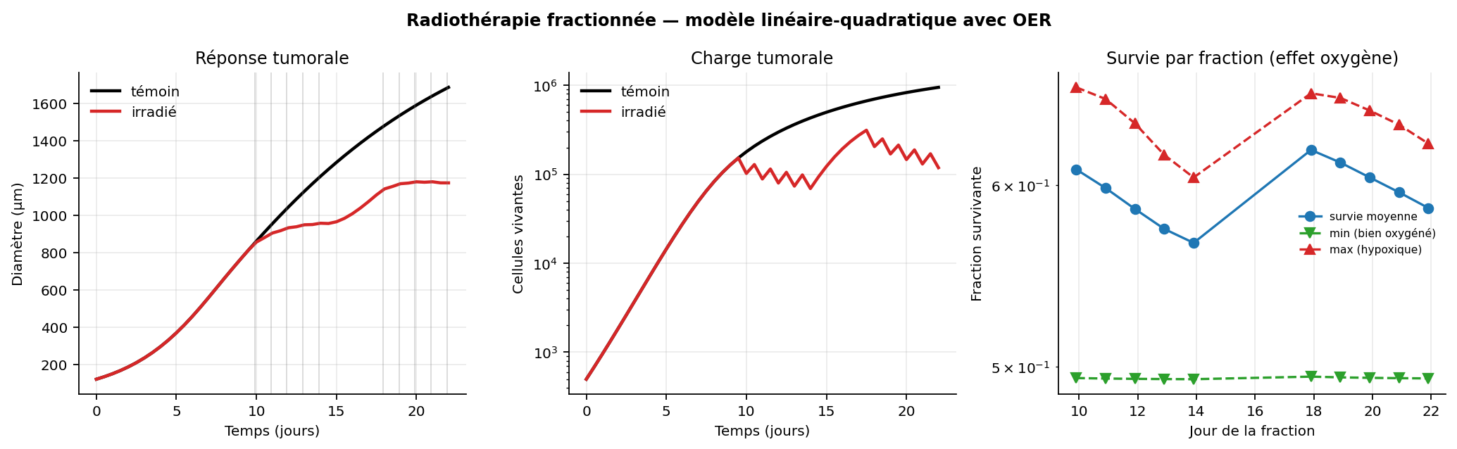
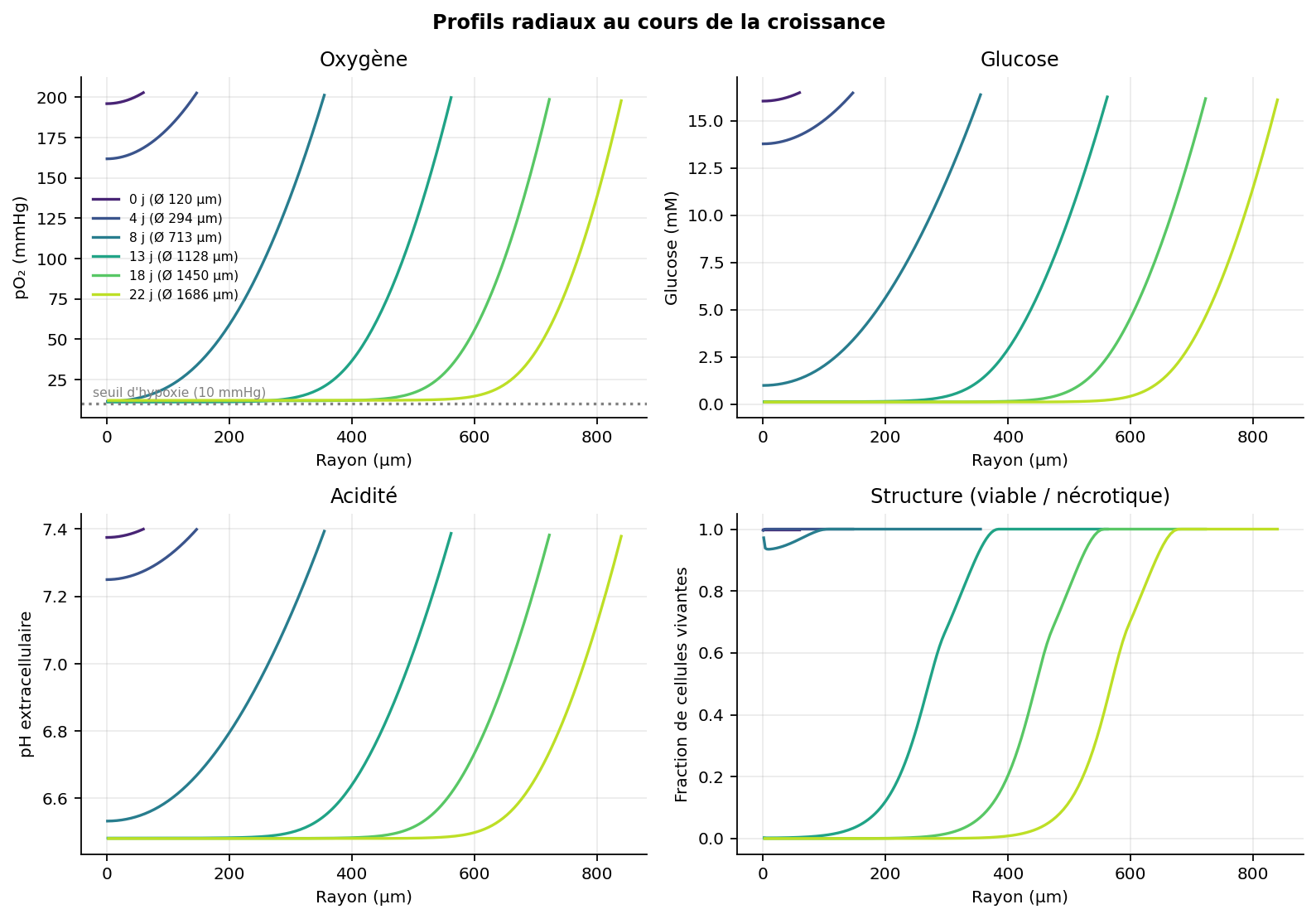
pO2 < 10 mmHg), where the absence of oxygen prevents radiation from fixing DNA damage—making central tumor cells up to 3x more resistant (Alper & Howard-Flanders, 1956). - Another may harbor expanding radioresistant subclones selected by overly aggressive doses (
Britten et al., 1996;Zhang et al., 2017), or sit next to a fragile organ such as the lung (TD50 = 24.5 Gy), where blind high-dose radiation triggers lethal toxicity before shrinking the tumor.
I built Tumor Lab to turn this hidden clinical reality into an interactive, transparent simulation: users step into the role of a clinician who must combine noisy scans, blood biomarkers, oxygen imaging, and biopsies to understand a tumor before deciding how—and whether—to treat it.
Supporting Beginners with Zero Biology Background (Acting on Mentor Feedback)
During preliminary feedback, mentors praised our factual biological modeling and scientific plots, and asked how we could support visitors with zero prior biology or oncology background. We redesigned the user journey on two levels—accessible in seconds for newcomers, while keeping every differential equation inspectable:
- Zero-Friction 1-Click Guest Access: Visitors launch the simulator immediately without creating an account.
- 5-Step Guided Diagnostic Tutorial: Walks first-time players step by step through ordering an Oxygen Scan (
pO2), reading hypoxia reports, delivering a safe2.0 Gyfraction, running a Needle Biopsy, and consulting the neural network advisor. - Plain-Language Mode (
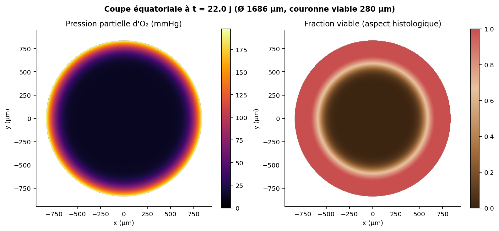
ONby default): Adds clear everyday explanations under technical metrics (µm,Gy,pO2,NTCP), displays a400 µmtarget ring and1 mmscale bar on the live cross-section, translates organ tolerance into plain risk levels (ResilienttoVery Fragile), and explains "What just happened?" after every turn. - 3-Minute Interactive Guide (
/learn): Features everyday visual analogies, a live Dose x Oxygen Radiobiology Sandbox driven by the Linear-Quadratic + OER equations, a jargon-free glossary, and a 3-question self-check quiz. - Plain-Language Figure Summaries & Bilingual (
EN/FR) Interface: All 9 scientific figures include an "In plain words" takeaway box, switchable in one click between English and French.
Diagnostic Tools & Partially Observable Clinical Environment (POMDP)
Tumor Lab formulates clinical decision-making as a Partially Observable Markov Decision Process (POMDP). The underlying PDE engine computes exact spatial concentrations and cell populations (truth_state), but never exposes them to the player or the AI. Instead, decisions rely on four realistic diagnostic channels:
- Morphological Size Scan (
±5%Gaussian measurement noise): Estimates outer tumor diameter (µm) at every turn relative to the< 400 µmdurable control target, without revealing internal necrosis or clonal makeup. - Serum Biomarker (
±20%log-normal noise): A blood-test proxy (analogous to PSA or ctDNA) that tracks whether the hidden viable cell burden is rising or falling across turns despite single-measurement fluctuations. - Quantitative Oxygen Imaging (
pO2Scan, costs 1.5 days): Maps central oxygen tension (mmHg) and the hypoxic fraction (%) of the tumor. Detecting severe hypoxia alerts the user that high single doses will waste healthy-organ tolerance unless fractionated sessions allow tumor shrinkage and reoxygenation (or vascular normalization in the vascular scenario). - Needle Biopsy (
n = 60sampled cells, costs 2.0 days): Draws a stochastic binomial sample from the tumor's internalResistanceDistribution(Alfonso & Berk, 2019) to estimate the percentage of surviving radioresistant clones (alpha_factor < 0.75) and average radiosensitivity, preventing blind dose escalation.
Real Trained Actor-Critic Neural Network (PolicyValueNet)
Rather than faking clinical AI with hardcoded if/else rules, we built and trained a genuine multi-head Actor-Critic neural network from scratch in pure NumPy (tumeur/neural_net.py, tumeur/ai.py, train_ai.py):
1. Network Architecture (5,575 Trainable Parameters)
PolicyValueNet takes a 14-dimensional normalized vector of strictly public clinical observations (x in R^14):
- Noisy diameter ratio, recent diameter velocity, noisy biomarker log-burden, normalized clinical day, cumulative delivered dose (
Gy), current LKBNTCPtoxicity, organ radiosensitivity (TD50 / sparing_factor), time elapsed since lastpO2scan, measured hypoxic fraction and centralpO2, time elapsed since last biopsy, measured resistant clone fraction, vascular regime indicator, and measured vascular activity.
These 14 diagnostic inputs pass through two dense hidden layers (14 -> 64 (tanh) -> 64 (tanh)) into three output heads:
- Discrete Policy Head
pi_theta(a | s): Masked Softmax over the 5 clinical actions (po2_imaging,biopsy,irradiate,antiangiogenic,wait), automatically maskingantiangiogenicin avascular modes. - Continuous Dose Head
mu_theta(s): Sigmoid output scaled to[0.5, 5.0] Gyper fraction. - State-Value Critic
V_theta(s): Linear head predicting expected normalized clinical return from the current diagnostic state.
2. Analytical Backpropagation & Two-Stage Training
- Finite-Difference Gradient Verification: Before training, analytical backpropagation through all three heads and action masks is verified against central finite differences (
epsilon = 1e-5), achieving5.44e-6max relative error on the supervised loss and8.29e-8on the A2C reinforcement learning loss. - Stage 1 — Counterfactual Supervised Pre-Training (140 Epochs, Mini-Batch Adam): Across 653 clinical states generated by counterfactual PDE rollouts (episode-split into 403 training / 250 held-out validation states), Adam (
lr = 2.5e-3, cosine decay) minimizes a composite objective:L = L_CE(pi_theta) + 2.5 * L_MSE(mu_theta) + 0.4 * L_MSE(V_theta) + 1e-4 * ||theta||_2^2Training loss drops from0.997to0.192(92.3%accuracy), while held-out episode validation loss converges from0.937to0.466(86.8%unseen-episode action accuracy). - Stage 2 — On-Policy Advantage Actor-Critic (A2C) Reinforcement Learning: Fine-tuned directly on live stochastic
TumorGametrajectories using standardized advantagesA_t = G_t - V_theta(s_t)and policy entropy regularization (-0.015 H(pi_theta)), teaching the network when ordering a diagnosticpO2scan or biopsy yields higher long-term survival than immediate irradiation.
3. Held-Out Clinical Benchmark & Input-Gradient Explainability
Evaluated on unseen random seeds across all four clinical scenarios at full PDE resolution (n_grid = 160):
PolicyValueNet(Trained Neural Net): 1617.8 mean score (Rank #1, beating every baseline in every scenario).- Hand-Crafted Clinical Heuristic:
1578.4mean score. - Aggressive
5.0 Gy/dayProtocol:1184.2mean score (causes-802.2lethal pulmonary toxicity in the Critical Organ lung scenario). - Random Policy:
1159.3mean score (-847.8on lung). - Untreated (
wait):1100.1mean score (25%control).
Every time a user clicks Ask Neural Net, the interface displays the network's live Softmax probability bars, continuous Gy dose output, Critic value V(s), and input-gradient saliency (|d z_a* / d x_i|)—highlighting in plain language which diagnostic signals (e.g., unmeasured pO2, rising resistant clones on biopsy, or tight lung TD50 margin) drove its decision.
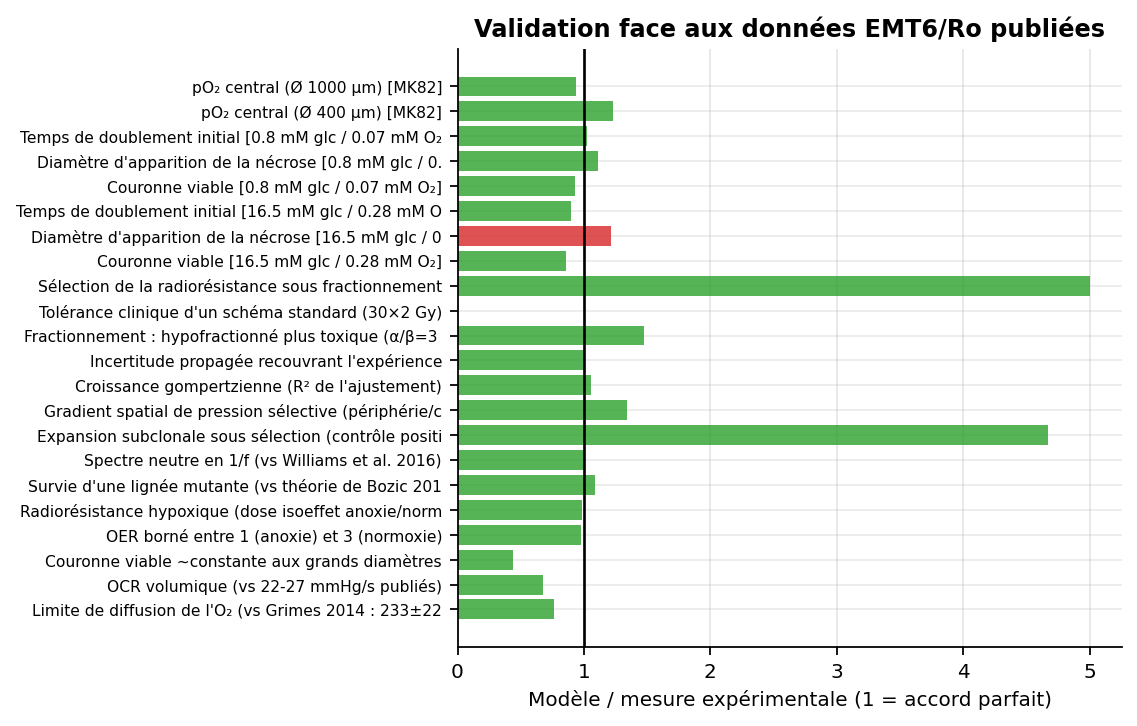
Mechanistic Biophysical Engine & Scientific Validation (31 / 32 Checks)
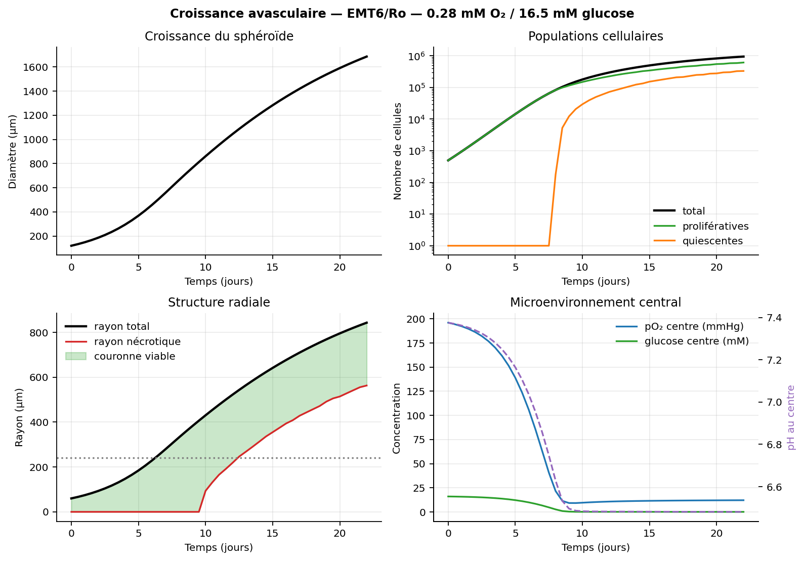
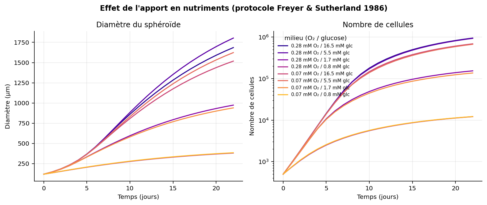
Underneath the web interface, Tumor Lab solves coupled biophysical equations calibrated from 30 peer-reviewed publications (Freyer & Sutherland 1986, Mueller-Klieser 1982, Ward & King 1997, Bertuzzi 2010, Bozic 2010, Williams 2016, Emami & Burman 1991):
- Conservative finite-volume reaction-diffusion PDEs for oxygen, glucose, lactate, and extracellular
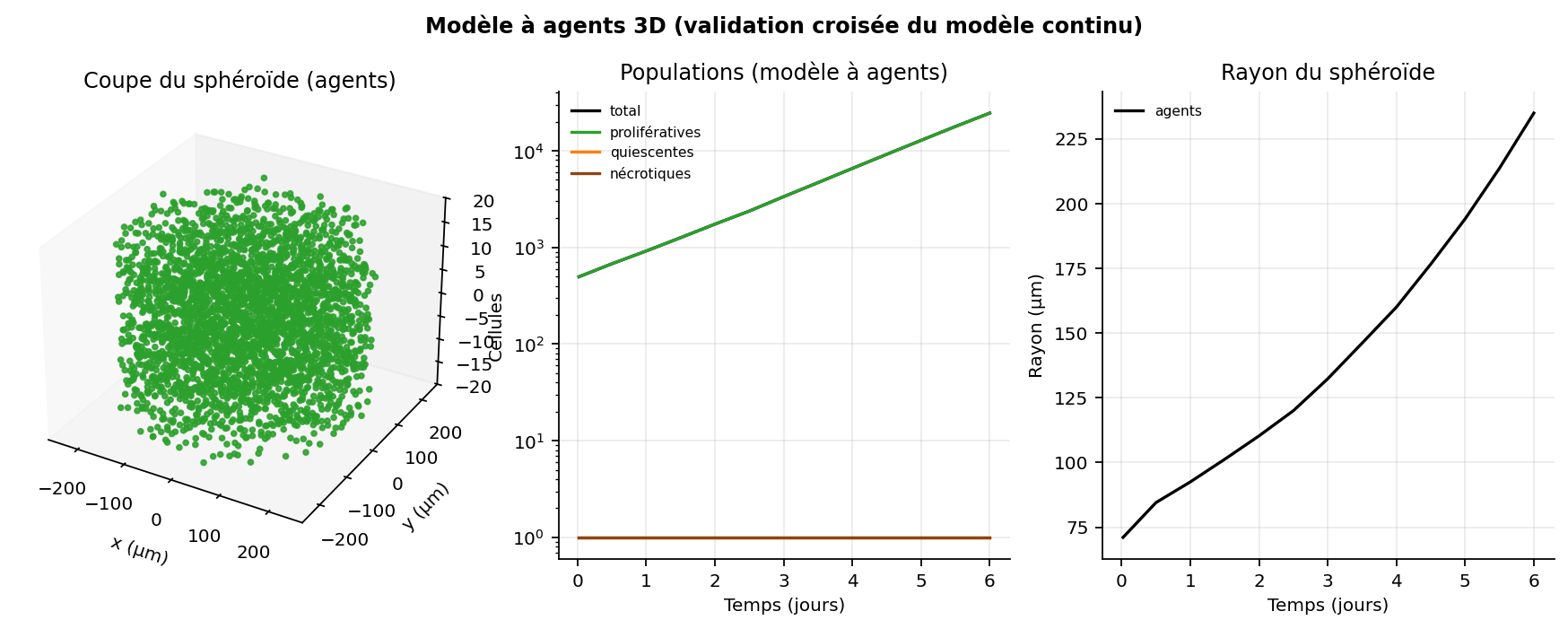
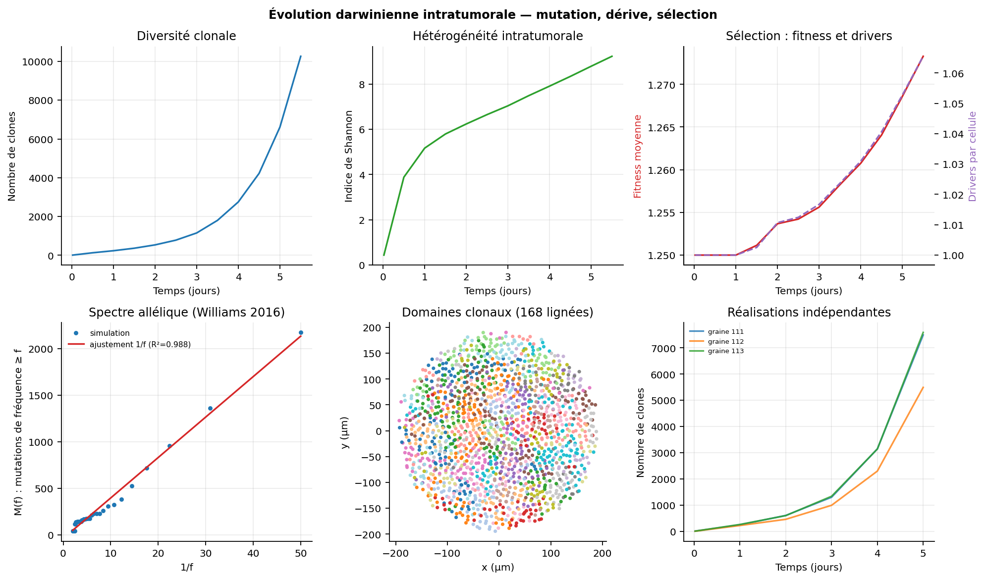
pH, coupled to free-boundary tumor growth (SSP-RK2+van LeerTVD advection) and ATP-threshold necrosis. - Cross-validation against a 3D center-based agent model (
Drasdo & Hohme 2005) and neutral1/fsubclonal evolution (R^2 = 0.988). - 31 out of 32 automated verification & experimental validation checks passed (9/9 numerical solver checks + 22/23 published lab benchmarks). The single discrepancy—rich-medium necrosis onset (
627 µmvs516 µm,Casciari 1992)—is kept and documented transparently without ad-hoc parameter fudging. - Clear scientific boundaries: Strictly separates experimentally validated avascular spheroid physics (
in_vitro_strict) from educational human vascular scenarios (vascular_human).
Medical Disclaimer: Tumor Lab is an educational and theoretical simulation platform. It is not a medical device and must never be used for clinical diagnosis or patient treatment decisions.


Log in or sign up for Devpost to join the conversation.