-
-

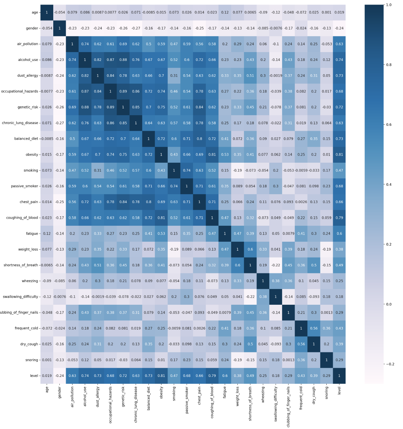
Correlation Plot Between Potential Lung Cancer Factors
-
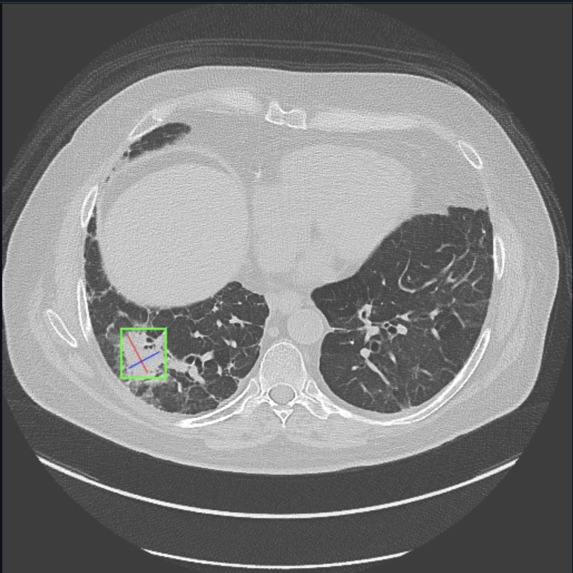
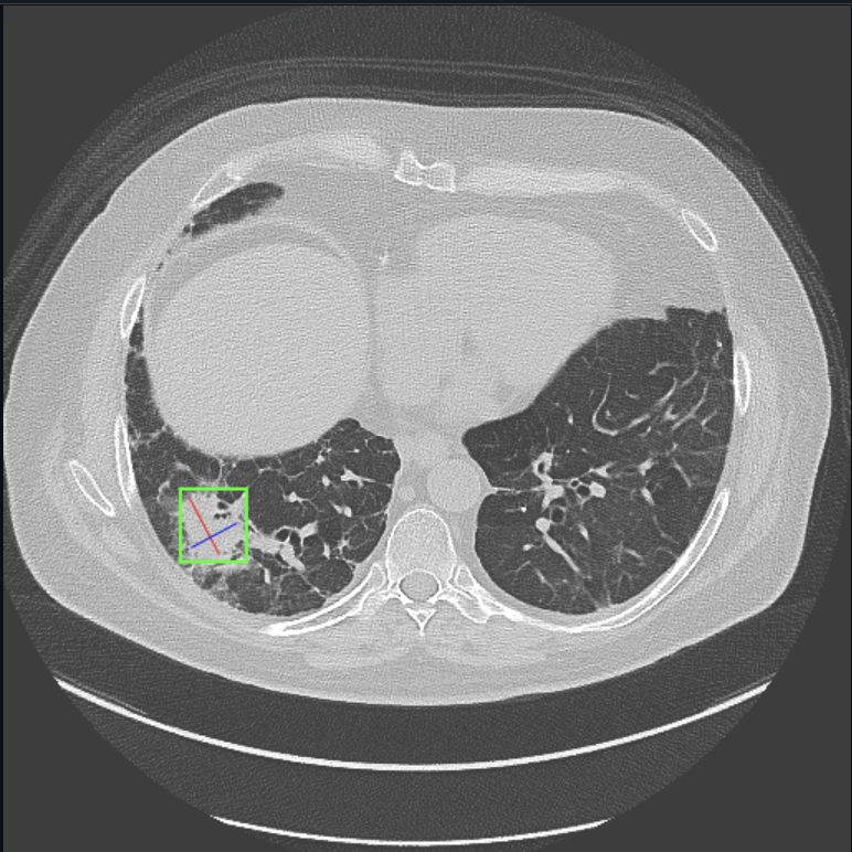
Example of Lung X-Ray Scan Used to Train Model
Inspiration
For this project, we found inspiration from the increasing global health concern surrounding lung cancer, one of the leading causes of cancer-related deaths. Early detection significantly improves survival rates, yet many diagnostic systems still rely on subjective analysis from radiologists. We wanted to create a tool that could assist in early detection using AI to reduce human error and provide a more reliable diagnosis through image detection and data analysis.
What it does
Our project uses AI to analyze X-ray scans/datasets to detect and predict lung cancer.
How we built it
The project was built using the NVIDIA AI Workbench and various AI/ML libraries such as Torch and Scikit-learn. We used an extensive lung cancer dataset from Kaggle (https://www.kaggle.com/code/guslovesmath/lung-cancer-ml-classification/input) to train the Random Forest model, as well as X-ray scans of healthy lungs and those with cancerous growths to train our deep learning model.
Challenges we ran into
One of the biggest challenges we ran into was locating appropriate datasets and X-ray scans to use. Some of our research resulted in medical datasets with class imbalances (i.e., fewer positive cases of lung cancer compared to negative ones) that could skew the model’s performance toward predicting non-cancerous cases, as well as missing data that has a strong correlation with lung cancer. We had to broaden our search and use techniques like data augmentation and weighted loss functions to remove low-correlated data and optimize it for training our model.
Accomplishments that we're proud of
We are proud of the project that we have put together. It took countless days of planning and communicating together, but we were ultimately able to create an AI model that can potentially assist real-time health professionals in detecting lung cancer early. Our product has the chance to save lives, and that is something that makes us hold our heads high.
What we learned
Throughout this project, we learned the intricacies of medical image analysis, particularly when dealing with x-ray scans. We gained a deeper understanding of how to preprocess image data, handle large datasets, and apply machine learning techniques such as convolutional neural networks (CNNs) for image classification. Additionally, we became more familiar with medical imaging datasets and the ethical concerns of using sensitive health data in AI applications. Moreover, we learned the importance of teamwork. By putting our heads together, we were able to boost progress and receive instant feedback for our ideas from one another. Debugging became easier as well as three heads were more likely to spot the error than one person would. Work also turned out to be more fun as we would spend countless hours working together while socializing.
What's next for PulmoAI
We are looking forward to receiving feedback from the judges and our peers on how we can improve our project. Our ultimate goal is for our product to be sophisticated enough to function in the real world and assist health professionals with their lung cancer diagnosis. However, there are some integration features that we were not able to implement due to time constraints, but we are ready to revisit those issues in the near future.
Built With
- csv
- git
- joblib
- jupyterlab
- nvidia
- png
- python
- scikit-learn
- torch


Log in or sign up for Devpost to join the conversation.