Inspiration
The inspiration behind STELLA.ai comes from a personal experience with my mom. She was diagnosed with a mild stroke and admitted to our provincial hospital. During her admission, she underwent a CT scan, and we were anxiously waiting for the radiologists' findings. These findings are crucial as they guide the neurologists in prescribing the appropriate medication. However, the process was slow because the segmentation and identification of stroke-affected areas in the brain CT scan were done manually. At our hospital, a single patient's brain CT scan could have up to six slices, depending on the machine used. This manual segmentation process took nearly two days. This delay made me realize the need for a solution to expedite the process. This situation has also occurred in other healthcare centers, especially those with limited equipment and technologies. Thus, I envisioned a system that Radiologists could use to make brain scan segmentation more time-efficient, allowing healthcare professionals to shift their focus from diagnosis to patient care. This idea eventually led to the development of STELLA.ai.
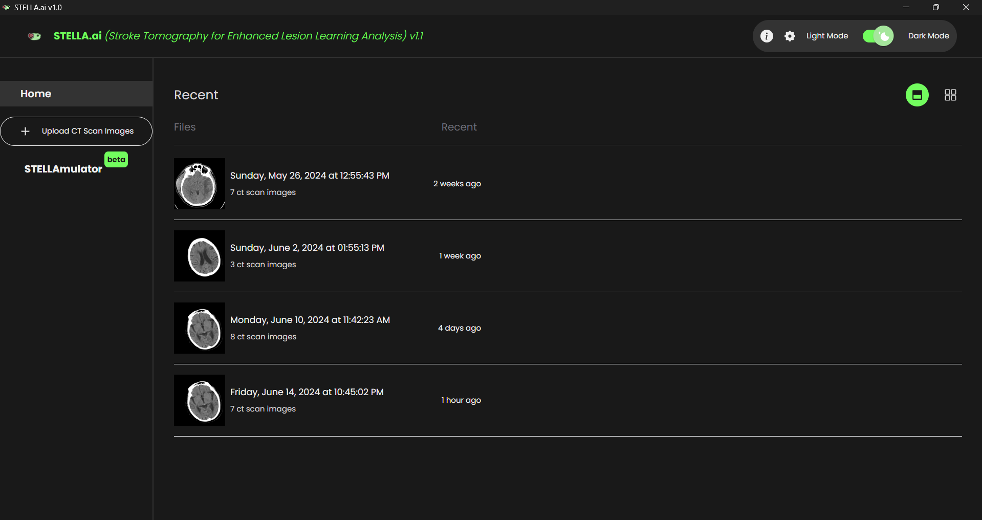
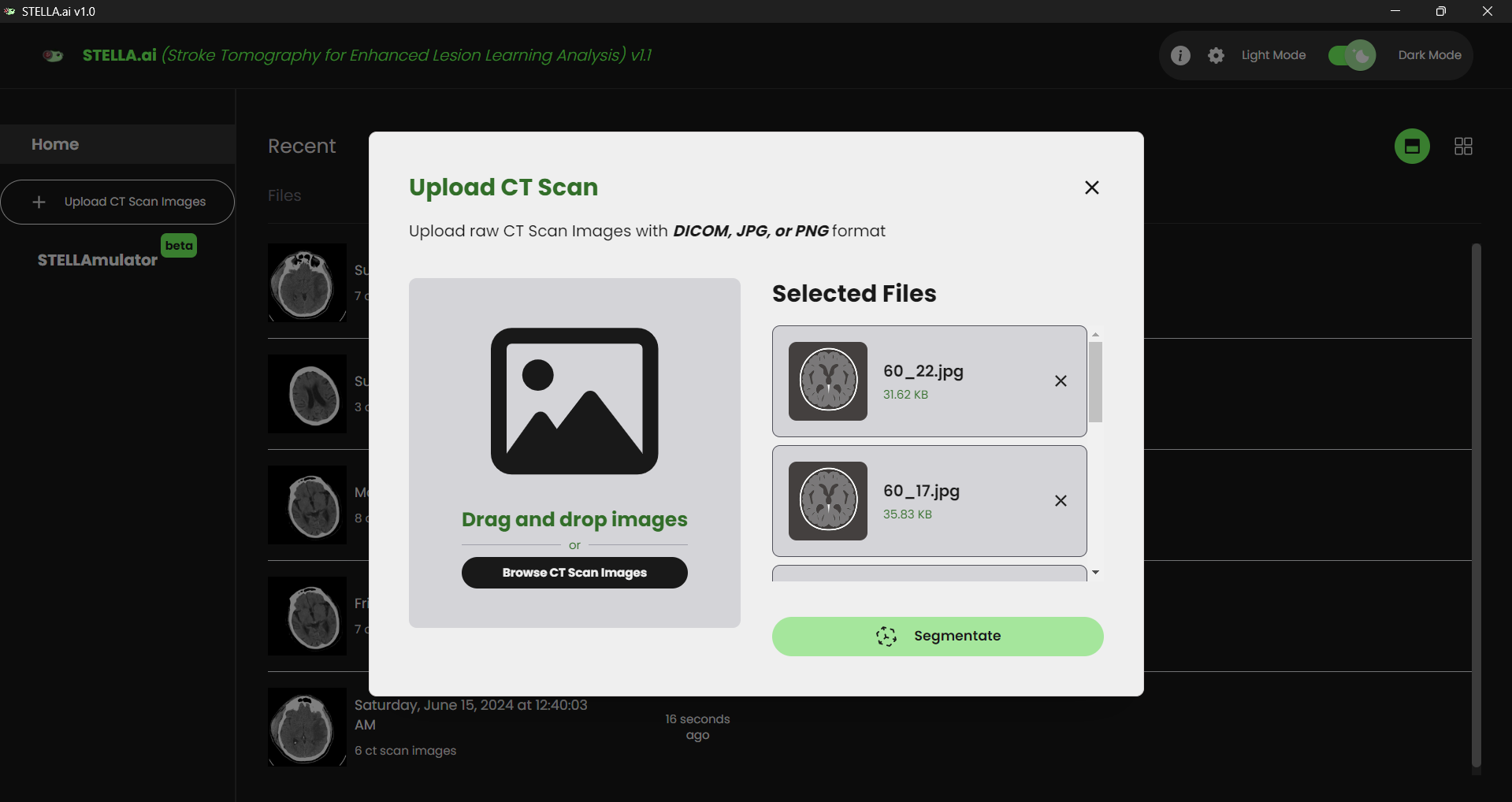
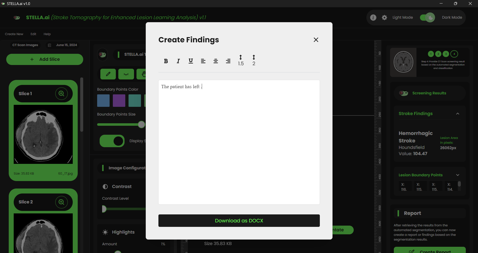
What it does
STELLA will process uploaded Brain CT scans, automatically segmenting brain lesions that could indicate a stroke. In just one click, it will classify the type of stroke, streamlining the diagnostic process.
How the developer built it
I researched various technologies to build a robust intelligent system for radiologists. During my search, I discovered that Electron-Vite is suitable for fast development and easy to use, as it integrates the three pillars of web development. For the backend, I chose FastAPI, a Python framework that efficiently handles server requests and machine learning computations. For training and preprocessing, I utilized OpenCV, NumPy, and PyTorch.
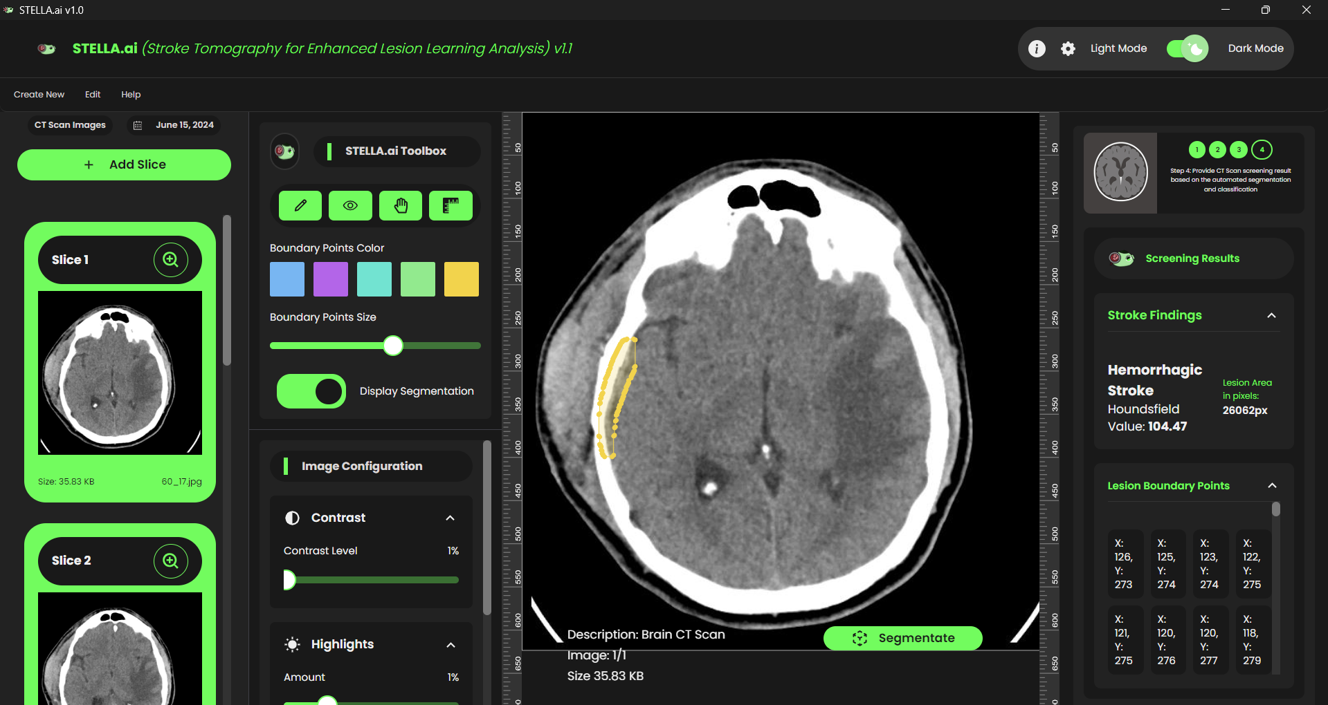
In classifying strokes, I utilized the Hounsfield Scale, a linear transformation of the original linear attenuation coefficient measurement. Here, the radiodensity of distilled water at standard pressure and temperature (STP) is set as 0 Hounsfield units (HU), while air at STP is defined as -1000 HU. This approach aids in determining the density value of lesion tissue by calculating the mean of the segmented area by summating all pixel values and substituting it as the Pixel Value for transforming the CT Scan into Hounsfield Unit using its formula, HU = Pixel Value x Slope x Intercept where the slope and intercept values, typically 1 and -1024, respectively.
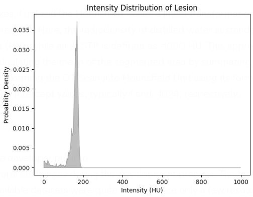
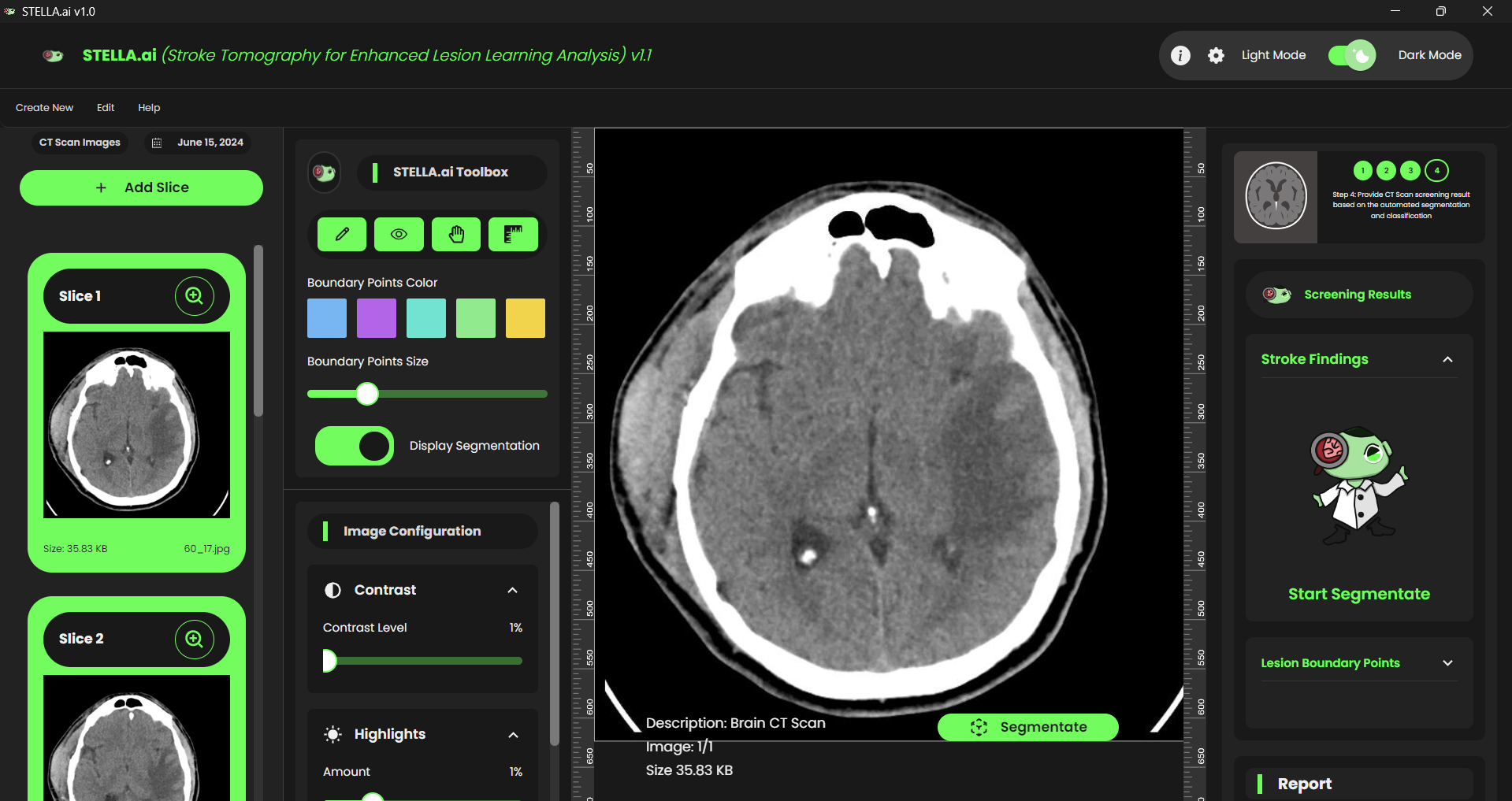
The diagram above shows the Hounsfield Scale sample based on the cropped segment on the CT Scan, which is also the Brain Lesion. It defines intensity thresholds for classifying strokes into ischemic or hemorrhagic types. It will then proceed to calculate the average intensity of the segmented lesion region by analyzing non-zero pixel values within the grayscale image, which represents the lesion. This average intensity is clipped to a specific range (0-1000 Hounsfield Units) to standardize the measurements and plot the intensity distribution, mapping the grayscale values to the lesion's intensity level.
If the intensity falls below the ischemic threshold, the stroke is classified as ischemic, with a confidence level inversely proportional to the intensity. If the intensity is above the hemorrhagic threshold, the stroke is classified as hemorrhagic, with confidence increasing with intensity. If the intensity falls between these thresholds, the stroke is considered unclassified. The classification results, including stroke type, density value, and confidence level, are then returned, providing a scientific basis for distinguishing between ischemic and hemorrhagic strokes based on the lesion's intensity.
Challenges the developer ran into
I encountered several challenges during the development process. The first was obtaining sufficient datasets for training, as the available datasets were quite limited since only a few resources are available specifically for Brain CT Scans that have annotated data for "Stroke." I searched extensively to find enough data for an effective training process. Secondly, my device had limited GPU power, which constrained the training to only 10 epochs.
Accomplishments that the developer we're proud of
Despite the challenges of limited datasets and constrained GPU power, I could still achieve high-quality results in testing and validation. Given the available datasets, the Mask RCNN has achieved an impressive 60% Intersection over Union (IoU) accuracy in segmenting brain lesions. The model produced accurate segmentation, demonstrating its effectiveness despite the training limitations.
What the developer earned
I've gained significant insights while developing this revolutionary system. From training the model to backend development and finally, to frontend integration, every step was a valuable learning experience. I thoroughly enjoyed the entire development process, and seeing the system come to life was incredibly rewarding. I honed my skills in machine learning, improved my understanding of backend frameworks, and enhanced my proficiency in front-end development. This project has expanded my technical knowledge and deepened my appreciation for the impact of intelligent systems in healthcare. Overall, it was a challenging yet fulfilling journey.
What's next for STELLA.ai
Looking ahead, the goal is to patent STELLA.ai and distribute it to healthcare centers. This will enable medical professionals to use it for faster and more efficient stroke diagnosis. STELLA.ai aims to improve patient care and outcomes by streamlining the diagnostic process.
Apart from that is there will be a feature that can be used by students who are studying radiologists for simulating the segmentation for stroke and the AI will assess their performance based on the time taken during segmentation and accuracy of the segmentation.

Log in or sign up for Devpost to join the conversation.