🧠 AI-Based Ophthalmoscopic Retinal Analysis for Myopia Detection
🌟 Inspiration
Myopia has become one of the most common vision problems worldwide, affecting billions of people.
Early detection can prevent severe complications such as retinal detachment and glaucoma,
but clinical diagnosis requires expensive equipment and access to ophthalmologists.
Our inspiration came from the idea of using AI and computer vision to make early myopia screening
more accessible to everyone — especially in regions where medical resources are limited.
We wanted to explore how a Python-based AI system could analyze ophthalmoscopic retinal images
and provide non-diagnostic insights to support vision care.
🧩 How We Built It
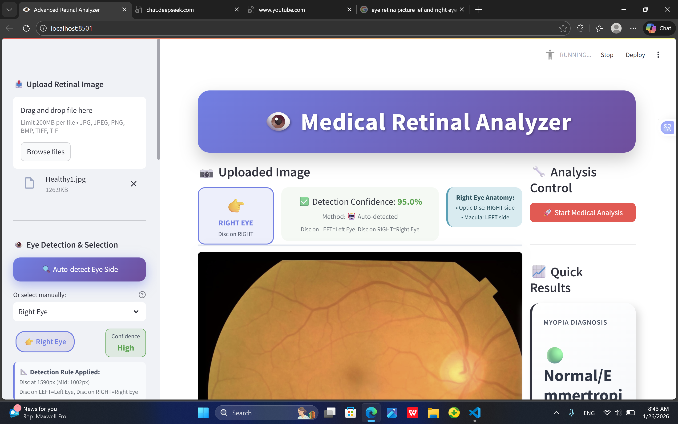
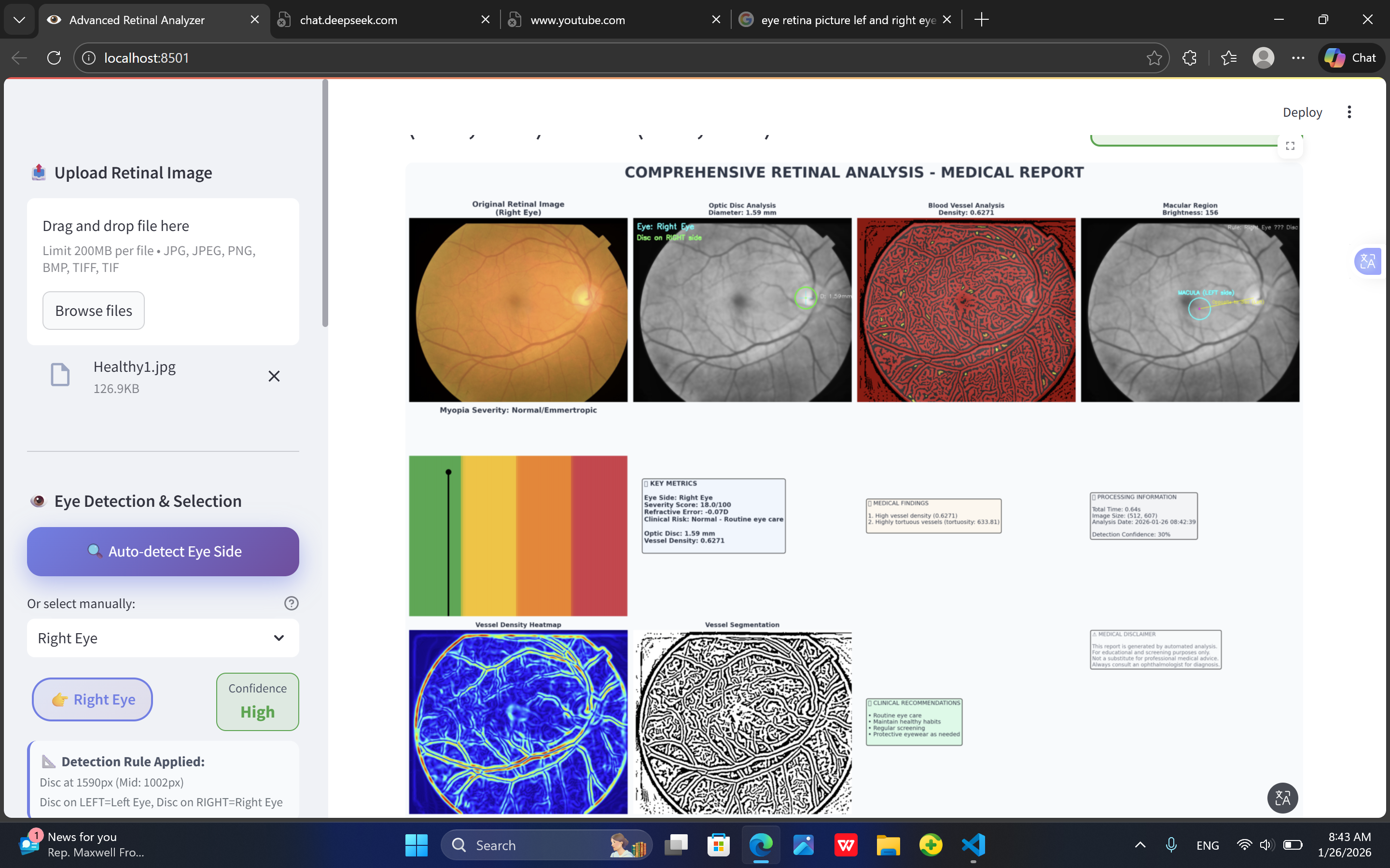
We developed a Python-based AI pipeline that can process and analyze retinal (fundus) images.
The workflow consists of the following stages:
Image Preprocessing
- Conversion to grayscale
- Histogram equalization
- Edge and vessel enhancement using filters such as Sobel and Gaussian
- Conversion to grayscale
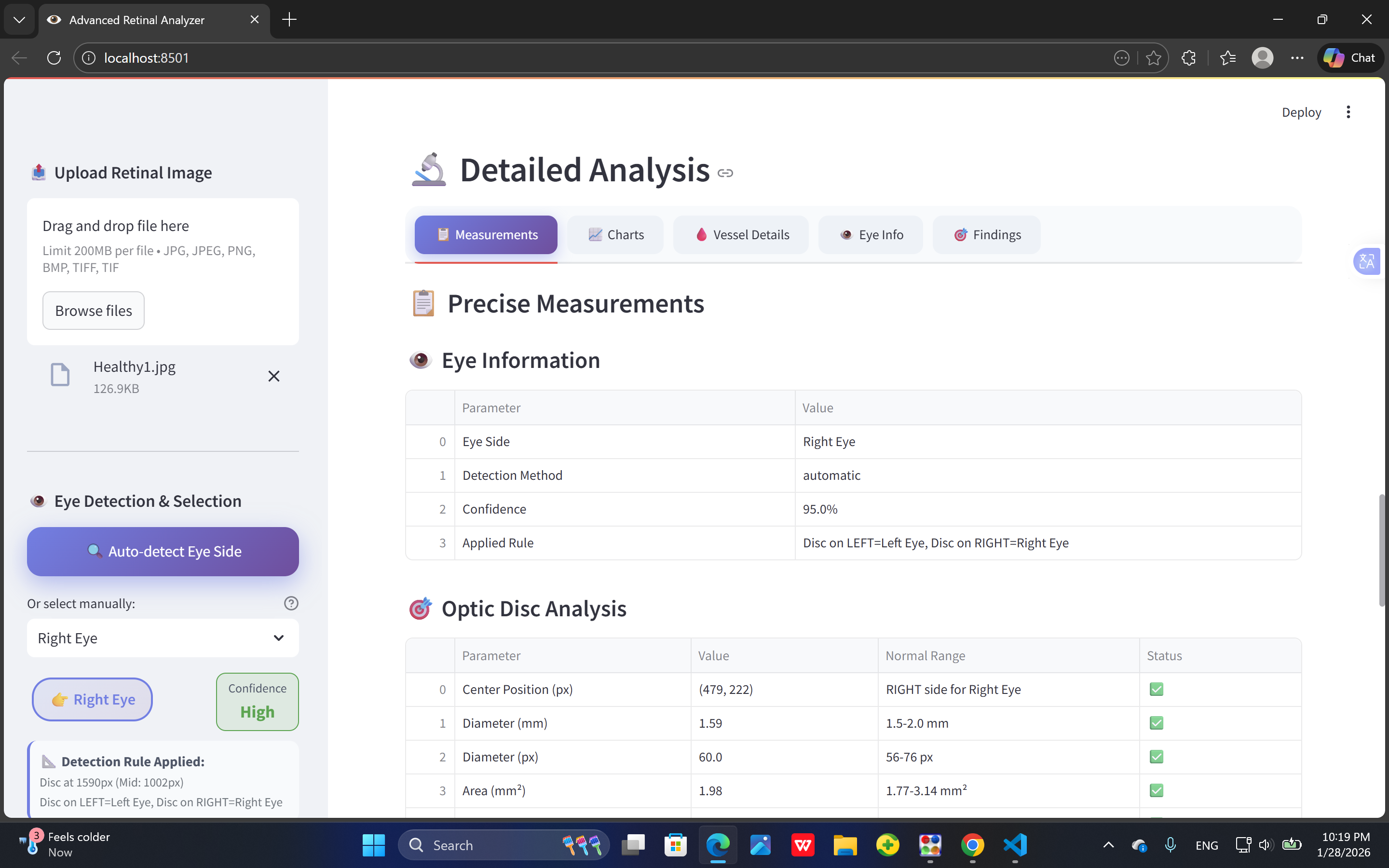
Feature Extraction
We extracted structural and texture-based features that correlate with myopia indicators:- Optic disc shape
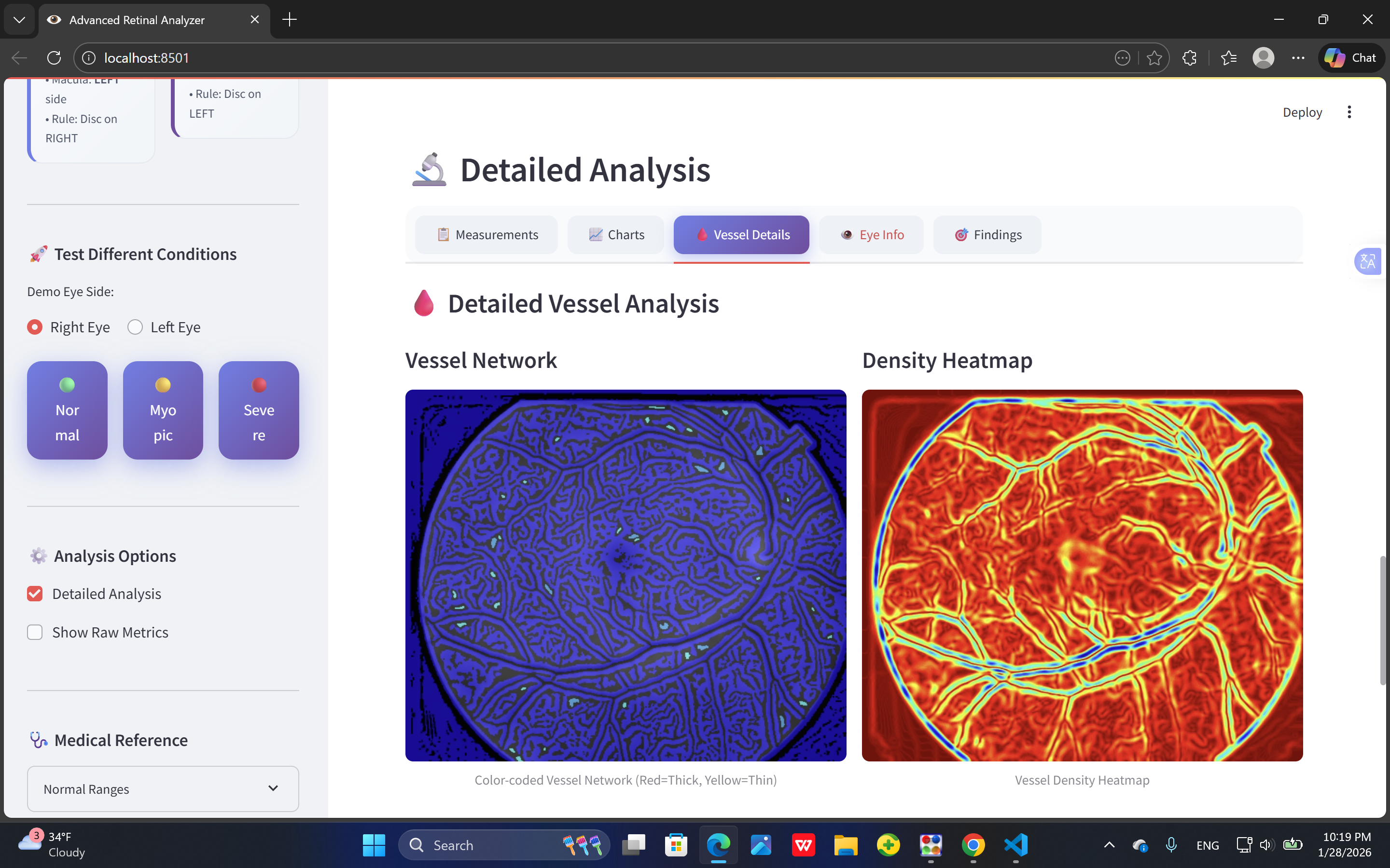
- Retinal vessel density
- Foveal pit curvature
- Optic disc shape
Mathematically, vessel density ( D_v ) was estimated as:
[
D_v = \frac{A_v}{A_t}
]
where ( A_v ) is the area of vessel pixels and ( A_t ) is the total image area.
AI Inference
Using a pretrained convolutional neural network (CNN) from the Python AI library,
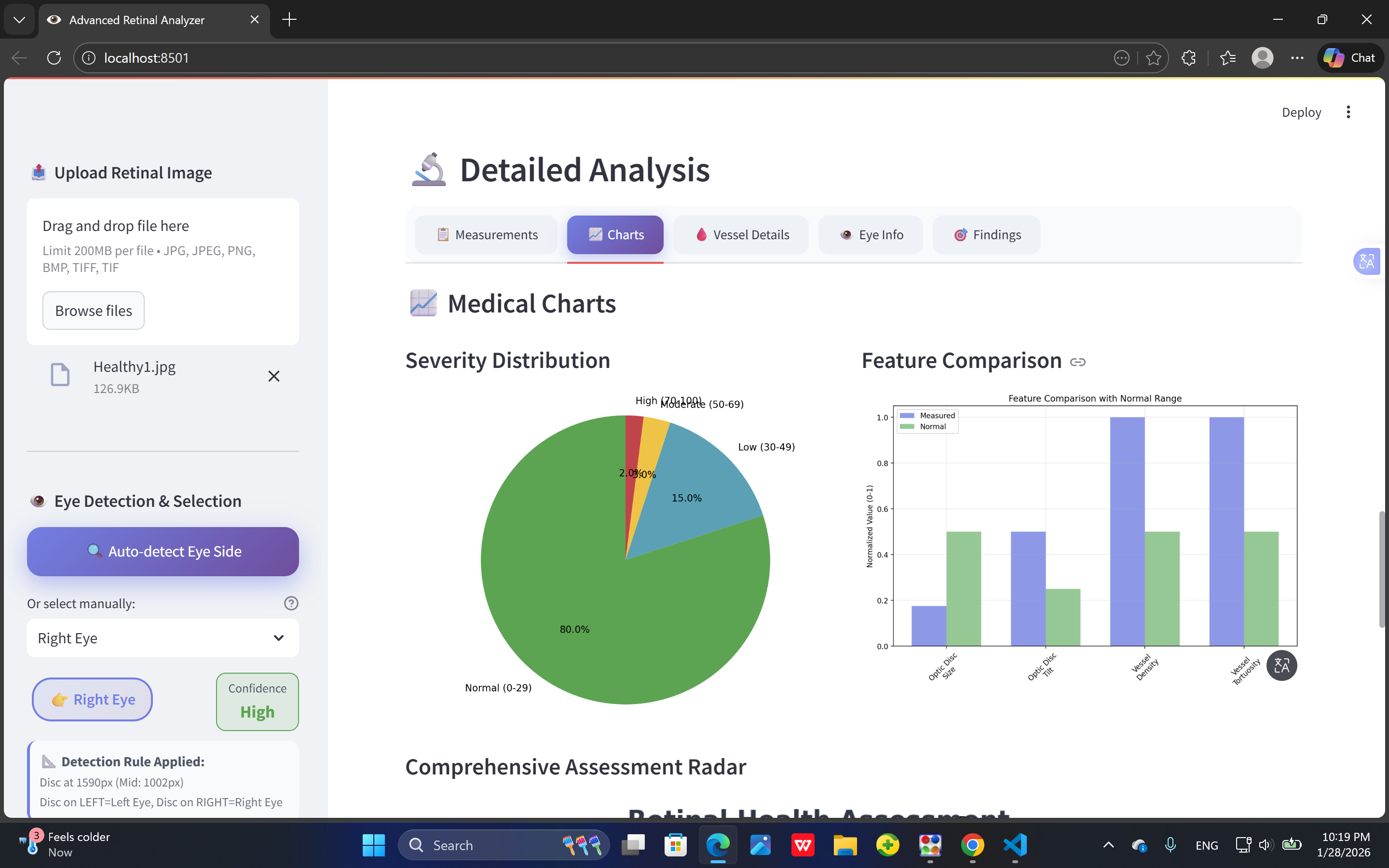
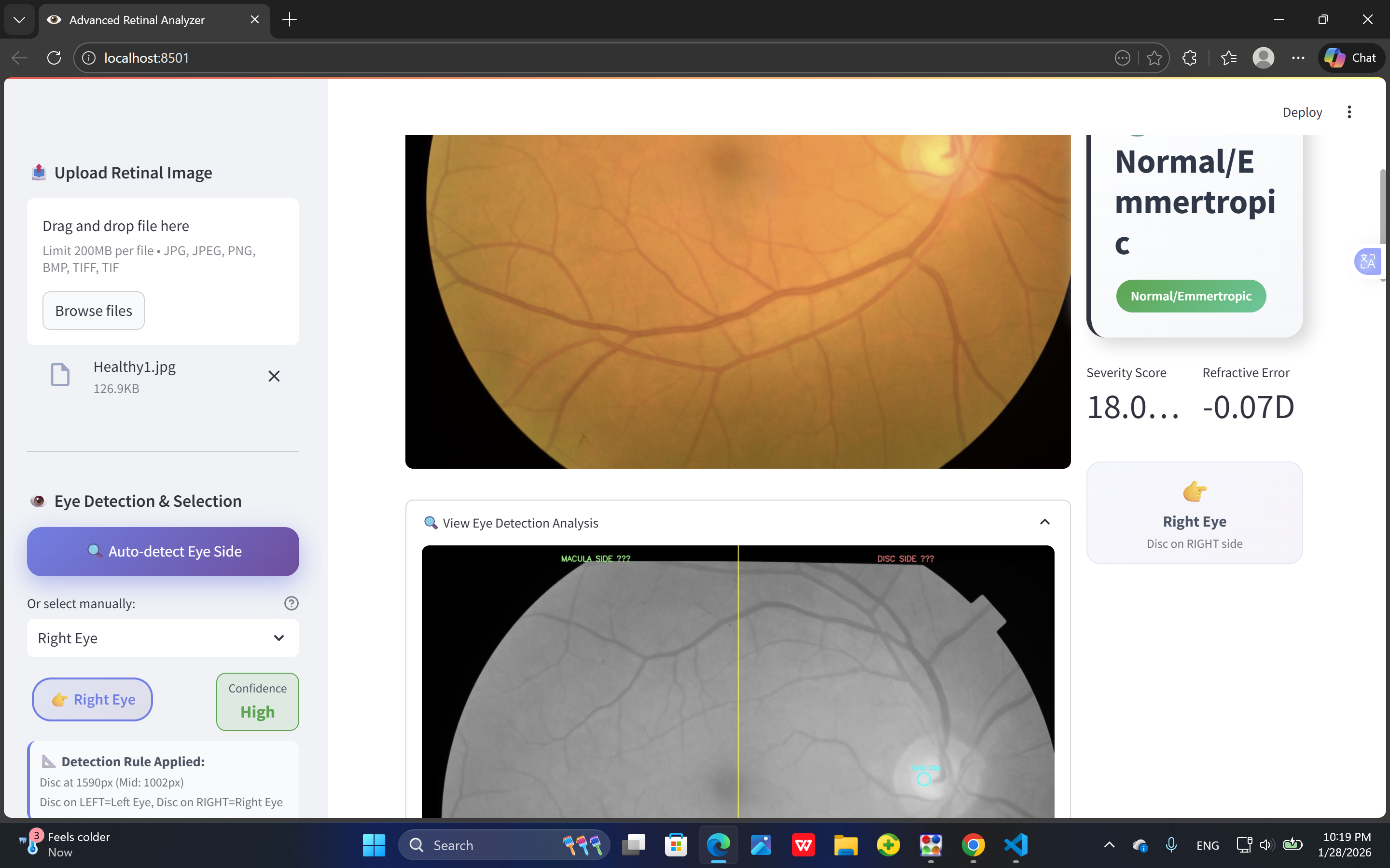
we classified the severity of myopia into three categories: mild, moderate, severe.Visualization
We used Matplotlib and OpenCV to visualize detection results
and display regions associated with potential myopic changes.
🚀 What We Learned
- How to apply AI and computer vision to real-world medical imaging problems.
- The importance of data preprocessing in improving the accuracy of predictions.
- How to integrate Python-based AI libraries with ophthalmic image data.
- The need for ethical AI development — especially when working with sensitive medical data.
We also learned how to interpret model results cautiously and ensure that our system
acts only as a screening support tool, not as a replacement for clinical diagnosis.
⚙️ Challenges We Faced
- Limited dataset: Obtaining high-quality retinal images with labeled myopia grades was difficult.
- Model generalization: Ensuring the AI performs consistently across different camera types and lighting conditions.
- Technical barriers: Managing large image files and fine-tuning preprocessing parameters.
- Ethical constraints: Avoiding medical misinterpretation and clearly defining this tool as non-diagnostic.
Despite these challenges, our project demonstrated that AI-powered ophthalmic screening
is possible even with limited resources.
📈 Future Work
We plan to expand the dataset and integrate deep learning models such as ResNet or Vision Transformer (ViT)
to improve detection accuracy.
We also aim to collaborate with ophthalmologists to conduct validation studies
and eventually develop a mobile or cloud-based platform for real-time retinal screening.
Developed using Python, OpenCV, NumPy, and AI libraries for medical image analysis.
Built With
- ai
- learning
- machine
- python


Log in or sign up for Devpost to join the conversation.