-
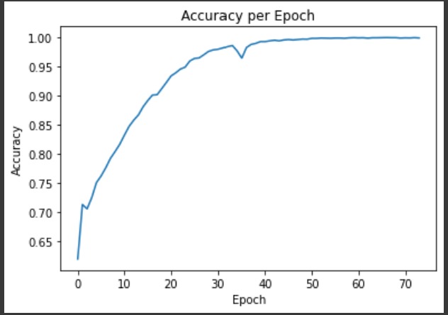
Training Accuracy per Epoch of our model
-
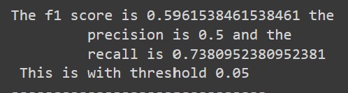
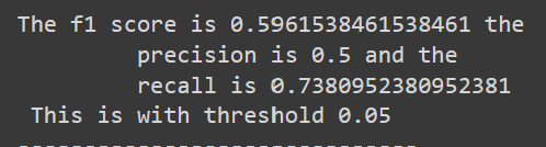
Performance measurements of our validation set of the CNN model

Inspiration
To help make more accurate medical diagnoses and improve the identification process of cancerous skin lesions
What it does
Predicts if a skin lesion is cancerous or non-cancerous utilizing a self-made CNN model that is implemented into a real-time photo application.
How we built it
Using TensorFlow and a skin lesion data set link[1][2], we built a CNN model to identify if a skin lesion is cancerous or non-cancerous. We used tkinter and pillow to build a real-time photo-application to use the model on photos taken and make predictions about it.
Challenges we ran into
The dataset we chose was heavily skewed towards non-cancerous lesions, which drove our model's validation accuracy low. We remedied this by adding more cancerous labeled images into our training and testing/validation sets to even the distribution more. The dataset we had was also too large for our computers to handle so we had to use a small subset of around half the images. Tuning hyper parameters and evening out the data distribution allowed us to increase our testing/validation accuracy from ~30% to ~59%. We noticed that almost if not all photos were of light complexion skin tones and suspect that darker skin tone samples will not perform well with our model due to its absence during training.
Accomplishments that we're proud of
We're proud that we were able to create a working model and save and load it. We were also proud that we got a working application. We spent a lot of time and effort on this project and we came out with knowledge and more skills. We were able to increase the training accuracy to a high level and the accuracy curve looked better than our expectations.
What we learned
We learned more about CNNs and data processing for machine learning. We also learned and engaged more with GUIs and application development.
What's next for Skin Cancer Detector
Project still needs to be finished. The GUI does not interact with the backend at the moment. This project seeks to diversify the dataset and ultimately increase the accuracy of our model to provide better results and prove more useful to healthcare professionals. For our application we foresee that it can be improved to make productions in real-time using video and not require photos.
References
[1] Noel Codella, Veronica Rotemberg, Philipp Tschandl, M. Emre Celebi, Stephen Dusza, David Gutman, Brian Helba, Aadi Kalloo, Konstantinos Liopyris, Michael Marchetti, Harald Kittler, Allan Halpern: “Skin Lesion Analysis Toward Melanoma Detection 2018: A Challenge Hosted by the International Skin Imaging Collaboration (ISIC)”, 2018; https://arxiv.org/abs/1902.03368
[2] Tschandl, P., Rosendahl, C. & Kittler, H. The HAM10000 dataset, a large collection of multi-source dermatoscopic images of common pigmented skin lesions. Sci. Data 5, 180161 doi:10.1038/sdata.2018.161 (2018).

Log in or sign up for Devpost to join the conversation.