-
-
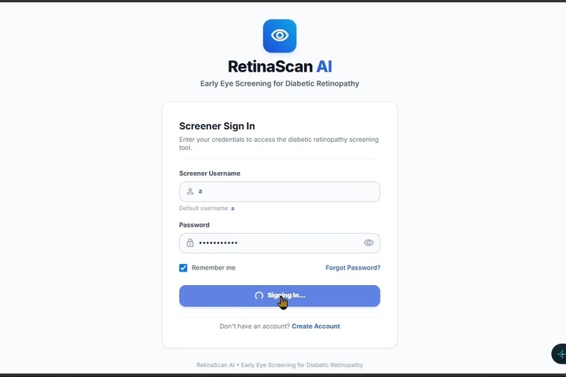
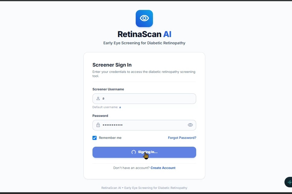
LOGIN PAGE
-
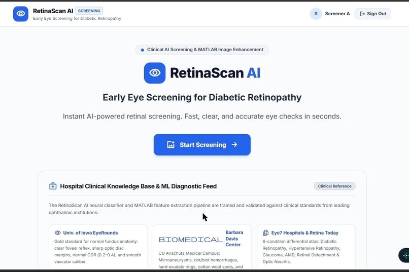
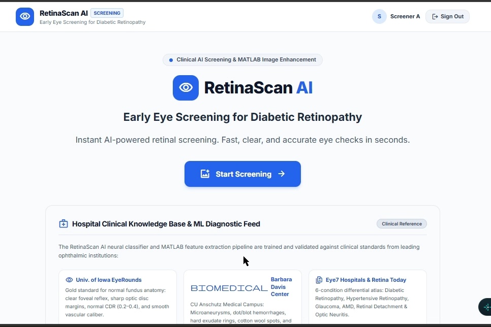
HOME PAGE 1
-
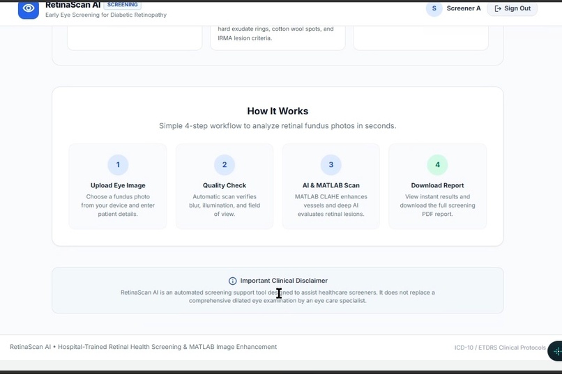
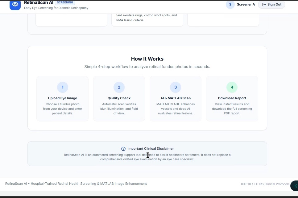
HOME PAGE 2
-
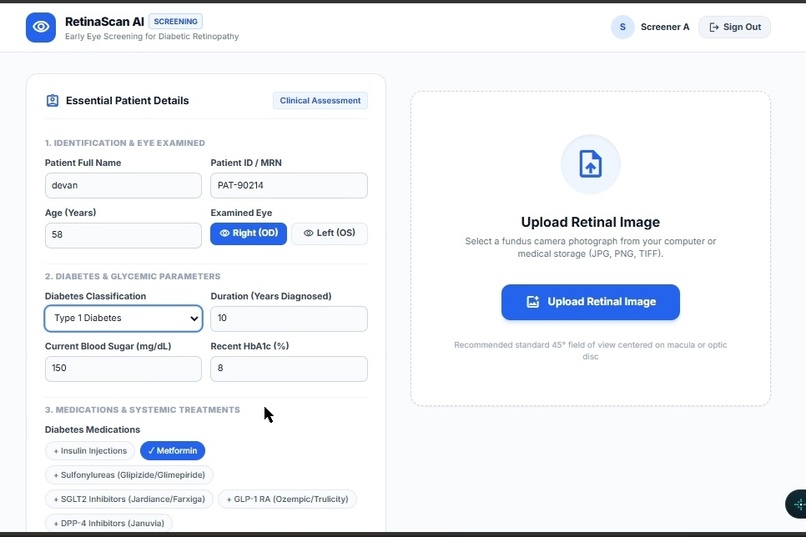
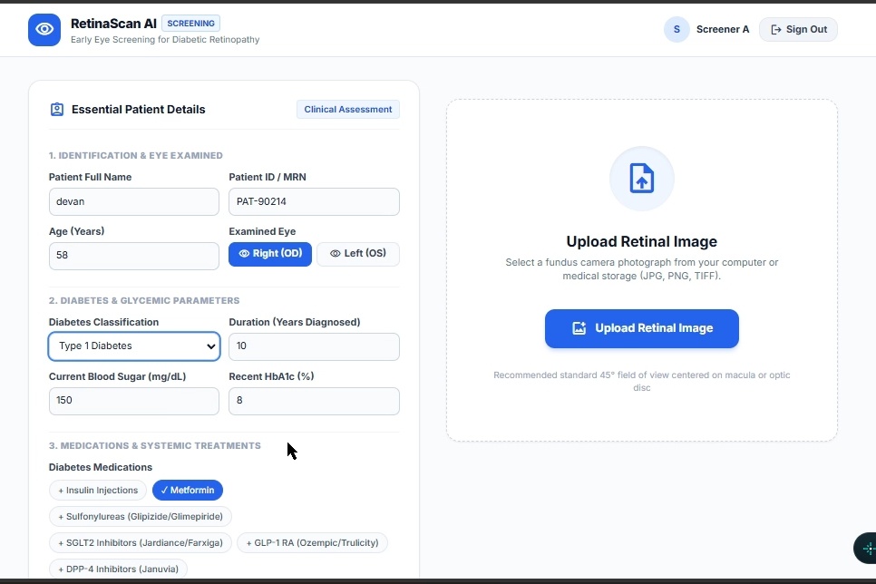
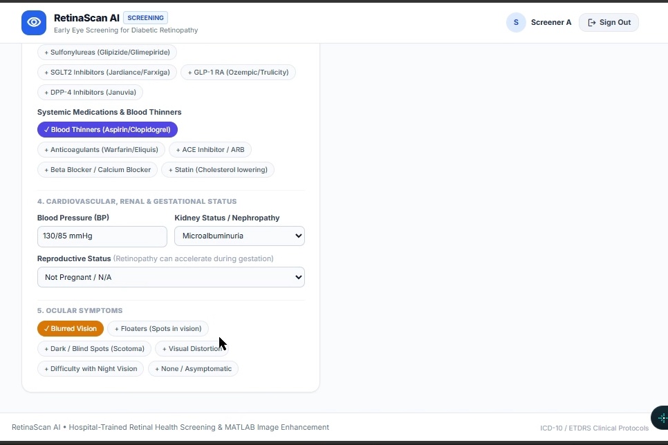
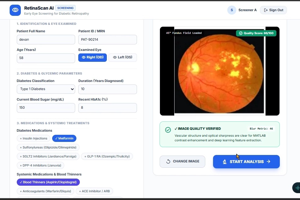
DETAILS FILLING 1
-
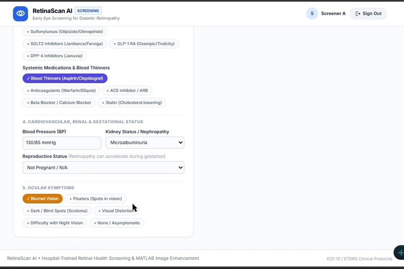
DETAILS FILLING 2
-
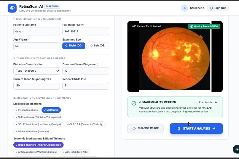
IMAGE ADDING
-
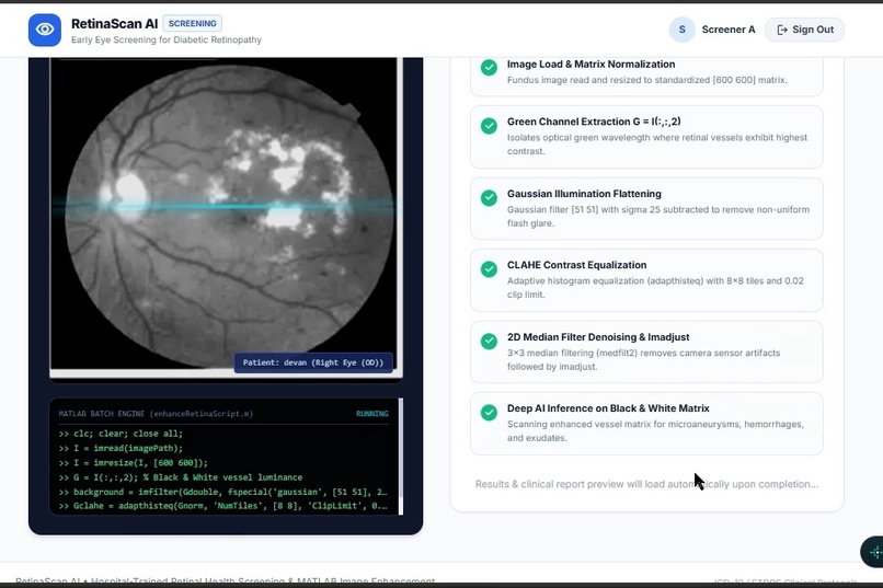
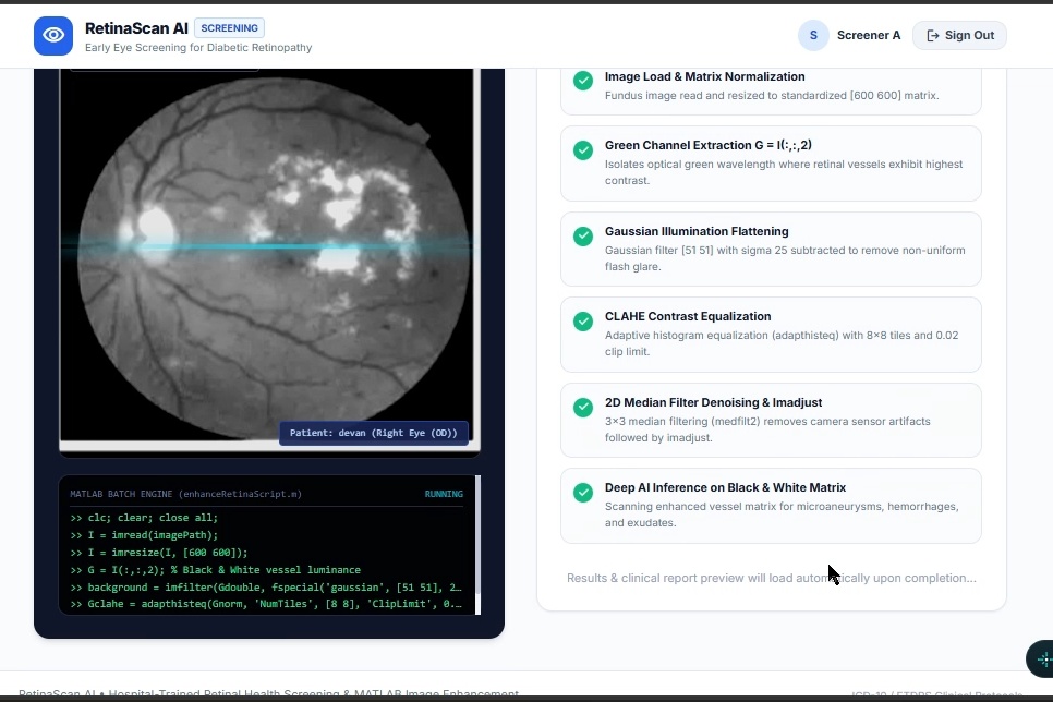
ML MODEL PREDICTING
-
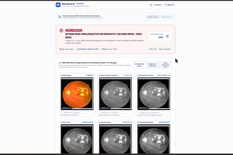
PREDICATION PERCENTAGE
-
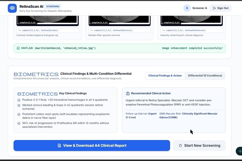
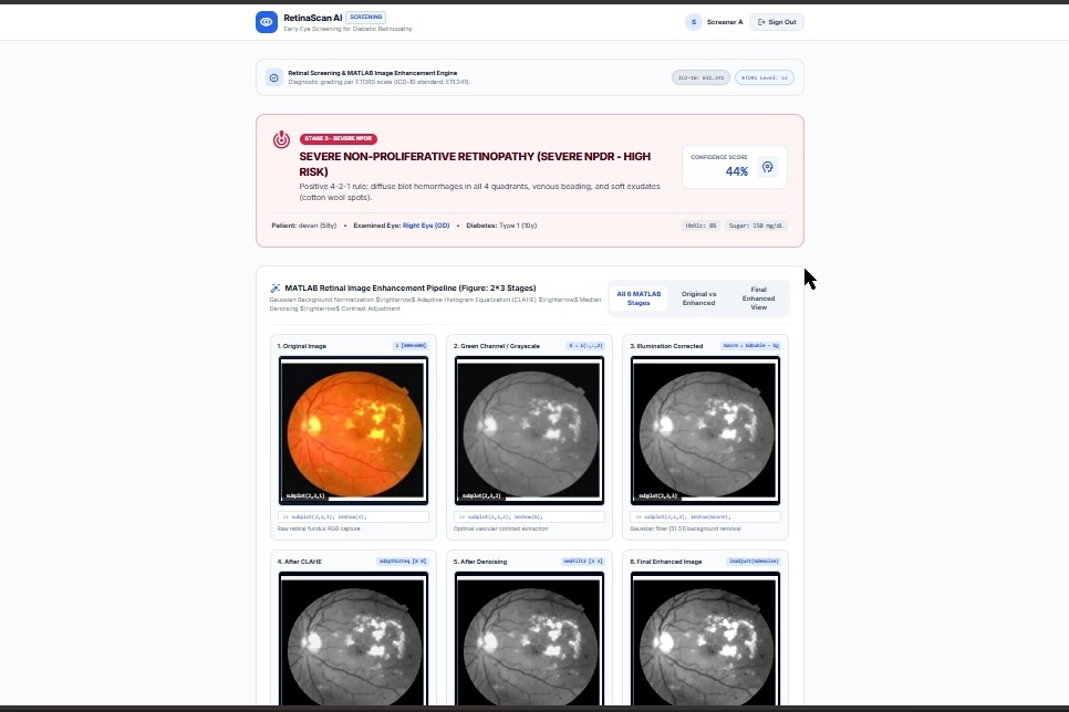
RESULT PAGE
-
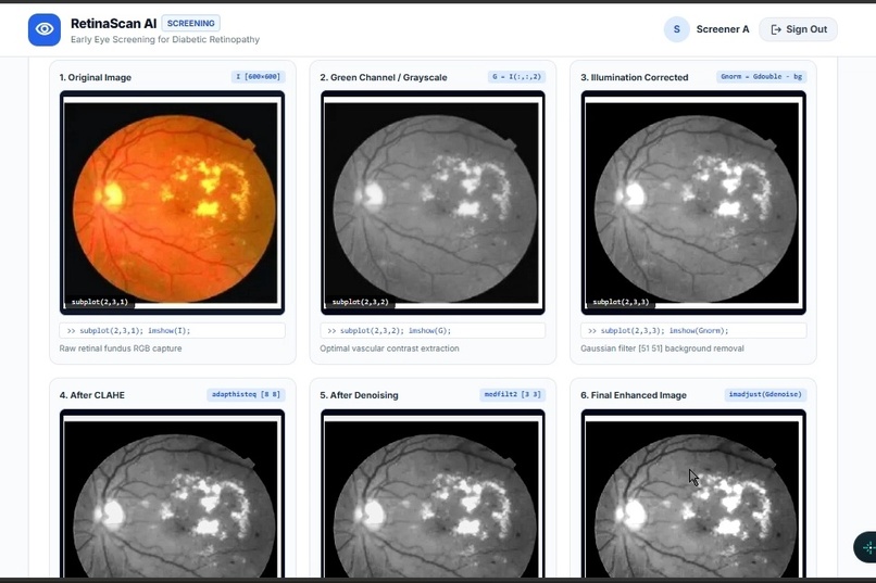
FINDING STAGES
-
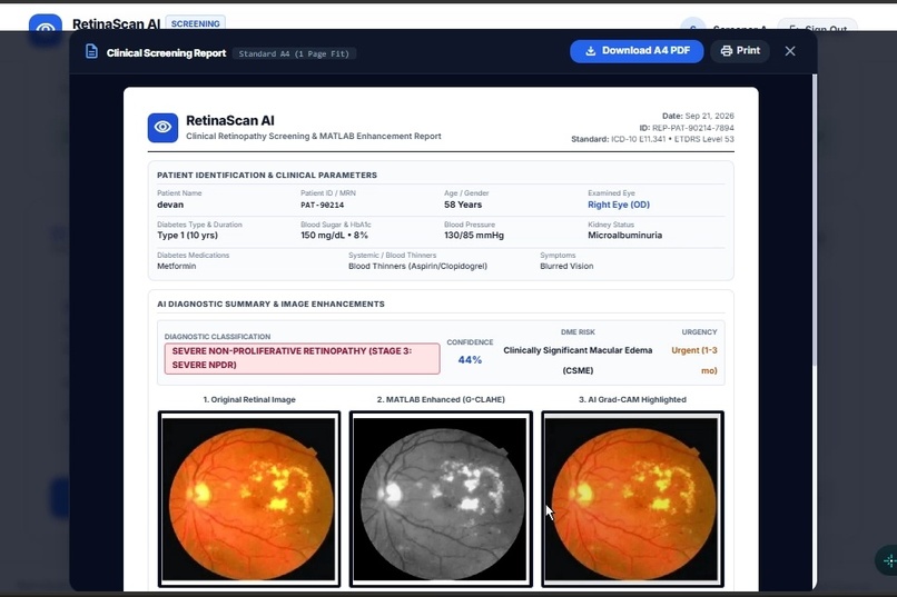
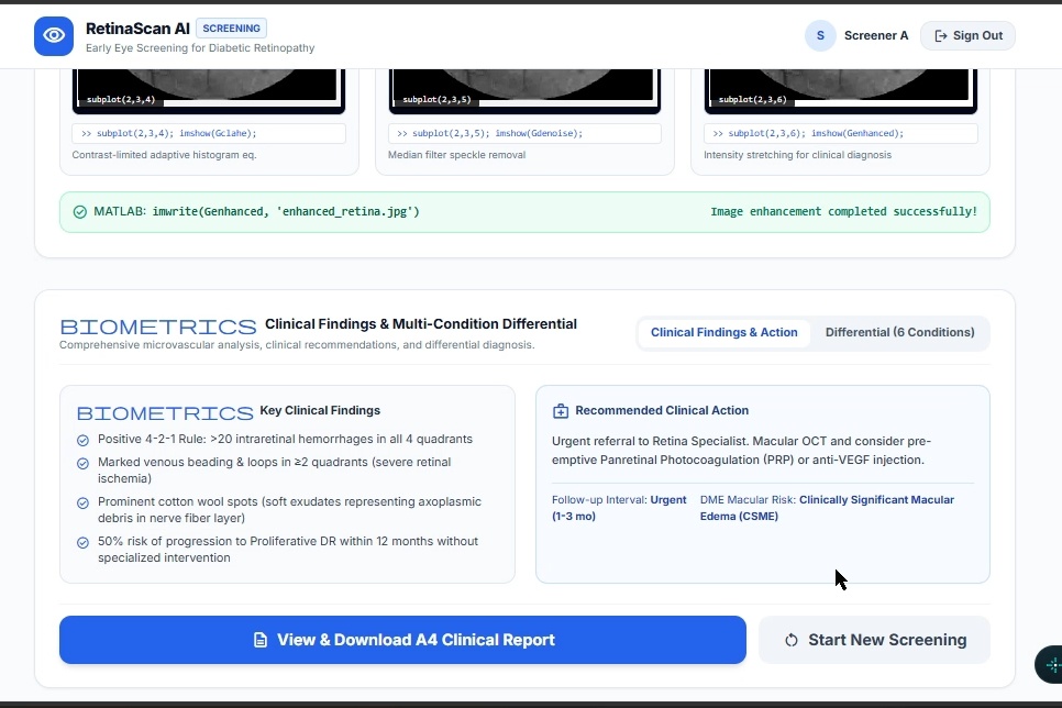
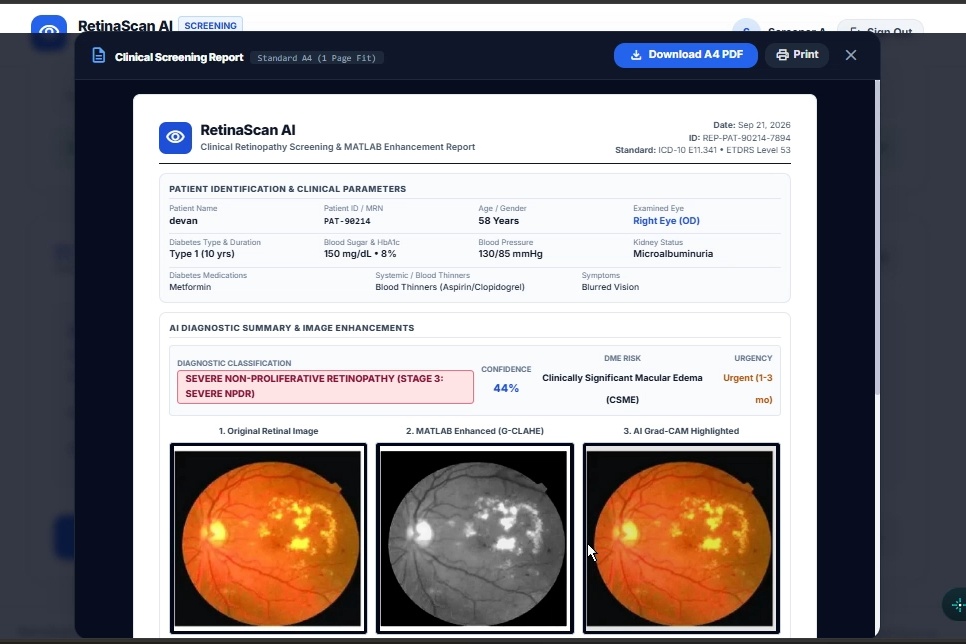
FINAL REPORT
-
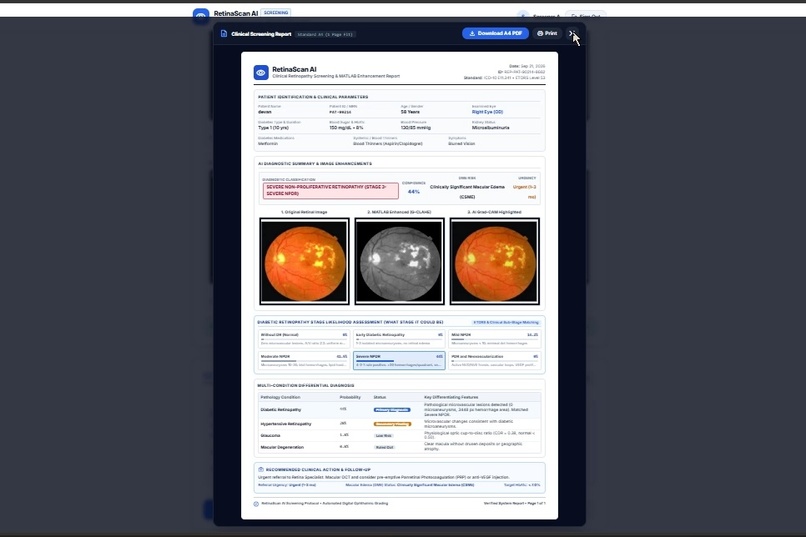
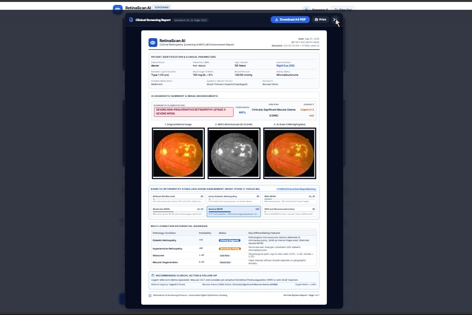
RESULT PDF
Inspiration
Diabetic Retinopathy (DR) can cause permanent vision loss when it is detected late. We were inspired by the difficulty faced by people in rural communities, where access to ophthalmologists and specialized eye-screening equipment can be limited. This led us to develop RetinaScan AI, an explainable AI-based solution for early diabetic retinopathy screening.
What it does
RetinaScan AI analyzes retinal/fundus images and detects signs of diabetic retinopathy. It classifies the condition into different severity levels:
Normal → Mild → Moderate → Severe → Proliferative
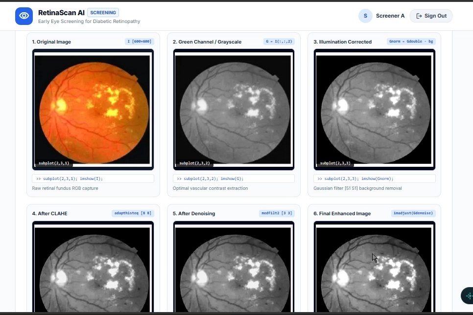
Unlike a system that only provides a prediction, RetinaScan AI also explains the result by highlighting the retinal regions that influenced the AI's decision. It provides a simple and understandable result and can recommend specialist referral when required.
How we built it
The proposed system follows this workflow:
Retinal Image → AI Model → DR Detection → Explainable Result → Severity & Referral
A retinal image can be captured using a low-cost fundus camera or a smartphone-assisted setup. The image is preprocessed and analyzed using a CNN/EfficientNet-based AI model. Grad-CAM is then used to highlight the retinal regions influencing the prediction.
The final output provides the detected condition, severity level, visual explanation, and referral recommendation.
Challenges we ran into
One of the major challenges was making the AI result understandable rather than providing only a disease prediction. We addressed this by incorporating Explainable AI using Grad-CAM.
Another challenge was designing the solution for rural environments, where expensive equipment, specialist availability, and reliable connectivity may be limited. This motivated our focus on a low-cost, accessible, and scalable screening approach.
Accomplishments that we're proud of
We developed a complete concept for an AI-powered diabetic retinopathy screening system that combines disease detection, severity classification, and explainability in a single workflow.
We are particularly proud of incorporating Grad-CAM so that the system can show the retinal regions influencing its prediction, making the result easier for healthcare workers and patients to understand.
What we learned
Through this project, we learned how AI and computer vision can be applied to medical image analysis. We explored CNN-based image classification, retinal image processing, severity classification, and Explainable AI using Grad-CAM.
We also learned that developing a healthcare AI solution requires more than accuracy. Accessibility, interpretability, and the ability to support healthcare workers are equally important.
What's next for RetinaScan AI
Our future plans include smartphone-based retinal screening, regional-language AI explanations, telemedicine and cloud integration, and offline/low-network support for remote areas.
We also aim to explore improved AI models for better accuracy and earlier-stage detection, with the long-term goal of making RetinaScan AI suitable for deployment in rural clinics, health camps, and mobile screening units.


Log in or sign up for Devpost to join the conversation.