-
-
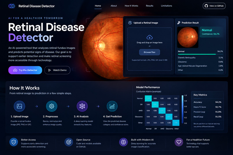
what Retinal Disease Detector actually looks like and how it works
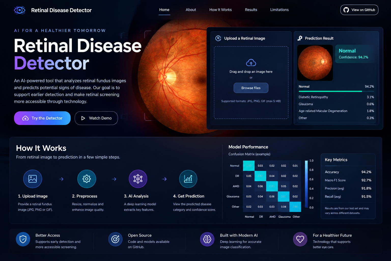
Inspiration
Retinal diseases can cause serious vision impairment, and identifying signs of disease in retinal images can support earlier clinical assessment. However, interpreting these images requires specialized knowledge and can be challenging in settings with limited access to ophthalmic expertise.
We developed Retinal Disease Detector to explore how artificial intelligence and deep learning can help analyze retinal images and support research into accessible, AI-assisted retinal screening.
What it does
Retinal Disease Detector is a deep-learning-based application that analyzes retinal fundus images and predicts the corresponding disease category.
The project combines an image-classification model with an interactive interface, allowing users to submit a retinal image and view the model's prediction. It demonstrates how computer vision can be applied to medical imaging and provides a foundation for further research into AI-assisted screening.
The application is an experimental prototype, not a clinically validated diagnostic system. Its predictions require appropriate clinical interpretation.
How we built it
We built the project using Python and deep-learning techniques for image classification. The workflow involves preparing retinal images, processing them into a format suitable for the model, training a convolutional neural network (CNN), and integrating the trained model into an interactive application.
We also created a live demonstration to make the model accessible beyond the development environment. The project is supported by a GitHub repository documenting its implementation.
Challenges we ran into
One of the main challenges was achieving reliable classification across different retinal disease categories. Medical-image datasets can contain variations in image quality, visual characteristics, and class distribution, all of which can affect model performance.
Another challenge was understanding the limitations of a model that can produce predictions without necessarily learning all the clinically relevant features. Evaluating performance beyond a single accuracy score and communicating the limitations of AI-generated predictions are essential for medical applications.
Accomplishments that we're proud of
We are proud to have developed a working AI-based retinal image classification prototype and made it accessible through an interactive demonstration.
The project allowed us to connect deep learning with a real biomedical engineering problem, moving beyond theoretical concepts to a practical medical-imaging application. It also established a foundation for further improvements in model evaluation, interpretability, and reliability.
What we learned
Through this project, we gained practical experience in applying deep learning to medical images, developing an image-classification workflow, and integrating a trained model into an application.
We also learned that medical AI requires more than a functioning model. Dataset quality, class-specific performance, generalization, interpretability, and responsible communication of limitations are all important when developing tools intended to support healthcare.
What's next for Retinal Disease Detector
Our next steps are to strengthen the model's evaluation, investigate class-specific performance and potential sources of bias, and improve its ability to generalize to images from different sources.
We also aim to explore explainability methods that can help users understand which image regions influence a prediction, improve the application's usability, and establish a more rigorous evaluation framework.
Ultimately, we want to develop Retinal Disease Detector into a more robust research prototype for AI-assisted retinal screening, with appropriate validation and clinical collaboration required before any real-world medical use.

Log in or sign up for Devpost to join the conversation.