Inspiration
It started with a simple goal: build a flat foot predictor that could help people. In India, most people pay little attention to their feet. My father lived with foot pain for years, so I spent time learning about foot types and the right shoes for each. That one small change made a huge difference to his foot health.
I've always wanted to build something in the med-tech space, so the idea grew from a consumer tool into something for clinics. An interview with Jensen Huang pushed me further. He said radiology would be one of the first fields AI changes. I believe humans should always have the final say, but it made me ask: what if AI took over the slow, repetitive part of reading an X-ray and left the decisions to the doctor?
What it does
Remi AI is an offline Windows desktop app for foot doctors.
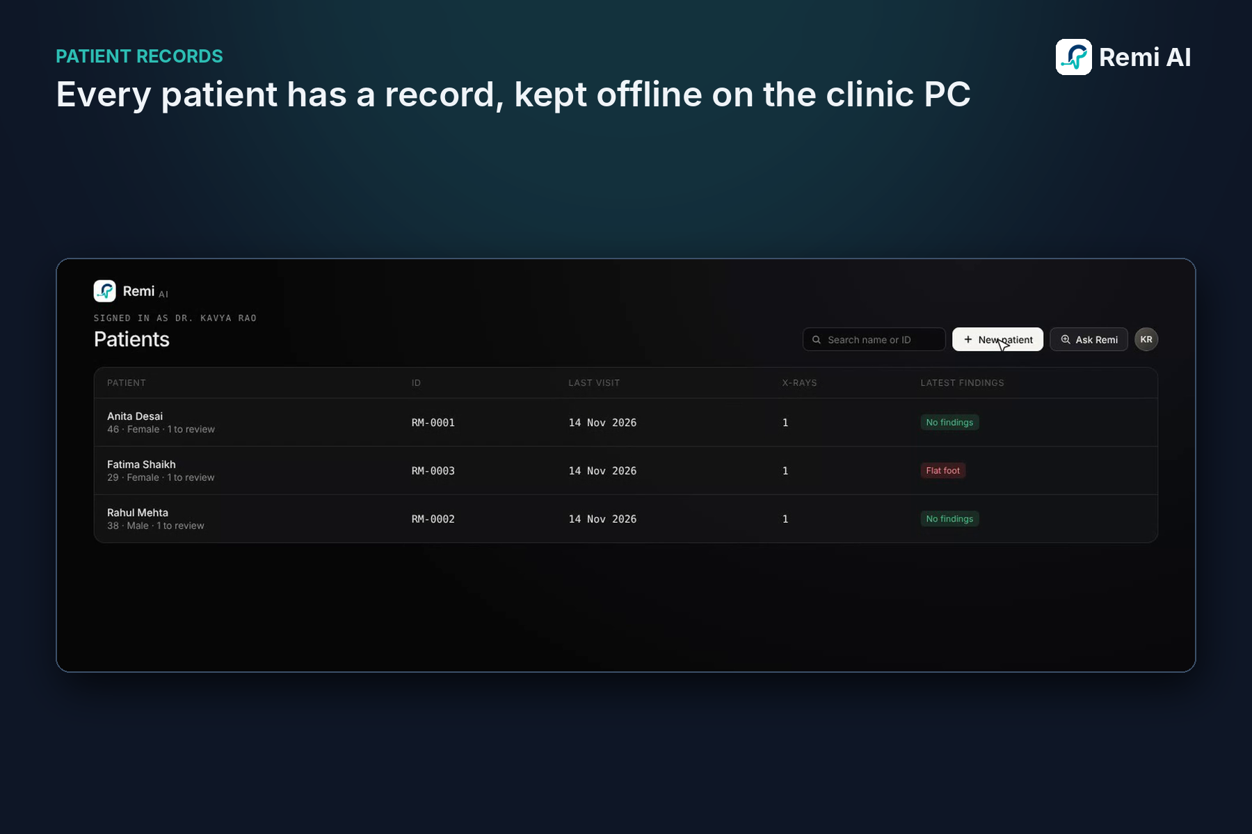
- Doctors sign in and manage a patient list, and each patient has their own record and history.
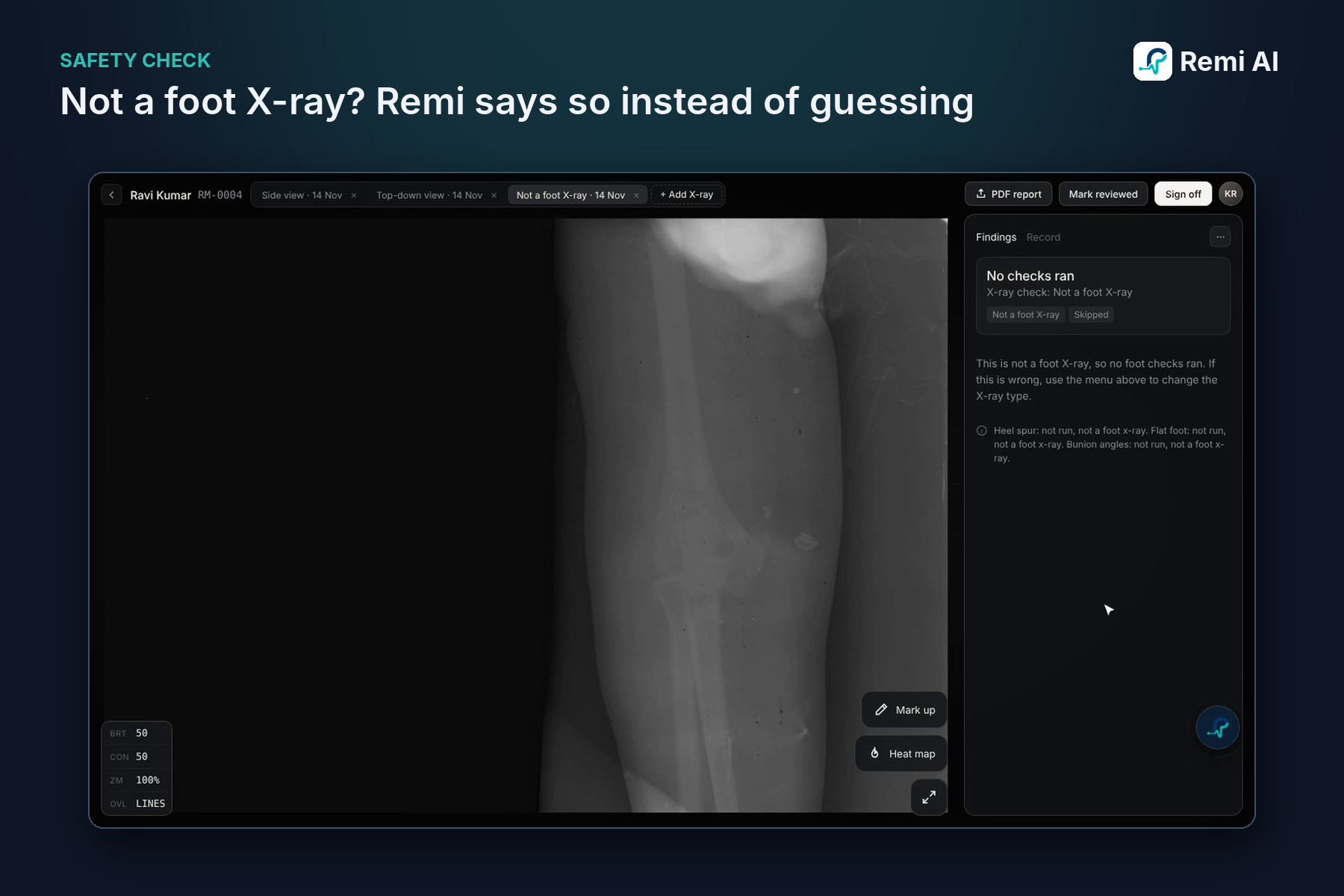
- When an X-ray is uploaded, Remi first checks what kind of view it is. Then it runs only the models that fit that view:
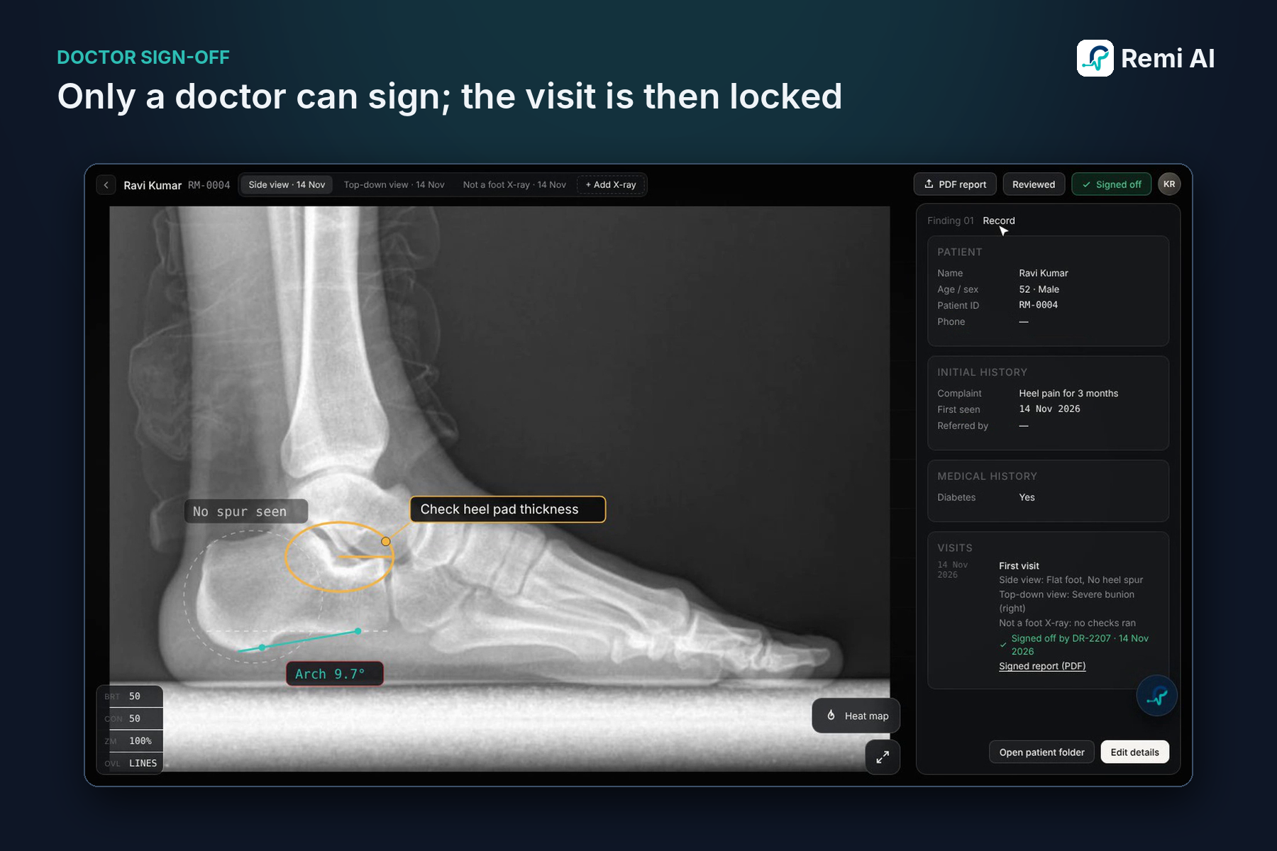
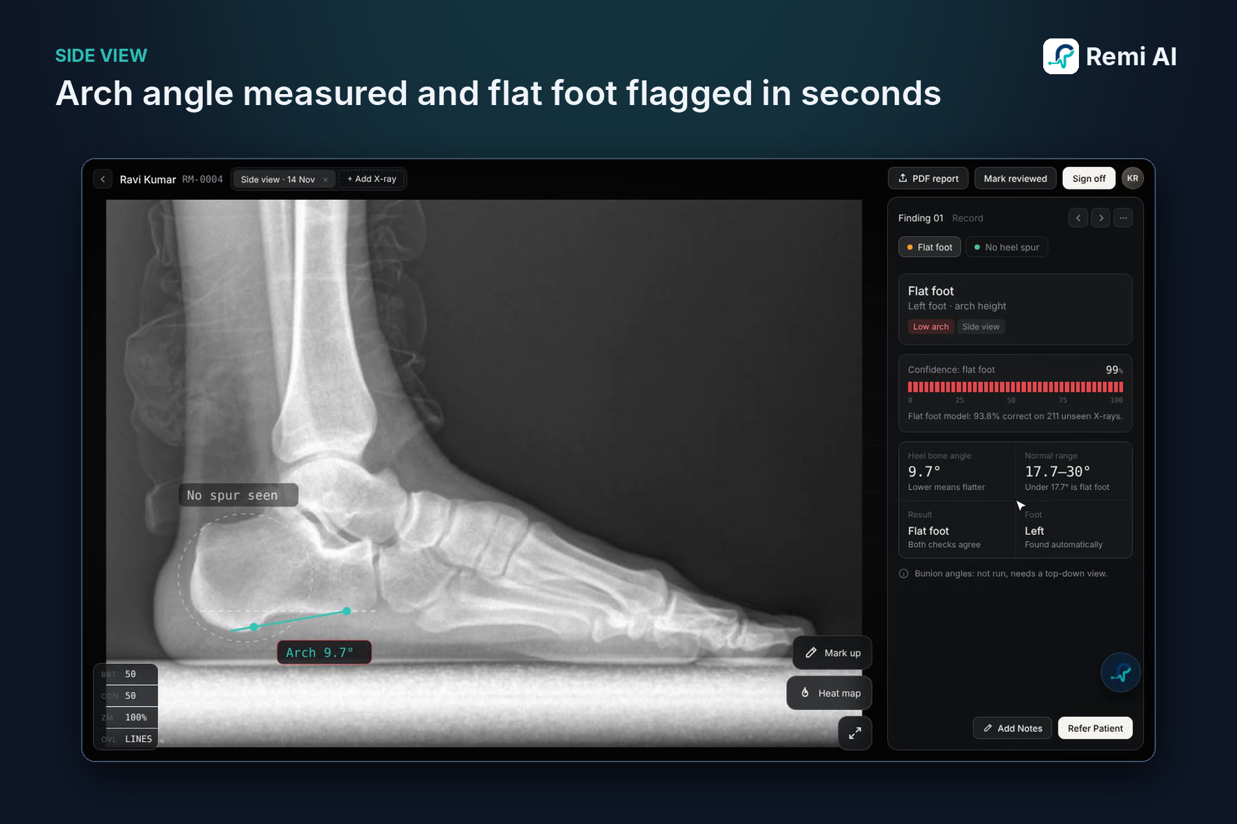
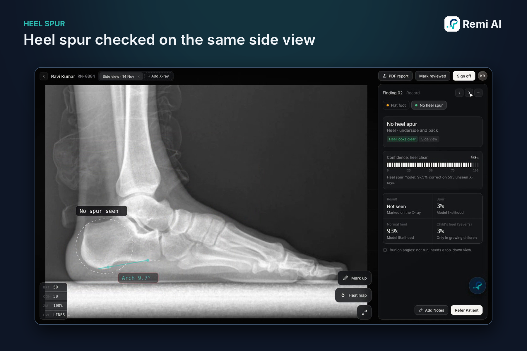
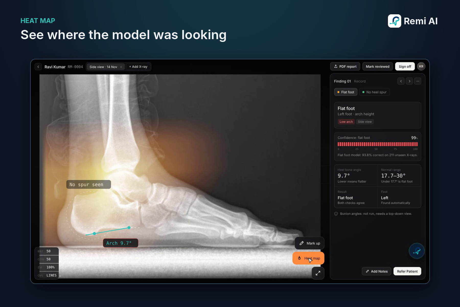
- Side view: it measures the arch angle to flag flat foot and checks the heel for a spur.
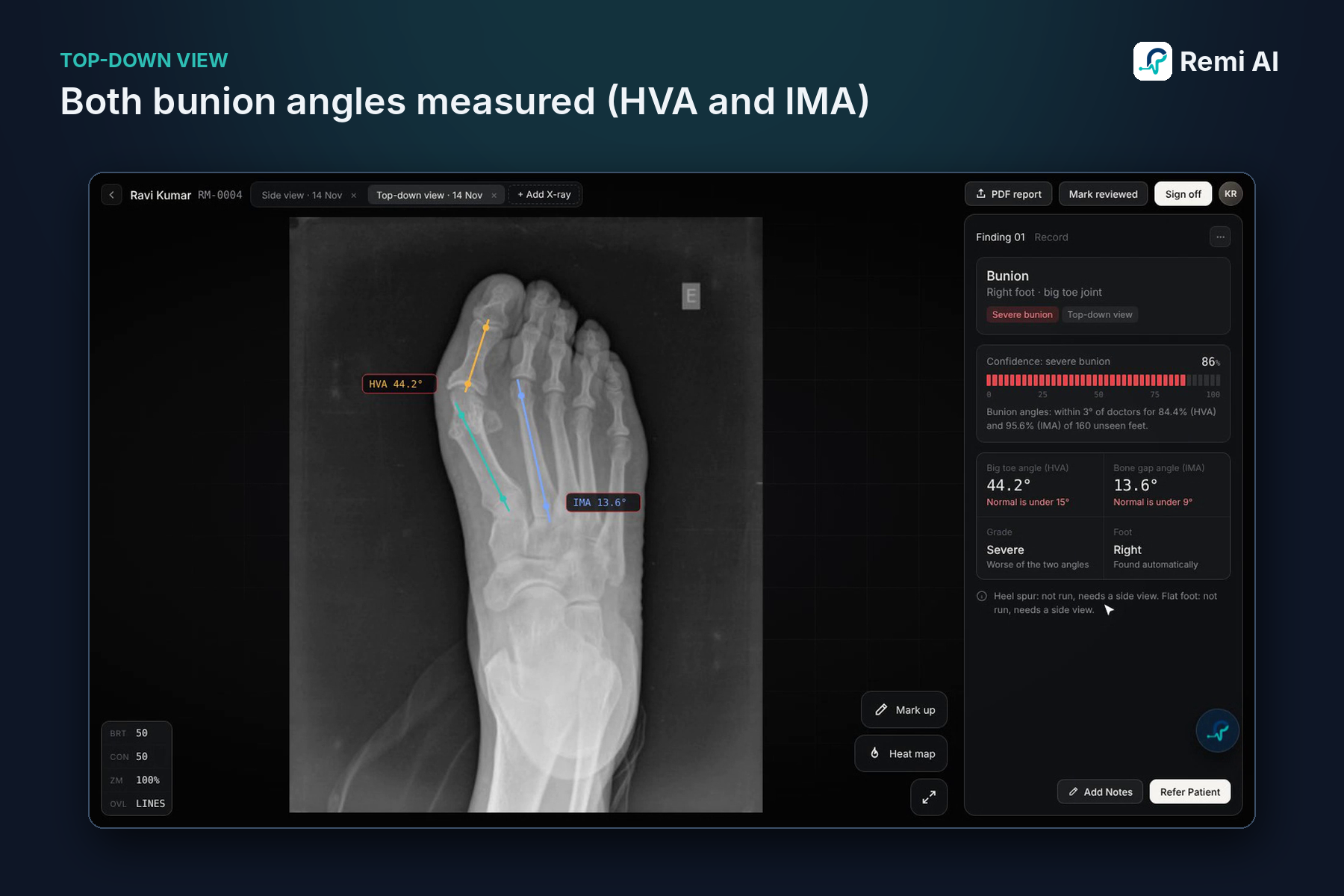
- Top-down view: it finds the foot and measures both bunion angles (HVA and IMA).
- Not a foot X-ray: it says so instead of guessing.
- Every measurement is drawn on the image. The doctor can drag any point and the angle updates live. A heat map shows where the model looked.
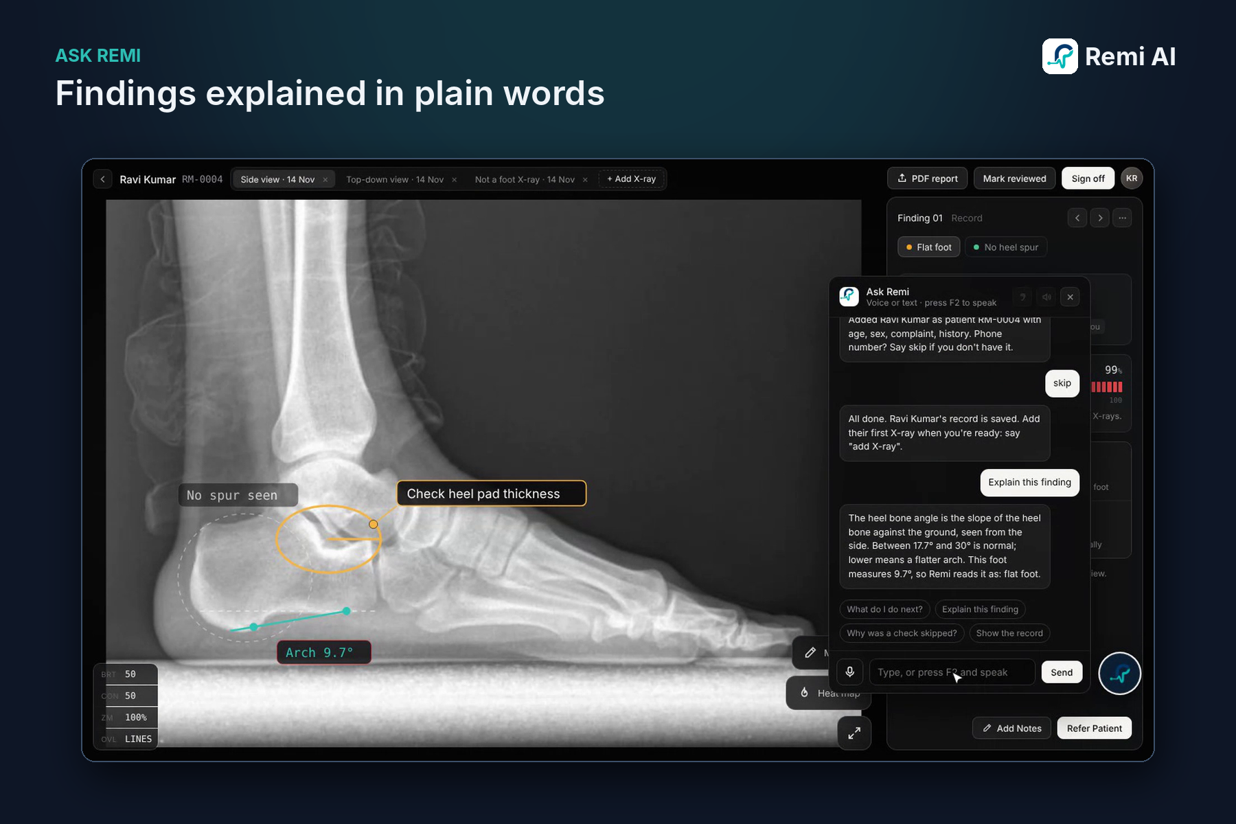
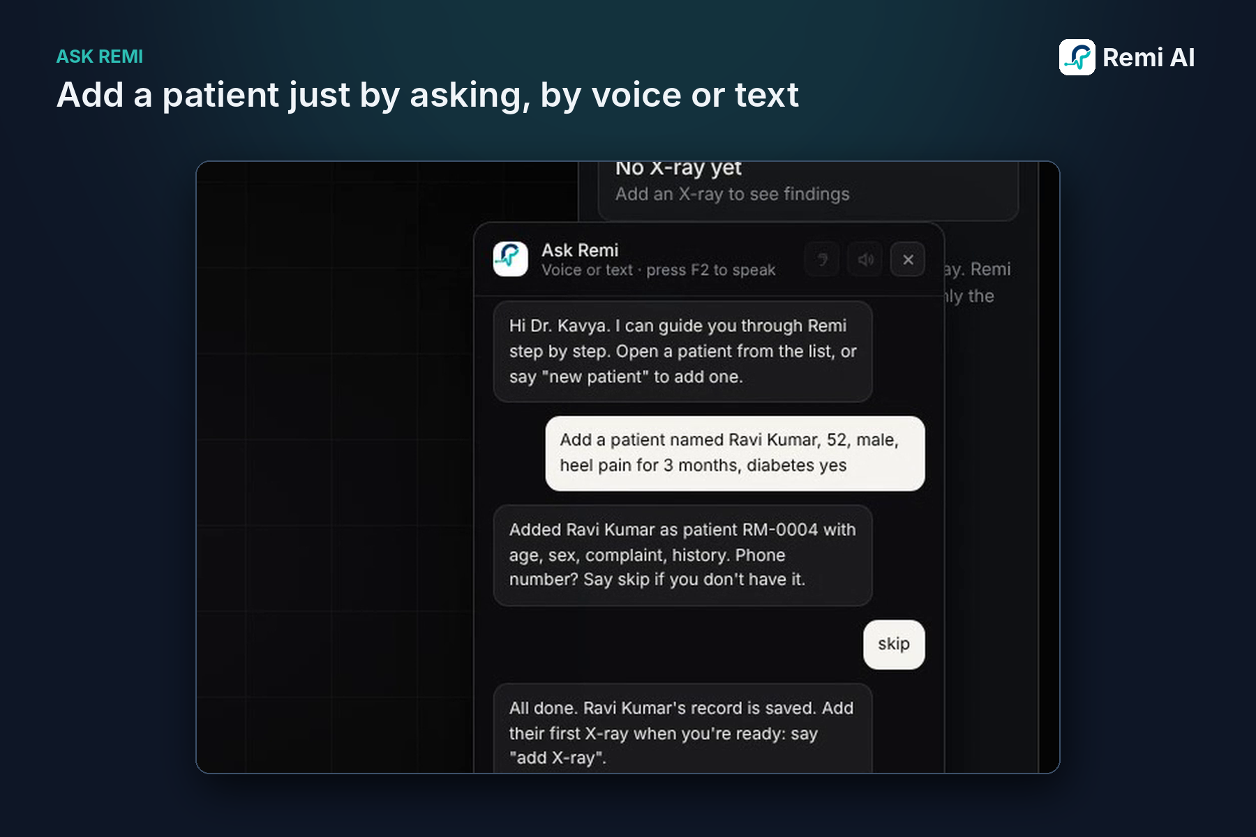
- Ask Remi is a built-in assistant that takes voice or text. It can add patients, explain results in plain words, and guide new users through each step.
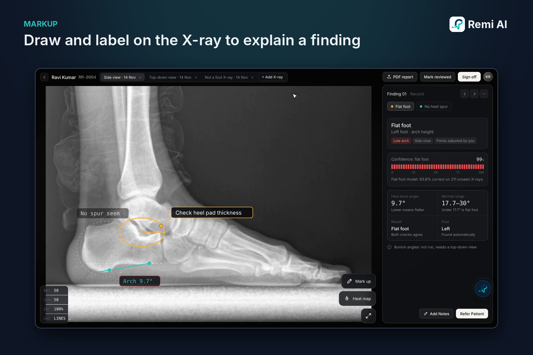
- Doctors can draw on the X-ray and place movable labels to explain a finding.
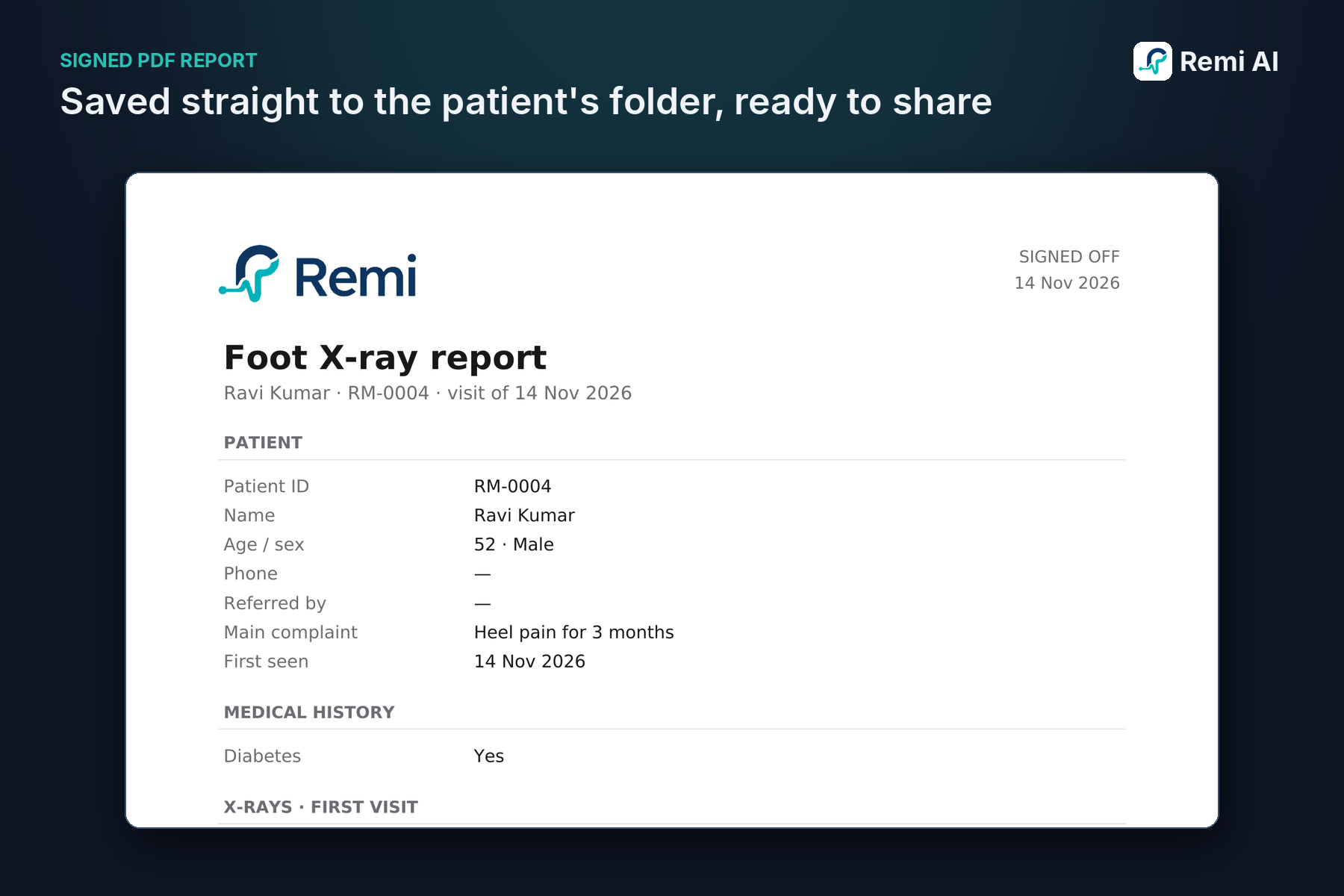
- When a doctor signs off, the visit is locked and a PDF report is saved to the patient's folder. Assistants can prepare reports, but only a doctor can sign.
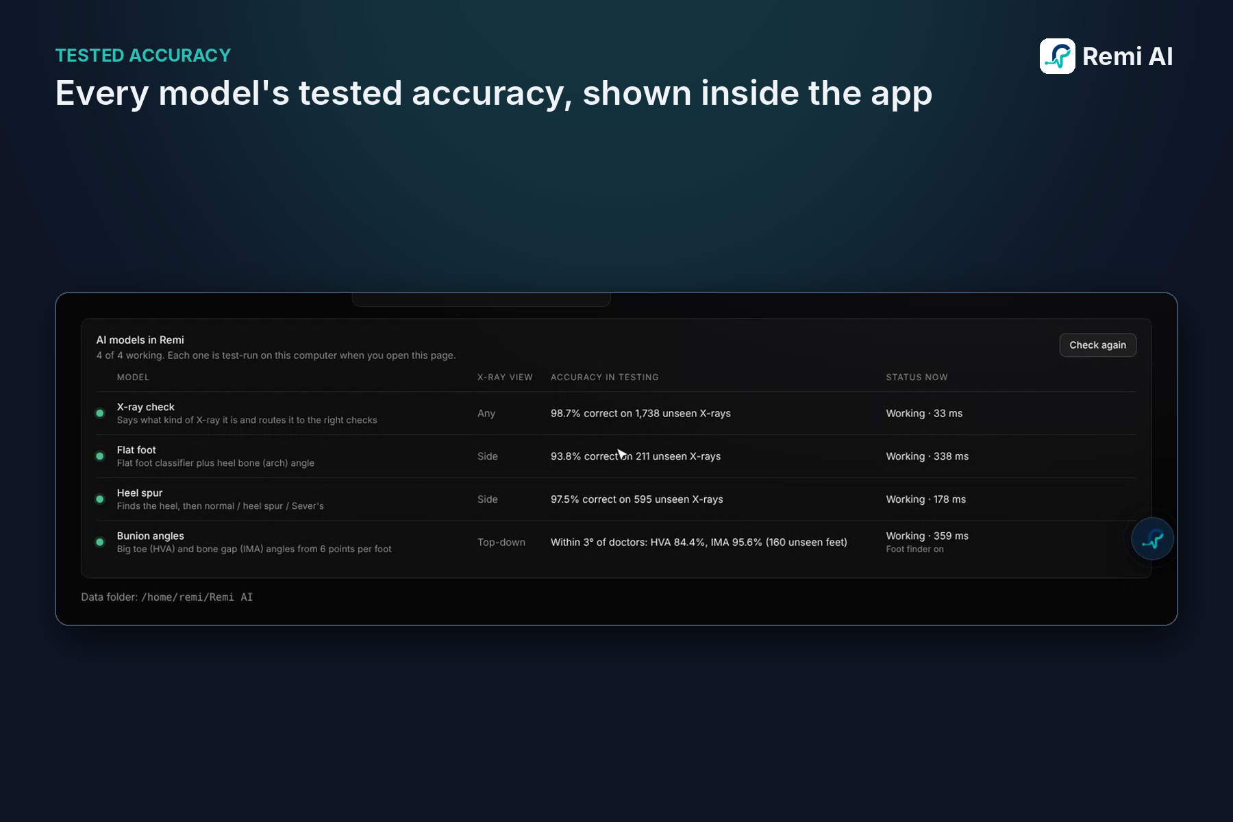
- A model status page shows each model's tested accuracy, so doctors know how far to trust it.
How I built it
- I trained separate models for each task: a view classifier, a flat foot arch-angle model, a heel spur detector, a foot finder and a bunion landmark model. All were exported to ONNX so they run fast offline on a regular clinic PC.
- I used public, openly licensed datasets from Mendeley, Kaggle and figshare, plus a published bunion dataset.
- I trained on my GTX 1650 locally and on cloud CPUs.
- I built the app in Python with pywebview, which gives a modern web-style interface inside a native Windows window. Patient records are stored locally.
- I wrote 71 automated tests and ran every model on images it had never seen.
Challenges I ran into
- Data: labeled foot X-ray datasets are rare. Several features I wanted, like extra angles, had no free dataset, so I focused on what I could measure honestly.
- Running the right model on the right X-ray: early versions ran heel checks on top-down X-rays. A view classifier that routes each X-ray to the right models fixed this.
- Hardware: a small GPU and a nearly full drive meant careful training runs.
- Honest accuracy: I test only on images the models never saw during training, and I pitch Remi as saving time, not as making diagnoses.
Accomplishments that I'm proud of
Tested on images the models had never seen:
- X-ray view check: 98.7%
- Flat foot: 93.8%
- Heel spur: 97.5%
- Bunion angles within 3°: HVA 84.4%, IMA 95.6%
I'm also proud of a full clinical workflow (patients, sign-off, reports and assistant roles) that runs completely offline, so patient data never leaves the clinic.
What I learned
- Data quality and licensing matter as much as the model.
- Doctors need to see why a result was given and be able to correct it. Editable measurements and heat maps build that trust.
- AI works best as an assistant that does the measuring, while the doctor makes the decision.
What's next for Remi AI
- Learn more and get better hardware.
- Validate with practising foot doctors and clinics in India.
- Add more angles once suitable labeled data is available.
- Train on more diverse patient data.
- Work towards the regulatory steps needed for clinical use (CDSCO in India).
