-
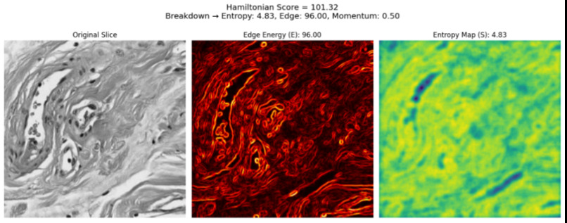
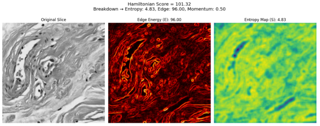
QADS output for histopathology slides, which provides the Hamiltonian Score and the values for each metric.
-
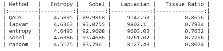
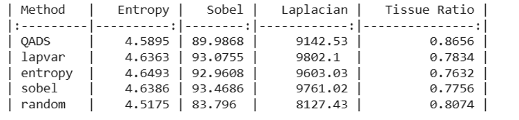
Comparative analysis between QADS and current methods in tissue ratio across top-10 histopathology slides.
-
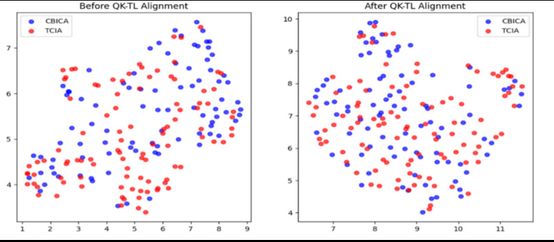
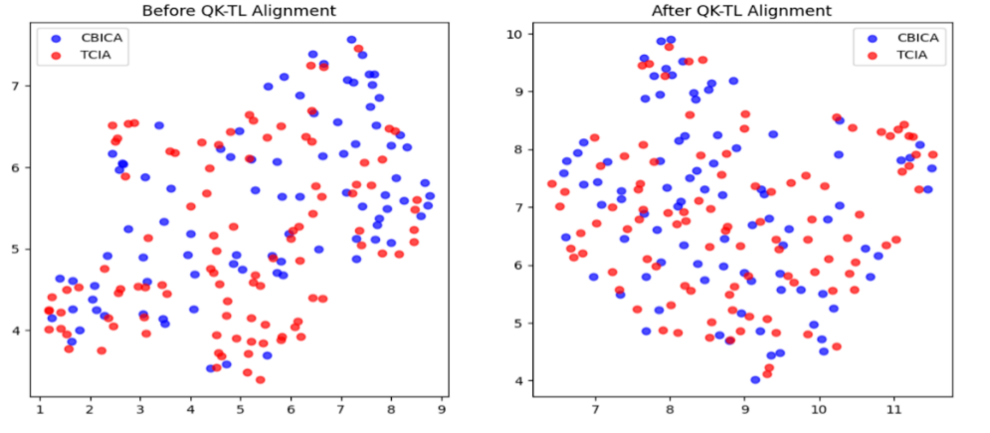
The overlap between blue (CBICA) and red (TCIA) points before and after the QK-TL alignment. (more overlap indicates better performance)
-
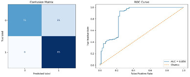
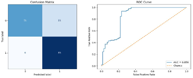
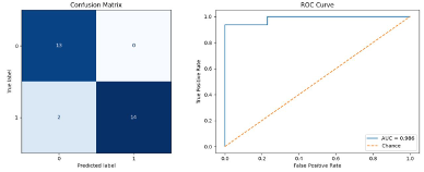
Confusion matrix and ROC curve illustrating the classification performance of the proposed model during the training stage.
-
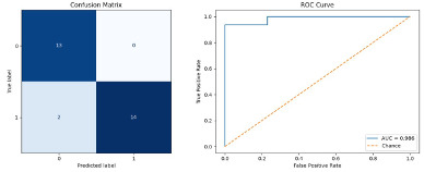
Confusion matrix and ROC curve illustrating the classification performance of the proposed model during the validation stage.
-
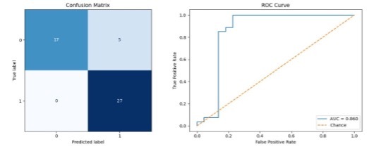
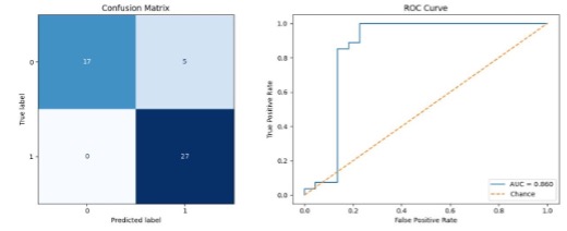
Confusion matrix and ROC curve illustrating the classification performance of the proposed model during the testing stage
-
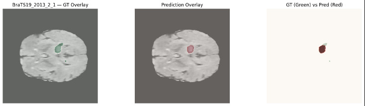
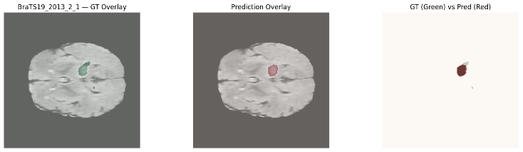
Segmentation model output and the overlap between the ground truth label and the predicted label.
-
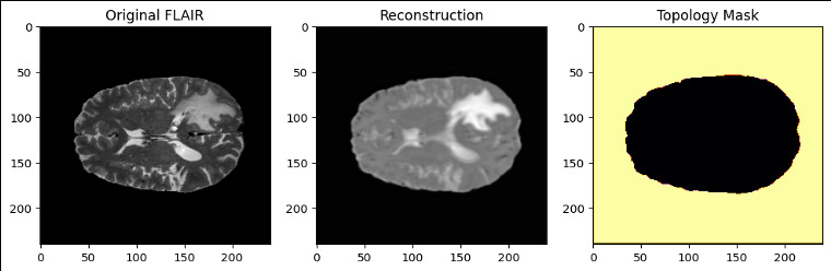
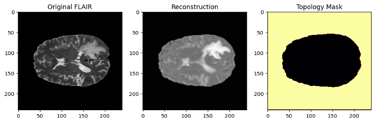
Reconstruction model output and topology mask.
Quantum-Inspired AI for Interpretable Brain Tumor Diagnosis
Inspiration
What if artificial intelligence could do more than identify a brain tumor? What if it could understand the relationships between different medical tests, reconstruct the tumor in three dimensions, and explain the evidence behind its predictions?
Brain tumors represent one of the most challenging problems in modern medical diagnosis. Accurate classification and characterization are essential because different tumor grades can require substantially different treatment strategies. However, brain tumors frequently exhibit overlapping imaging characteristics, heterogeneous tissue structures, and complex molecular profiles, making reliable diagnosis difficult.
The consequences of diagnostic uncertainty can be significant. A systematic review of glioma diagnosis found substantial variation in reported diagnostic errors, with misgrading rates reaching 47% in certain studies. Misclassification can influence treatment decisions, create unnecessary interventions, or delay appropriate care.
Artificial intelligence has emerged as a promising approach for analyzing medical images, yet important limitations remain.
First, many existing systems rely primarily on MRI, even though imaging alone cannot reveal the complete biological characteristics of a tumor. Histopathological examination provides microscopic information about tumor architecture, while molecular biomarkers reveal additional characteristics that can help distinguish tumor behavior.
Second, AI models frequently struggle when applied to data from different hospitals. Variations in MRI scanners, image acquisition protocols, staining techniques, and patient populations can introduce distributional differences that undermine model generalization.
Third, many high-performing systems operate as black boxes. Although they produce classifications or segmentation masks, they often provide limited insight into which regions or sources of evidence contributed to their conclusions.
These challenges inspired QIM-AI, a quantum-inspired multimodal artificial intelligence framework that integrates MRI, histopathology, and molecular biomarkers to investigate a central question:
Can principles inspired by quantum mechanics, information theory, and computational topology enable more accurate, generalizable, and interpretable brain tumor analysis?
Rather than building another conventional image classifier, we aimed to develop a framework that addresses multiple barriers to trustworthy medical AI: information selection, cross-institutional variation, multimodal reasoning, and three-dimensional tumor understanding.
Our broader motivation is to help move medical AI beyond isolated predictions toward systems that can provide clinicians with more comprehensive and understandable diagnostic evidence.
What it does
QIM-AI is a quantum-inspired multimodal AI framework that combines complementary medical information to classify brain tumors, improve feature consistency across datasets, and reconstruct tumor regions in three dimensions.
The framework is designed around four interconnected computational capabilities.
Intelligent medical information selection. Rather than analyzing every image region equally, QIM-AI identifies tissue regions containing potentially important diagnostic information. Its adaptive sampling strategy evaluates local image complexity, structural boundaries, and stochastic variation, helping prioritize meaningful areas while reducing attention to redundant information.
Cross-institutional domain alignment. Medical images collected at different hospitals can differ substantially, even when representing similar biological conditions. QIM-AI uses kernel-based representations and optimal transport to reduce these differences and produce more consistent feature spaces.
Interpretable multimodal tumor classification. QIM-AI combines MRI, histopathology, and MGMT biomarker information through a novel triplet-attention architecture. This enables the classification model to capture three-way relationships between different evidence sources rather than treating each modality independently.
Three-dimensional tumor segmentation and reconstruction. QIM-AI uses topology-aware reconstruction and quantum-inspired feature transformations to identify tumor regions, preserve spatial relationships, and produce volumetric segmentation outputs.
These capabilities were evaluated through dedicated experiments on publicly available medical datasets.
The multimodal classification model achieved 89.8% accuracy, 0.860 ROC–AUC, and 100% recall for high-grade gliomas within the evaluated test set, while the segmentation model achieved a Dice score of 0.8481 and an intersection-over-union score of 0.7465.
A particularly important feature of QIM-AI is its emphasis on interpretability. Instead of providing only a classification result, its architecture supports examining influential medical image regions and the relative contributions of different modalities.
QIM-AI is currently a research prototype evaluated on retrospective datasets, rather than a clinically approved diagnostic system. Its intended future application is to support radiologists, pathologists, and neuro-oncology teams by providing complementary evidence for tumor assessment and treatment planning.
How we built it
We developed QIM-AI using Python and PyTorch, integrating four quantum-inspired models into a unified computational research framework. Each component was designed to address a different technical limitation of conventional medical AI.
1. Quantum Adaptive Diffusion Sampling (QADS)
Medical images contain substantial amounts of redundant or diagnostically irrelevant information. Traditional patch-selection techniques commonly prioritize isolated image properties, such as entropy or edge strength, which can sometimes select noise instead of meaningful tissue.
To address this limitation, we developed QADS, a physics-inspired adaptive sampling algorithm based on a Hamiltonian scoring formulation.
The algorithm combines three complementary signals: Shannon entropy to characterize local information density, edge energy to highlight structural transitions, and stochastic momentum to introduce controlled variation into the selection process.
These signals are integrated into a single score used to rank image regions and prioritize informative MRI slices and histopathology patches.
QADS achieved a tissue relevance ratio of 0.8656, outperforming several conventional selection strategies. Its scoring process also provides a transparent explanation of why particular regions were prioritized.
2. Quantum Kernelized Transfer Learning (QK-TL)
A major obstacle in medical AI is domain shift, where differences between data sources reduce the consistency of learned representations.
We developed QK-TL to improve cross-domain feature alignment using quantum-inspired kernel representations and barycentric optimal transport.
The approach maps features into a shared mathematical representation, preserves relationships between similar samples, and reduces discrepancies between feature distributions using Maximum Mean Discrepancy and additional alignment objectives.
We evaluated QK-TL using feature distributions from the CBICA and TCIA data sources.
The model reduced MMD from 0.100 to 0.001, while barycenter loss decreased from 0.146 to 0.008. It also achieved a centroid distance of 0.033, outperforming the tested alignment baselines on this measure.
These results demonstrate effective statistical feature alignment, providing a foundation for further investigation of robustness across medical institutions.
3. Quantum-Inspired Multimodal Entanglement Network (QIMEN)
Conventional multimodal AI models frequently combine medical information through simple concatenation or pairwise attention. Although these approaches can be effective, they may not explicitly capture interactions involving three different clinical modalities simultaneously.
We developed QIMEN to address this limitation using a quantum-inspired triplet-attention fusion mechanism.
The model processes MRI images through a 2.5D ResNet-18 encoder, histopathology images through a hierarchical EfficientNet-B0 encoder, and MGMT biomarker information through a lightweight neural network.
These representations are then projected into a shared feature space, where the triplet-attention mechanism learns interactions among all three modalities.
The resulting representation is passed to a prototype-based classification head, supporting tumor grading while retaining information about relative modality contributions.
On the evaluated classification test set, QIMEN achieved 89.8% accuracy, 0.860 ROC–AUC, 0.9153 F1-score, and 100% recall.
Importantly, experiments removing individual modalities or the triplet-fusion mechanism demonstrated measurable decreases in performance, supporting the contribution of multimodal integration.
4. Quantum 3D Masked Image Modeling (Q-3D-MIM)
Brain tumors are inherently three-dimensional, and their structural characteristics cannot always be represented adequately by isolated 2D images.
To address this challenge, we developed Q-3D-MIM, which combines topology-aware masked reconstruction with a lightweight volumetric segmentation architecture.
The reconstruction component learns spatial relationships within MRI volumes by masking selected regions and reconstructing them using surrounding anatomical information. An uncertainty-aware objective helps regulate the reconstruction process.
The segmentation component introduces quantum-inspired wave mixing and dual-phase attention to refine volumetric representations and capture complex structural patterns.
The model was trained on 3D MRI patches using a PyTorch training pipeline with memory-efficient optimization strategies.
Q-3D-MIM achieved a Dice score of 0.8481, IoU of 0.7465, and HD95 of 8.97, with an average inference time of approximately 2.12 seconds per sample.
Experimental evaluation
We conducted comparative evaluations and ablation experiments using publicly available BraTS and TCGA resources.
The experiments examined classification accuracy, recall, precision, F1-score, ROC–AUC, segmentation overlap, boundary consistency, domain alignment, computational efficiency, and the contribution of individual architectural components.
Patient-level splitting and fixed random seeds were used in the documented training workflows to support reproducibility and reduce data leakage.
Importantly, the classification, domain-alignment, and segmentation results come from their respective evaluation settings; they do not represent a single prospective, end-to-end clinical trial.
Challenges we ran into
Developing QIM-AI required addressing several challenges at the intersection of medical imaging, machine learning, computational mathematics, and practical implementation.
Integrating fundamentally different medical modalities
MRI images, histopathological slides, and molecular biomarkers have different structures, spatial resolutions, and information content. Combining these inputs required modality-specific feature extractors and a fusion mechanism capable of learning meaningful cross-modal relationships.
Rather than simply concatenating features, we designed QIMEN around higher-order triplet attention, allowing information from three modalities to influence the resulting representation.
Handling differences between medical institutions
A model trained on one dataset may encounter substantially different image characteristics when evaluated on another.
To investigate this problem, we developed QK-TL to align feature distributions while preserving relationships between biologically similar samples. Finding a useful balance between reducing domain differences and retaining meaningful information was a significant modeling challenge.
Processing large three-dimensional medical images
High-resolution MRI volumes can require substantial computational memory, particularly when training volumetric neural networks.
We addressed these limitations through patch-based training, mixed-precision computation, lightweight network components, and controlled training configurations.
Balancing interpretability with predictive performance
High classification accuracy does not necessarily make an AI system useful to clinicians. We wanted the framework to provide insight into how regions and modalities contribute to its predictions.
This motivated the development of transparent Hamiltonian-based region scoring, attention-based fusion, and prototype classification.
Determining whether novel components actually improved performance
A complex architecture is not necessarily a better architecture.
We therefore conducted ablation experiments to evaluate the contributions of entropy, edge energy, domain alignment, triplet fusion, topology-aware masking, and quantum-inspired feature transformations.
These experiments were especially important because they allowed us to distinguish architectural ideas that measurably improved results from features that merely increased complexity.
Working with limited retrospective data
Paired multimodal patient datasets are substantially more difficult to obtain than individual image datasets. This constrained the size and diversity of the evaluated multimodal cohort.
Consequently, our findings should be understood as experimental evidence supporting the framework's potential, with larger external and prospective studies still required.
Accomplishments that we're proud of
Our greatest accomplishment is not any single metric, but the development and evaluation of multiple complementary computational innovations addressing different limitations of medical AI.
1. Developed four complementary quantum-inspired computational models
We designed and implemented QADS, QK-TL, QIMEN, and Q-3D-MIM, applying concepts from Hamiltonian dynamics, kernel methods, optimal transport, multimodal interaction modeling, and topology-aware reconstruction.
The framework demonstrates how mathematical principles inspired by quantum systems can be translated into practical AI methods using classical computing hardware.
2. Achieved 89.8% multimodal brain tumor classification accuracy
QIMEN achieved 89.8% accuracy when distinguishing high-grade and low-grade gliomas, alongside an ROC–AUC of 0.860.
This demonstrates promising performance for combining MRI, histopathology, and biomarker information in a single classification model.
3. Achieved 100% recall for high-grade gliomas in the test set
The classification model achieved 100% sensitivity, correctly identifying all high-grade glioma cases within the evaluated test set.
This result is especially relevant because underestimating tumor grade can affect clinical decision-making. However, the result is specific to the evaluated cohort and does not establish perfect sensitivity in future patients.
4. Demonstrated a measurable advantage from triplet-attention fusion
Ablation experiments showed that removing the triplet-fusion mechanism reduced classification accuracy from 89.80% to 79.59%.
The full model therefore achieved a 10.21-percentage-point improvement over the tested no-triplet variant.
This provides direct experimental evidence supporting the proposed multimodal interaction strategy.
5. Demonstrated the importance of combining multiple medical modalities
The MRI-only model achieved 59.18% accuracy, while the histopathology-only variant achieved 77.55%.
In comparison, the full multimodal QIMEN architecture achieved 89.80% accuracy.
These experiments indicate that integrating complementary imaging, tissue, and biomarker information can improve classification compared with relying on the tested individual image modalities.
6. Achieved strong three-dimensional tumor segmentation performance
Q-3D-MIM achieved:
- Dice coefficient: 0.8481
- Intersection-over-Union: 0.7465
- 95th-percentile Hausdorff Distance: 8.97
- Expected Calibration Error: 0.0011
- Average inference time: 2.12 seconds per sample
Together, these metrics demonstrate promising tumor-region overlap, boundary localization, prediction calibration, and computational efficiency.
7. Outperformed tested lightweight segmentation baselines
Q-3D-MIM achieved a Dice score of 0.8481, compared with 0.8161 for DDUNet, 0.7777 for Fast-SCNN, and 0.7742 for LIU-NET under the reported comparison.
The model also achieved a lower HD95 than these tested baselines, indicating improved boundary agreement in the evaluated setting.
These results support the potential benefit of combining topology-aware learning with quantum-inspired segmentation components.
8. Reduced cross-domain Maximum Mean Discrepancy by 99%
QK-TL reduced MMD from 0.100 to 0.001, representing a 99% reduction in the reported alignment metric.
The model also reduced barycenter loss from 0.146 to 0.008 and achieved the lowest centroid distance among the evaluated domain-alignment methods.
This is a significant technical result because differences between medical datasets remain an important obstacle to robust AI development.
9. Demonstrated the effectiveness of adaptive region selection
QADS achieved a tissue relevance ratio of 0.8656, compared with 0.8074 for random selection in the reported evaluation.
Removing both entropy and edge energy reduced the tissue ratio to 0.3410, highlighting the importance of combining meaningful structural and information-based signals.
These results demonstrate that the model's performance depends on its proposed selection principles rather than stochastic sampling alone.
10. Validated important architectural choices through ablation studies
We evaluated the contribution of individual components by systematically removing or modifying them.
For example, the reconstruction model achieved an MSE of 0.0008, compared with 0.0552 when topology-aware masking was removed.
Similarly, removing key domain-alignment losses increased centroid distance, while removing triplet attention substantially reduced classification performance.
These experiments provided evidence that the proposed modeling choices contributed to the observed results.
11. Developed a computationally efficient classification model
The QIMEN model required approximately 63.2 MB of model storage and achieved an average CPU inference latency of 2.65 seconds per patient on the evaluated Apple M4 system.
This is particularly meaningful because computational efficiency is an important consideration when developing AI systems that may eventually be used in environments with limited specialized hardware.
12. Independently conducted the research and implementation
QIM-AI was developed as an independent student research project.
We carried out the problem formulation, methodology development, dataset preprocessing, model implementation, training, benchmarking, ablation experiments, and interpretation of experimental results.
Completing the work without access to an institutional laboratory demonstrated that technically ambitious medical AI research can be pursued using publicly available scientific resources, personal computing equipment, and cloud-based computation.
13. Connected technical innovation with an important healthcare problem
Beyond the numerical achievements, QIM-AI investigates how AI can produce more comprehensive and interpretable information about brain tumors.
Its combination of multimodal classification, explainable information selection, domain alignment, and volumetric segmentation creates opportunities for future tools that support clinicians in evaluating tumor characteristics and understanding the evidence behind model predictions.
What we learned
Developing QIM-AI taught us that meaningful innovation in healthcare AI requires more than achieving a high accuracy score.
First, we learned that multimodal integration is a reasoning problem, not simply a data-combination problem.
MRI, histopathology, and molecular biomarkers describe different aspects of tumor biology. Their usefulness depends on whether an AI model can capture relationships between these signals rather than treating them as unrelated features.
The QIMEN ablation results reinforced this lesson by demonstrating substantial improvements when multimodal information was integrated using triplet attention.
Second, we learned that mathematical ideas from different scientific disciplines can inspire useful machine-learning methods.
Concepts from Hamiltonian dynamics helped inform region selection, optimal transport provided a method for aligning feature distributions, and topology-aware learning helped guide 3D image reconstruction.
This demonstrated that quantum-inspired approaches do not require quantum hardware to motivate computational architectures.
Third, we learned that experimental validation must be part of innovation.
Developing a novel algorithm is only the first step. Understanding whether it contributes to performance requires meaningful comparisons, controlled experiments, and ablation studies.
By removing components and comparing the resulting models, we learned which design choices were supported by measurable evidence.
Fourth, we learned that efficient implementation matters.
Medical imaging often involves large datasets and computationally intensive operations. Through patch-based training, efficient neural network architectures, and careful optimization, we explored how sophisticated models could remain computationally practical.
Finally, we learned that medical AI must be evaluated with particular scientific responsibility.
Strong results on retrospective datasets cannot automatically be interpreted as clinical readiness. Models intended for healthcare must also demonstrate reliability across populations, robustness under changing conditions, transparent limitations, and meaningful validation in realistic clinical workflows.
This changed our understanding of success: the objective is not simply to build an AI model that makes predictions, but to develop one whose behavior can be understood, tested, and eventually trusted.
What's next for Quantum-Inspired AI for Interpretable Brain Tumor Diagnosis
Our next goal is to transform QIM-AI from a collection of experimentally evaluated computational components into a more comprehensively validated and accessible medical AI research platform.
Interactive medical AI visualization platform
We aim to develop an interactive interface where users can explore MRI scans, visualize selected diagnostically relevant regions, examine three-dimensional tumor segmentation outputs, and understand how different information sources contribute to model predictions.
Such a platform would make QIM-AI's research outputs more accessible to medical students, researchers, and eventually clinical professionals.
Larger and more diverse clinical datasets
We plan to expand validation across additional institutions, scanner protocols, tumor types, and patient populations.
This would enable more rigorous testing of domain generalization and help determine whether the observed performance is preserved under conditions different from the original development datasets.
Stronger uncertainty estimation and interpretability
We aim to extend the framework's uncertainty-aware methods so that predictions can be accompanied by clearer confidence information and explanations.
A particularly important objective is to help identify cases where the system's predictions may be uncertain and additional medical review would be appropriate.
Additional brain tumor categories
The present work focuses primarily on glioma grading and tumor segmentation. Future research could evaluate additional tumor categories, including metastatic lesions and other intracranial tumors.
This would provide a broader assessment of the adaptability of the quantum-inspired architecture.
Personalized prognosis and treatment support
A longer-term direction is to incorporate longitudinal medical records, molecular profiles, treatment history, and clinical outcomes to investigate survival prediction and treatment-response modeling.
Rather than focusing exclusively on classifying an existing tumor, this could eventually help clinicians understand how different patient characteristics relate to prognosis and treatment decisions.
Clinical collaboration and prospective validation
Before any real-world clinical deployment, QIM-AI would require independent external testing, prospective clinical evaluation, and collaboration with medical experts.
These steps are essential for assessing safety, clinical utility, bias, and the limitations of the system in real diagnostic settings.
Our long-term vision is to develop medical AI that does not merely classify tumors, but integrates complementary evidence, explains its reasoning, and supports a deeper understanding of disease.
By connecting quantum-inspired computational innovation with practical challenges in neuro-oncology, QIM-AI aims to contribute toward a future in which artificial intelligence makes medical information more understandable, diagnostic support more reliable, and advanced healthcare technology more accessible.
