-
-
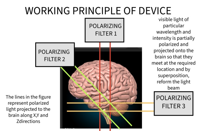
working principle explained- PART 1
-
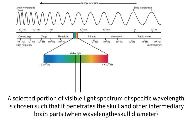
PART 2
-

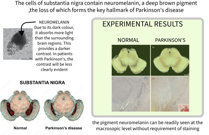
PART 3
-
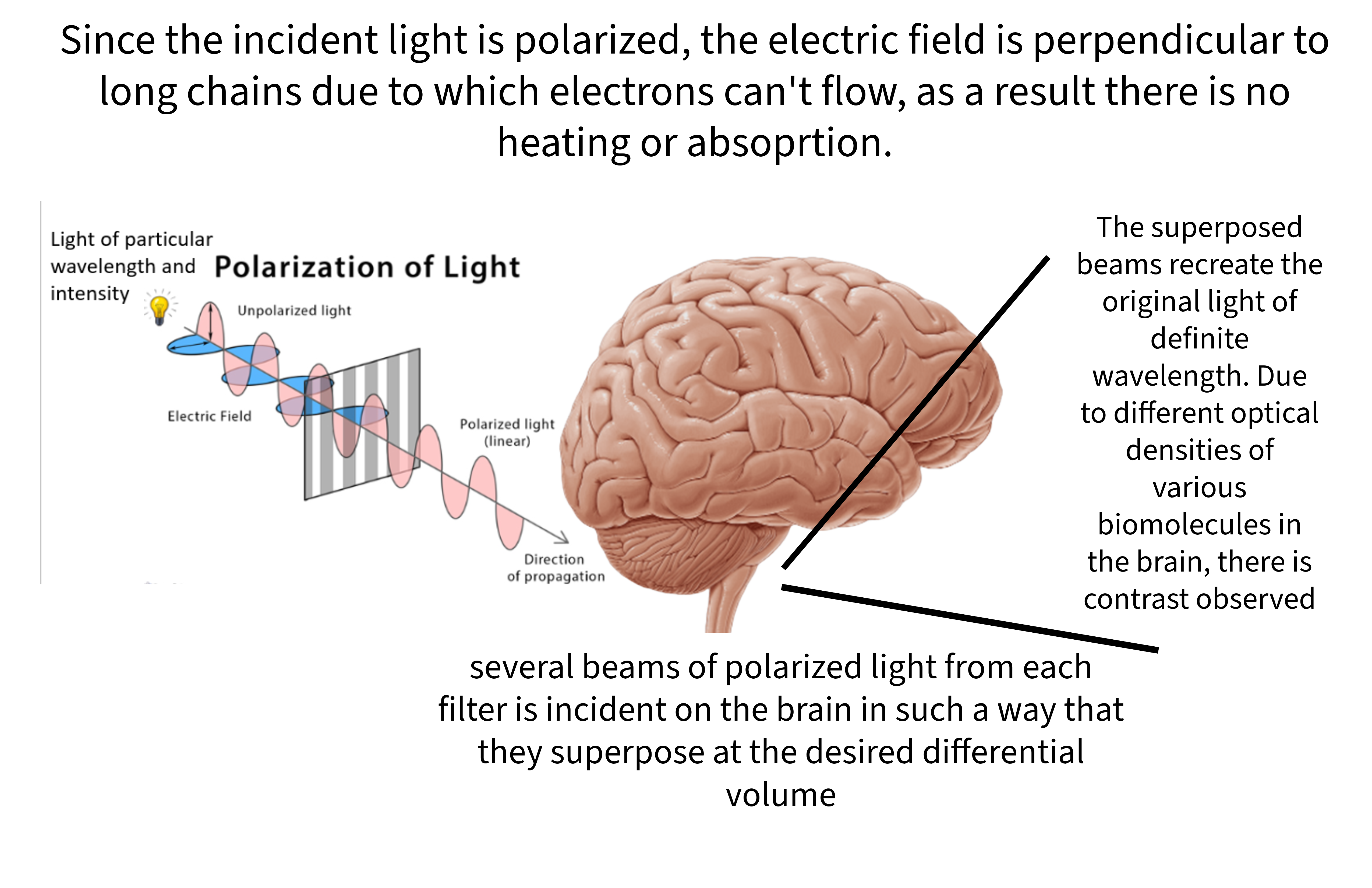
PART 4
-

PART 5


Inspiration:
*INSTANCE 1: * I have been a victim of anxiety and depression for the past 7 years. While I was prescribed medicines to manage my condition, I realized that unlike other physical health conditions, mental health issues had no definite diagnostic method, and hence the exact mental condition and the appropriate dosage was also only "estimated" by the doctor. *INSTANCE 2: * While my parents were undergoing a full-body checkup as part of their routine survey, I noticed that there was no test to monitor mental health and brain functionality. This was another key moment which made me realize the importance of diagnosing mental health issues and monitoring brain health. As a consequence, I researched thoroughly about the scientific breakthroughs in neuroscience and found that most neurobiological phenomena have been very less understood. Imaging tests are almost inconclusive when it comes to diagnosing brain diseases. Of those brain diseases I researched, the most documented study was about the neurodegenerative disorder Parkinson's disease (although the exact underlying cause as well as molecular mechanism are yet unknown). This is an incurable disease which manifests itself after almost 80% neurodegeneration has occurred, making it hard to detect and impossible to cure. It struck me that if a method of early diagnosis is found, it has the potential to save many lives. Additionally, if the extent of neurodegeneration is known, then there are better chances of treating the disease. This project is thus aimed at diagnosing the extent of neurodegeneration of dopamine neurons in the substantia nigra, located in the midbrain.
What it does
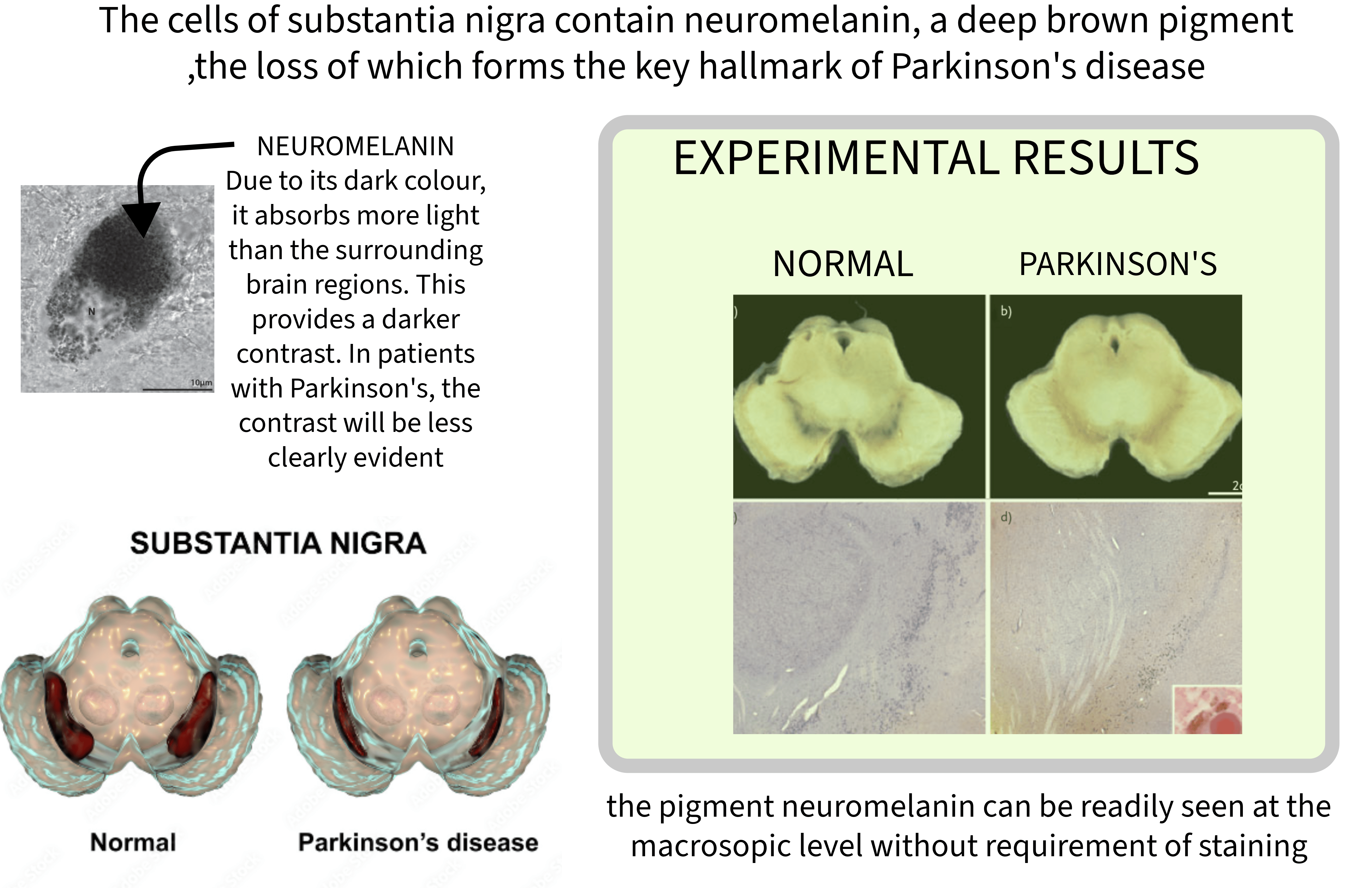
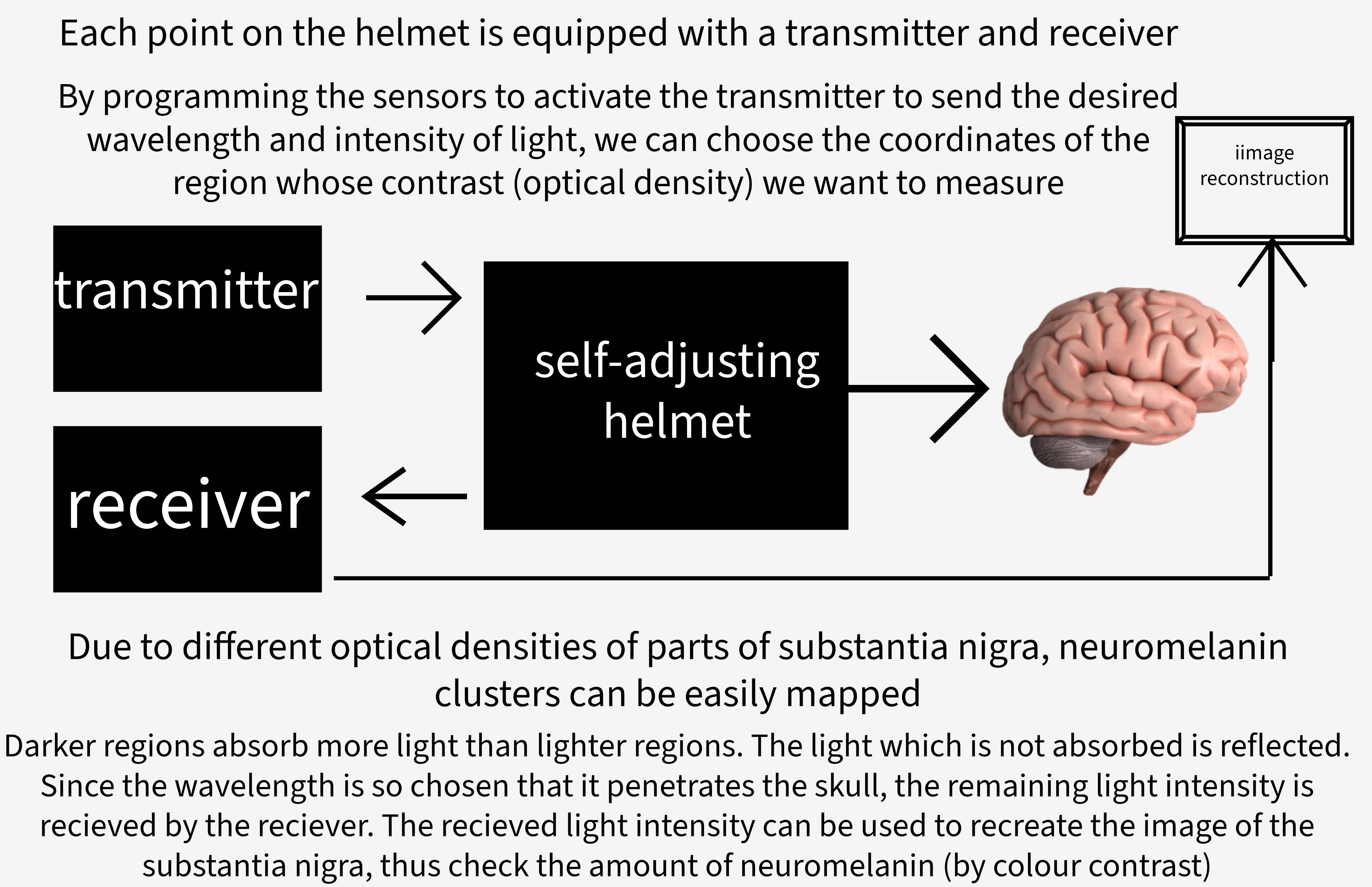
Parkinson's disease results from the neurodegeneration of dopaminergic neurons in the substantia nigra pars compacta, a structure located in the basal ganglia. A distinctive feature of dopaminergic neurons is the presence of deep brown pigment neuromelanin (which is subsequently depigmented in Parkinson's disease). This device uses mathematical models to set the volume of the brain whose optical density is to be quantified. A flexible helmet which adjusts according to the shape of the patient's head is equipped with sensors (transmitter and receiver). Once the coordinates of the brain region to be surveyed is determined, the transmitters in those regions are activated to send partial polarized light along the 3 dimensions. Once the polarized light beams meet within the brain, they superpose to form the light of characteristic wavelength and intensity (from which the initial polarized beams were derived). Due to the deep pigmentation of neuromelanin, it absorbs most of the incident light. The rest of the light which is not absorbed gets reflected from the brain and reaches the receiver. By integrating the intensities of light received, image reconstruction takes place. A healthy brain will have proper contrast due to the deep pigment neuromelanin while people with Parkinson's disease have very less contrast. By incorporating huge number of sensors on the helmet, the clarity of image reconstruction can be enhanced such that Parkinson's disease can be diagnosed even before symptoms occur (based on minute changes in contrast observed)
How we built it
Fundamental concepts of technology (IoT) and modern science (polarization of light) have been primarily used in the project. Application of AI may be used for image reconstruction, although it hasn't been used in this project. C coding language has been used to code for activating transmitters at desired coordinate locations to allow polarized light to be incident on the brain. Polarizing filter has been used to polarize light of a particular wavelength and intensity in such a way that all polarized beams recombine by superposition at the desired volume of the brain. Transmitter and receiver units are present on the helmet for producing high quality images of the desired brain region.
Challenges we ran into
The structure, synthesis and metabolism of neuromelanin are yet unknown. What has been documented so far is that neuromelanin comprises of deep pigmented granules, predominantly concentrated in the substantia nigra. With limited known properties, it was difficult initially to decide on the further course of the project. Designing multiple miniature transmitters and receptors was quite difficult and expensive. (However, if this technique is restricted to hospital use, then the issue of design and cost can be mitigated)
Accomplishments that we're proud of
Without the need to know the structure, physical and chemical properties of neuromelanin, we have developed a means of quantifying neuromelanin levels in the substantia nigra. This device makes use of fundamental concepts of the various phenomena of light (polarization, reflection, absorption, image construction). With this method, various molecular properties of the brain can be easily studied. This device can be supplied to hospitals and its application beyond Parkinson's disease diagnosis can be studied.
What we learned
While preparing for this project, I gained a much better understanding about the complexity of the brain and the way in which the myriad variety of brain cells communicate in order to maintain harmony in the body. Additionally, I also got to explore the working of various IoT devices and sensors. It was my first time working with IoT on a problem that had no sophisticated solution yet. The complexity of the brain makes it very difficult to diagnose and cure mental health conditions, nonetheless, this project is an attempt to help doctors and scientists with Parkinson's disease diagnosis with sufficient accuracy.
What's next for Prognosing Parkinson's disease
This method can be extended to survey several other brain regions by means of measuring the optical density of various tissues and cells. The advantage of measuring neuromelanin levels was that the deep pigmentation observed even with the naked eye allowed the use of fewer number of sensors (transmitters and receivers) for image reconstruction. This method can also be extended in order to image different regions of the body, since it is a non-invasive and non-toxic method (polarized light doesn't produce any heating effect or ionization, thus safe for cells). In order to enhance the accuracy of Parkinson's disease diagnosis, AI models for image reconstruction can be used.
Built With
- c
- filter
- image
- iot
- sensors

Log in or sign up for Devpost to join the conversation.