-
-

working principle- image 1
-
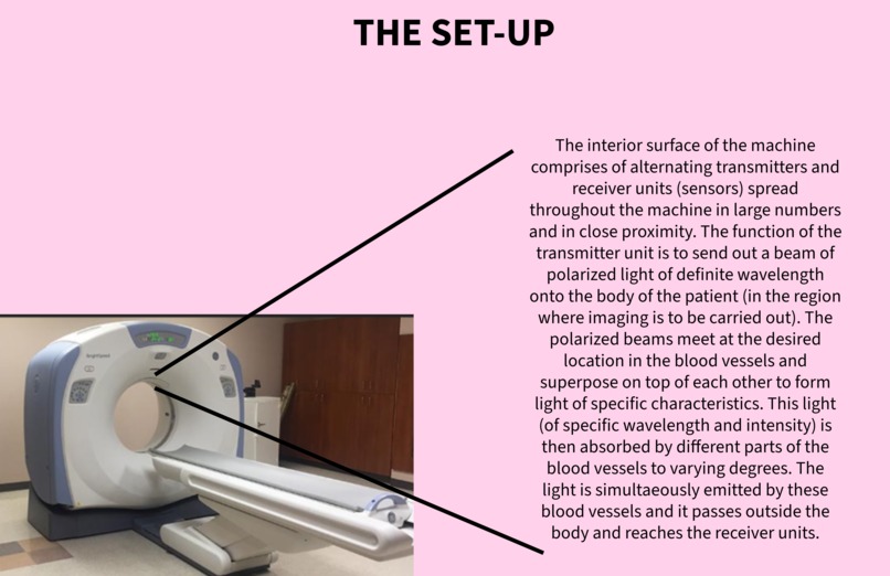
image 2
-

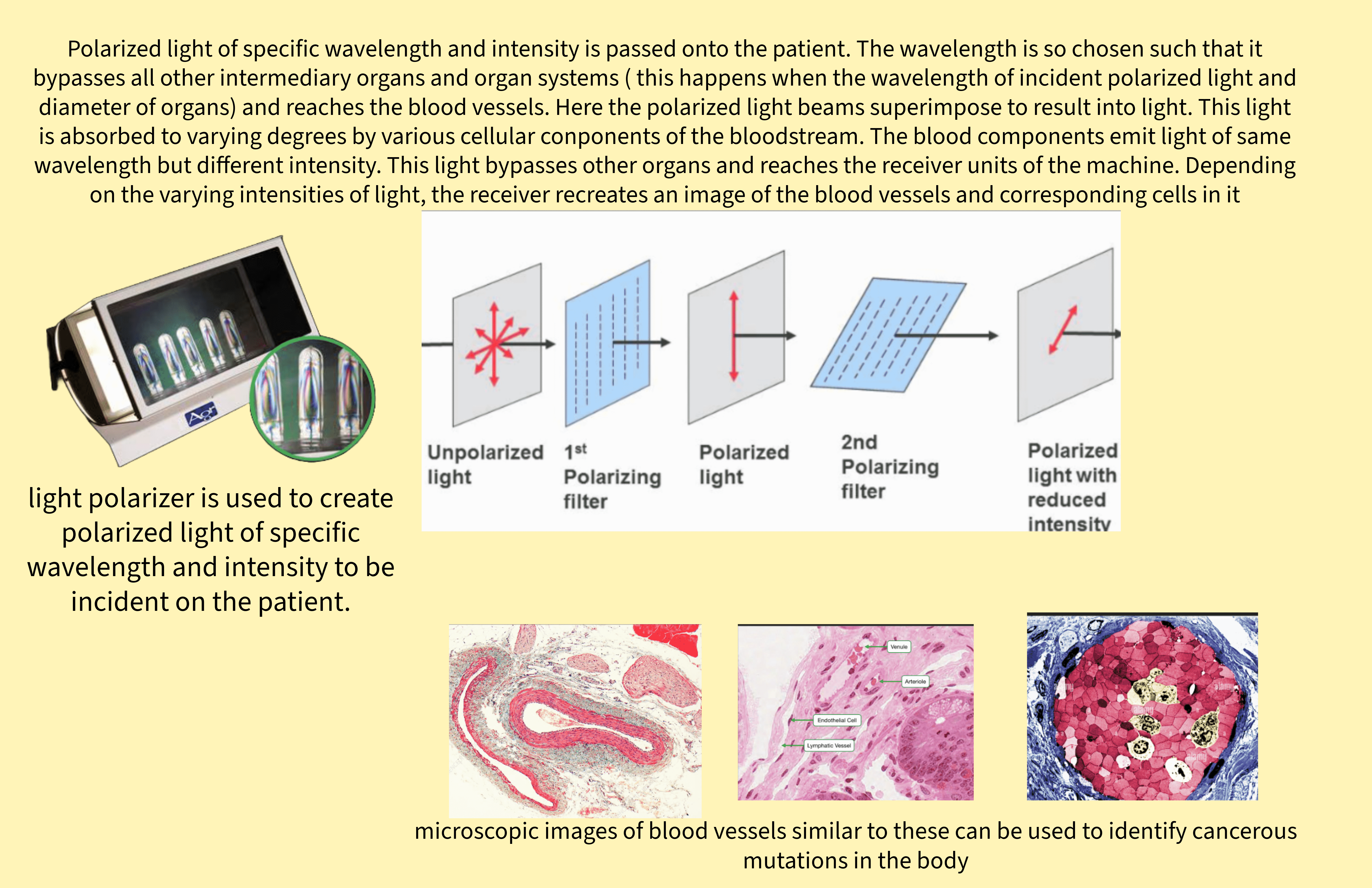
image 3
-
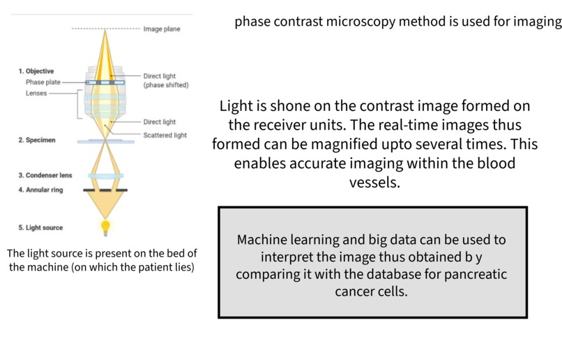
image 4


Inspiration
In 2022, approximately 20 million new cancer cases were diagnosed worldwide, and 9.7 million people died from the disease. By 2050, the number of cancer cases is projected to increase to 35 million due to population growth. Globally, cancer is a leading cause of death, accounting for nearly 10 million deaths in 2020, which is about one in six deaths. Due to its late diagnosis and rapid spread, it is highly crucial to develop high accuracy, non-toxic and non-invasive methods of diagnosis. While the method proposed in this project can be used to detect various forms of cancer, it is particularly used to detect pancreatic cancer (due to the location of the pancreas, which makes it difficult to study via imaging methods and also due to the rapid metastasis of pancreatic adenocarcinoma).
What it does
This device is used to form high quality images of the cellular components of blood vessels surrounding the pancreas. This device uses phase contrast microscope principle to form real-time cellular-level images of the blood vessels and compares the results with standard database of cancerous cell hallmarks by using ML models. If the results obtained by the test match with the database of malignancies, then it is indicative of pancreatic cancer or other gastrointestinal cancers (since we've carried out the test on those blood vessels surrounding the pancreas and recorded the images instantaneously). The targeted blood vessels are mainly those that are localized to the pancreas or other gastrointestinal organs. In addition to using multiple sensors (transmitters and receivers) to send out polarized light and to receive contrasted images, we have used phase plate and condenser annulus to form high resolution images. Since we've used polarized light, it doesn't impart any energy (in the form of heat) to the cells. Thereby it is a safe and non-invasive method of diagnosis.
How we built it
I've used the principles of microscopy (especially phase contrast microscopy) to build this device. This device also makes use of sensors, light polarizers and ML models to accurately determine the hallmarks of pancreatic cancer in blood in real time. The sensors use Raspberry pi model to form IoT interface.
Challenges we ran into
1)The location of the pancreas made it difficult for me to use microscopy principles directly on it, instead I had to choose the surrounding blood vessels to check for cancer hallmarks. This method is less efficient than would have been if I had directly conducted my study on the pancreas. 2) While using large volumes of data (by using ML and big data models), it is highly essential to choose the right information to obtain high accuracy of results (dealing with large amount of data is complex) 3) Due to the use of a number of complex components, the machine becomes bulky and expensive. Thus, it cannot be aggregated into homes but can be used in hospitals. This method however can also be extended to different cancers (not just pancreatic cancer) and also to detect other disease hallmarks (slight modifications to the machine with similar framework will be required). Thus, it can become an essential component of hospitals for disease diagnosis.
Accomplishments that we're proud of
Since this method uses principles of microscopy, this eliminates the need to use AI image reconstruction models to obtain high quality images. Lenses of desired focal lengths are sufficient to produce high resolution images. The location of the pancreas will not hinder the diagnosis, since we are tracking for cancerous cells within the blood vessels surrounding the pancreas. Additionally, this is a non-invasive method and hence can be preferred over invasive methods of diagnosis.
What we learned
This project enabled me to appreciate the various technological tools used in the medical industry and also to understand the shortcomings of each method. Despite the advances made in science and technology, there are various barriers when it comes to healthcare, since accuracy is of paramount importance. By using the concepts of physics and amalgamating it with technology, I came up with the idea highlighted in this project. It really helped me gain a better understanding of the human body and the intricacy with which all the organs work together in order to sustain us.
What's next for (Pancreatic) Cancer Predictor
This method of diagnosis can be used to identify any cancer hallmarks in blood vessels and is not only restricted to the cells of the pancreas (although it is particularly beneficial in this case, since the location of the pancreas makes it difficult to image via MRI and CT scans). With advancements in this method of diagnosis (by exploring new configurations of positioning lenses and choosing specific focal lengths and wavelengths of light beams), we can extend this method to targeting the pancreatic cells (or specific cells of the body) directly instead of surrounding blood vessels. This will furthermore increase the accuracy of diagnosis of pancreatic cancer (or any other organ/tissue-related cancers).
Built With
- bigdata
- microscopy
- ml
- polarizer
- raspberry-pi
- sensors


Log in or sign up for Devpost to join the conversation.