Welcome to PancreAI 🧬💡
Your AI-Powered Ally for Early Pancreatic Cancer Detection
🚨 The Silent Killer
Pancreatic cancer is one of the deadliest forms of cancer, often detected too late. Every year, it silently claims thousands of lives. But what if detection could happen earlier? What if AI could be the difference?
Enter PancreAI—a powerful, intelligent solution built to detect pancreatic cancer risks early, provide explainable insights, and empower patients and clinicians alike with timely, life-saving information.
🌟 How PancreAI Works
PancreAI works its magic through four core capabilities:
- 🧪 Risk Assessment: Rapid evaluation of pancreatic cancer risk using demographic, lifestyle, and clinical data.
- 🔍 Explainable Insights: Uses Cohere’s Retrieval-Augmented Generation (RAG) to clearly break down contributing risk factors.
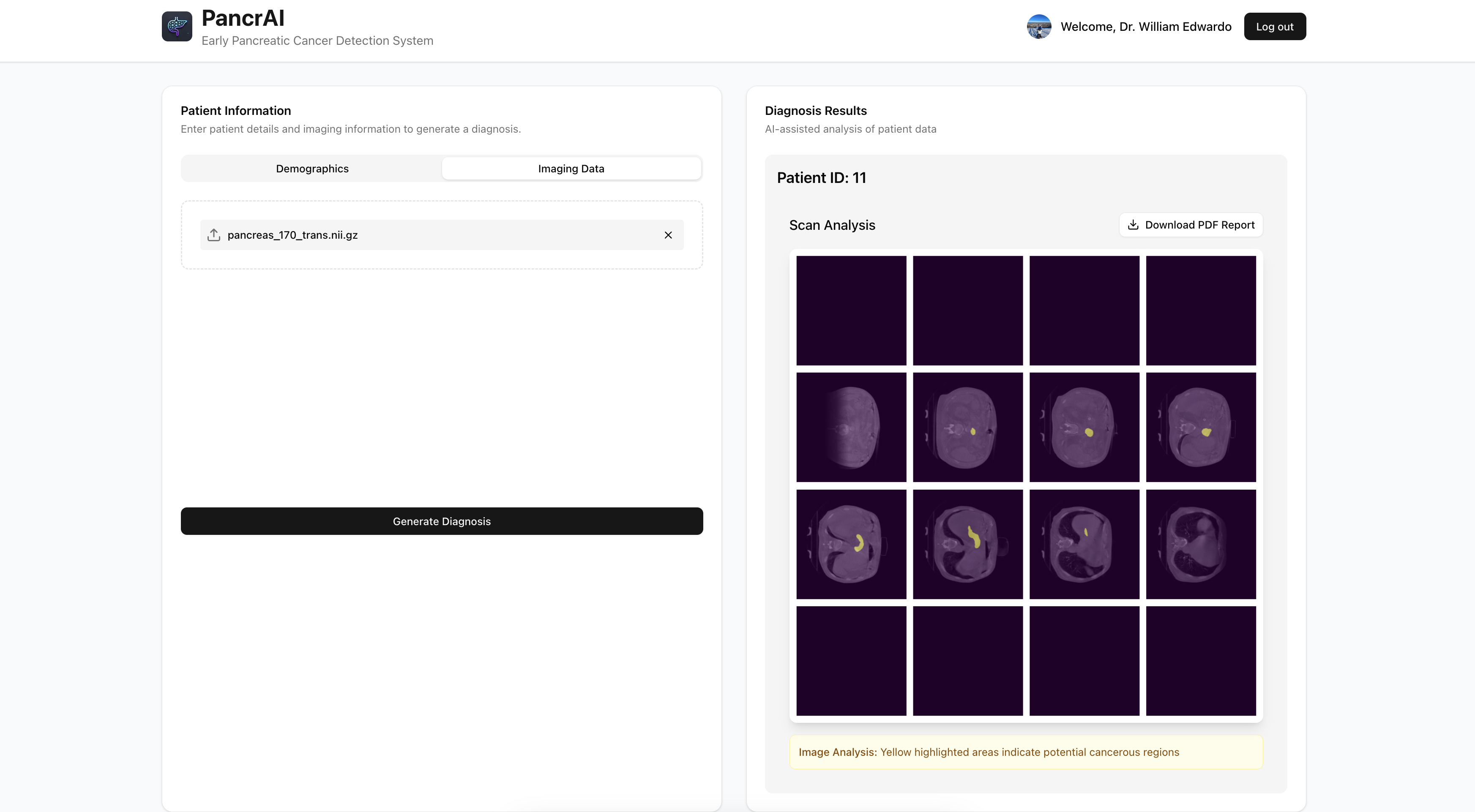
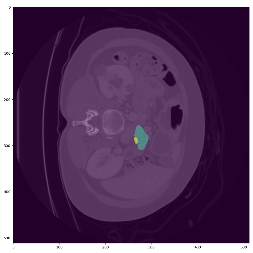
- 🖼️ Advanced Imaging Analysis: Enhances diagnostic accuracy through integrated CT scan analysis using Nvidia MONAI [1].
- 💡 Actionable Recommendations: Offers personalized, data-backed advice to encourage timely medical follow-ups.
🔧 Behind the Tech
PancreAI is built on a solid, intelligent tech foundation:
- 📊 Data: Sourced from specialized imaging datasets such as the National Institute of Cancer's TCIA. Four such datasets are used [2,3,4,5].
- 🧠 AI Modeling: Optimized LightGBM models combined with Cohere’s RAG for interpretability, coupled with Nvidia MONAI.
- 🧬 Synthetic Profiles: Synthetic patient data to enrich model explanations and diversity.
- 🌐 Frontend & Backend: Built using React, FastAPI, and Django, a fully deployed and accessible multicloud solution.
🚧 Challenges Conquered
- Balancing limited cancer patient data with synthetic generation.
- Merging high-accuracy prediction with AI explainability.
- Integrating advanced or unfamiliar tech components within a short timeframe.
- Communication under high time pressure and handling new solutions.
🎓 What We Learned
- Practical implementation of AI explainability in healthcare.
- Effective team collaboration and rapid prototyping.
- Deep understanding of medical data complexity and ethical tech development.
- Conversion and processing of over 200K DICOM and NIfTI frames [2,3,4,5].
🚀 Looking Ahead
- Partner with medical institutions for clinical validation [2,3,4,5].
- Expand to multilingual support to reach global users.
- Introduce deeper diagnostic features and real-time feedback.
- DevOps consideration for future development and compatibility.
✨ PancreAI isn’t just a hack—
It’s a lifeline.
Bibliography
[1] He, Y., Yang, D., Roth, H., Zhao, C. and Xu, D., 2021. Dints: Differentiable neural network topology search for 3d medical image segmentation. In Proceedings of the IEEE/CVF Conference on Computer Vision and Pattern Recognition (pp. 5841-5850).
[2] Roth, H., Farag, A., Turkbey, E. B., Lu, L., Liu, J., & Summers, R. M. (2016). Data From Pancreas-CT (Version 2) [Data set]. The Cancer Imaging Archive. https://doi.org/10.7937/K9/TCIA.2016.tNB1kqBU
[3] Hong, J., Reyngold, M., Crane, C., Cuaron, J., Hajj, C., Mann, J., Zinovoy, M., Yorke, E., LoCastro, E., Apte, A. P., & Mageras, G. (2021). Breath-hold CT and cone-beam CT images with expert manual organ-at-risk segmentations from radiation treatments of locally advanced pancreatic cancer [Data set]. The Cancer Imaging Archive. https://doi.org/10.7937/TCIA.ESHQ-4D90
[4] National Cancer Institute Clinical Proteomic Tumor Analysis Consortium (CPTAC). (2018). The Clinical Proteomic Tumor Analysis Consortium Pancreatic Ductal Adenocarcinoma Collection (CPTAC-PDA) (Version 15) [dataset]. The Cancer Imaging Archive. https://doi.org/10.7937/k9/tcia.2018.sc20fo18
[5] Chen, L., Wang, W., Jin, K., Yuan, B., Tan, H., Sun, J., Guo, Y., Luo, Y., Feng, S.-ting, Yu, X., Chen, M.-hu, & Chen, J. (2022). Prediction of Sunitinib Efficacy using Computed Tomography in Patients with Pancreatic Neuroendocrine Tumors (CTpred-Sunitinib-panNET) (Version 1) [Data set]. The Cancer Imaging Archive. https://doi.org/10.7937/SPGK-0P94


Log in or sign up for Devpost to join the conversation.