-
-
-
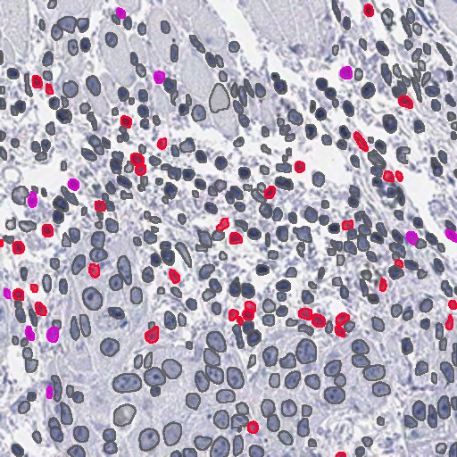
Co-Expression in sequential staining of a serial section (re-stained)
-
-
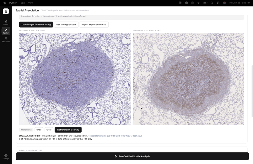
Landmark certification (passes)
-
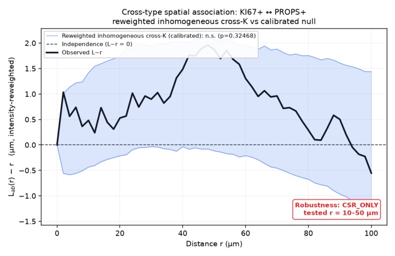
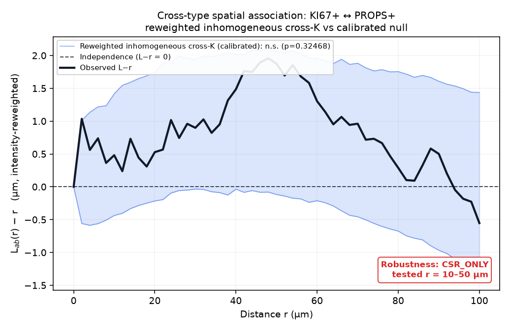
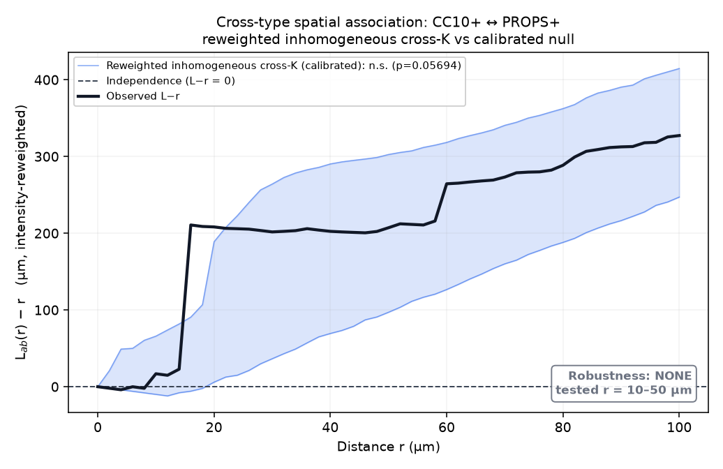
L−r null plot
-
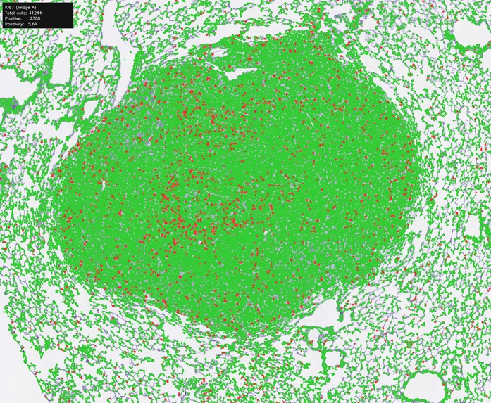
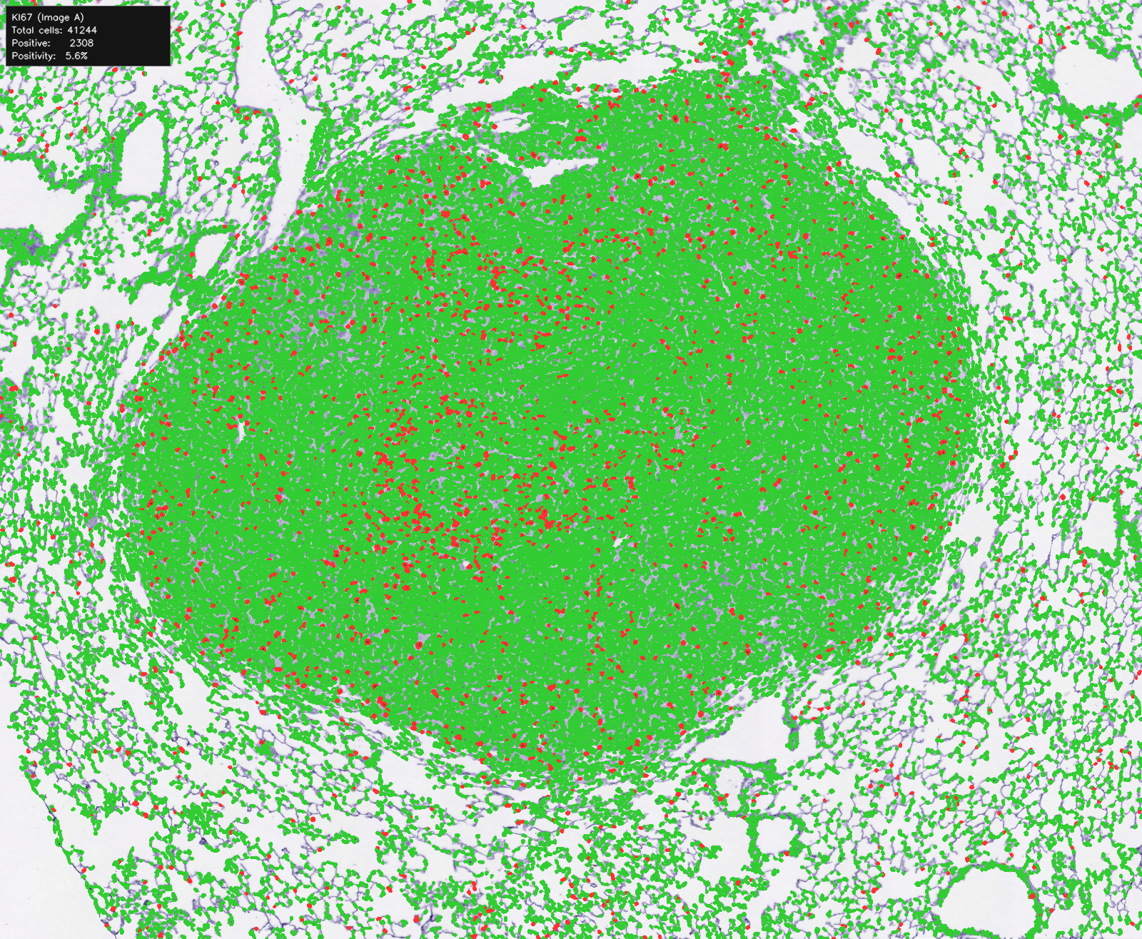
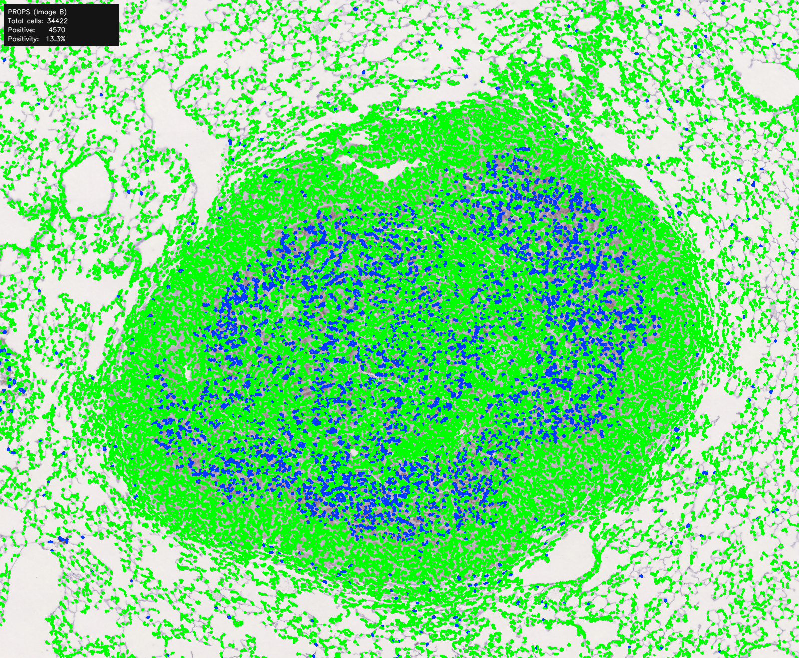
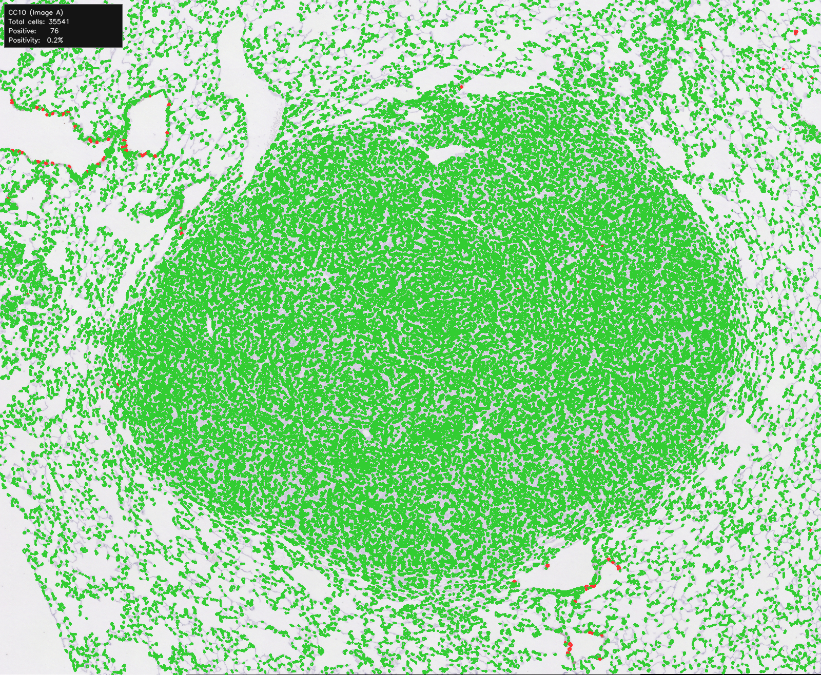
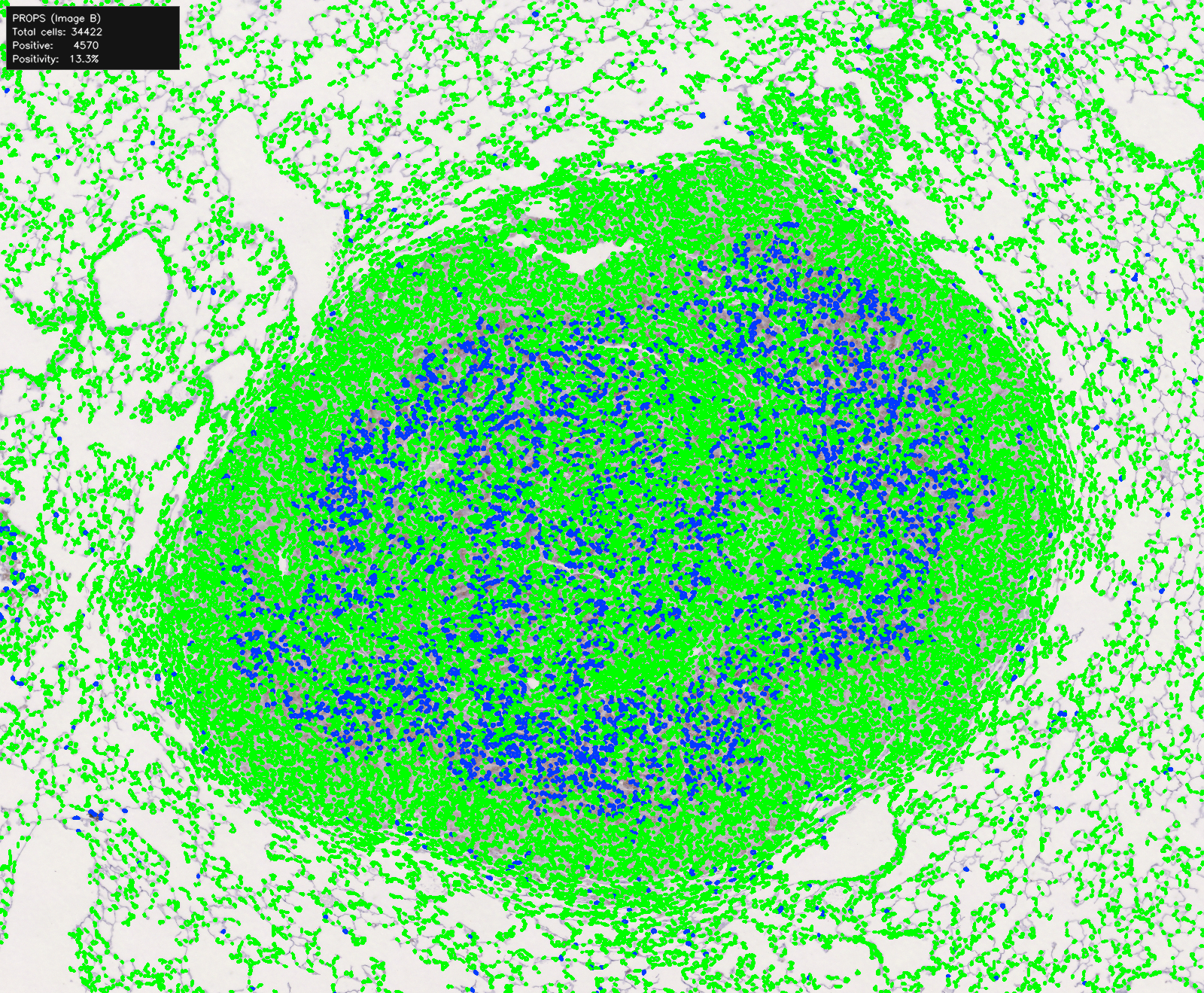
Spatial association: lung-lesion segmentation overlays
-
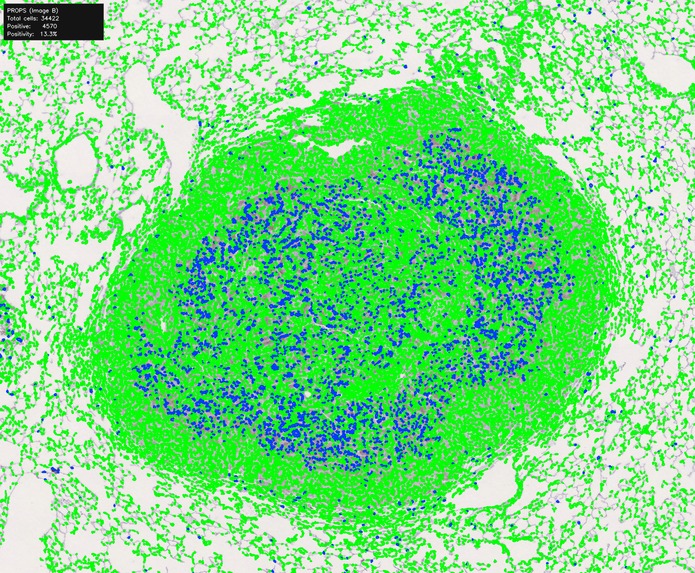
Spatial association: lung-lesion segmentation overlays
-
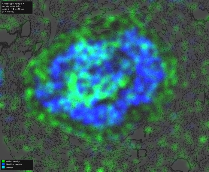
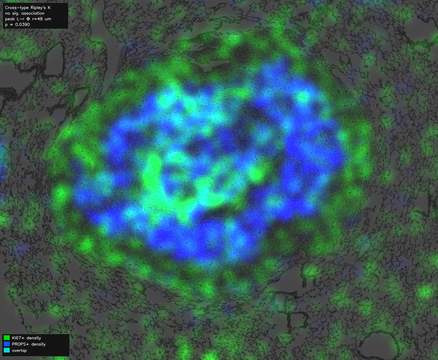
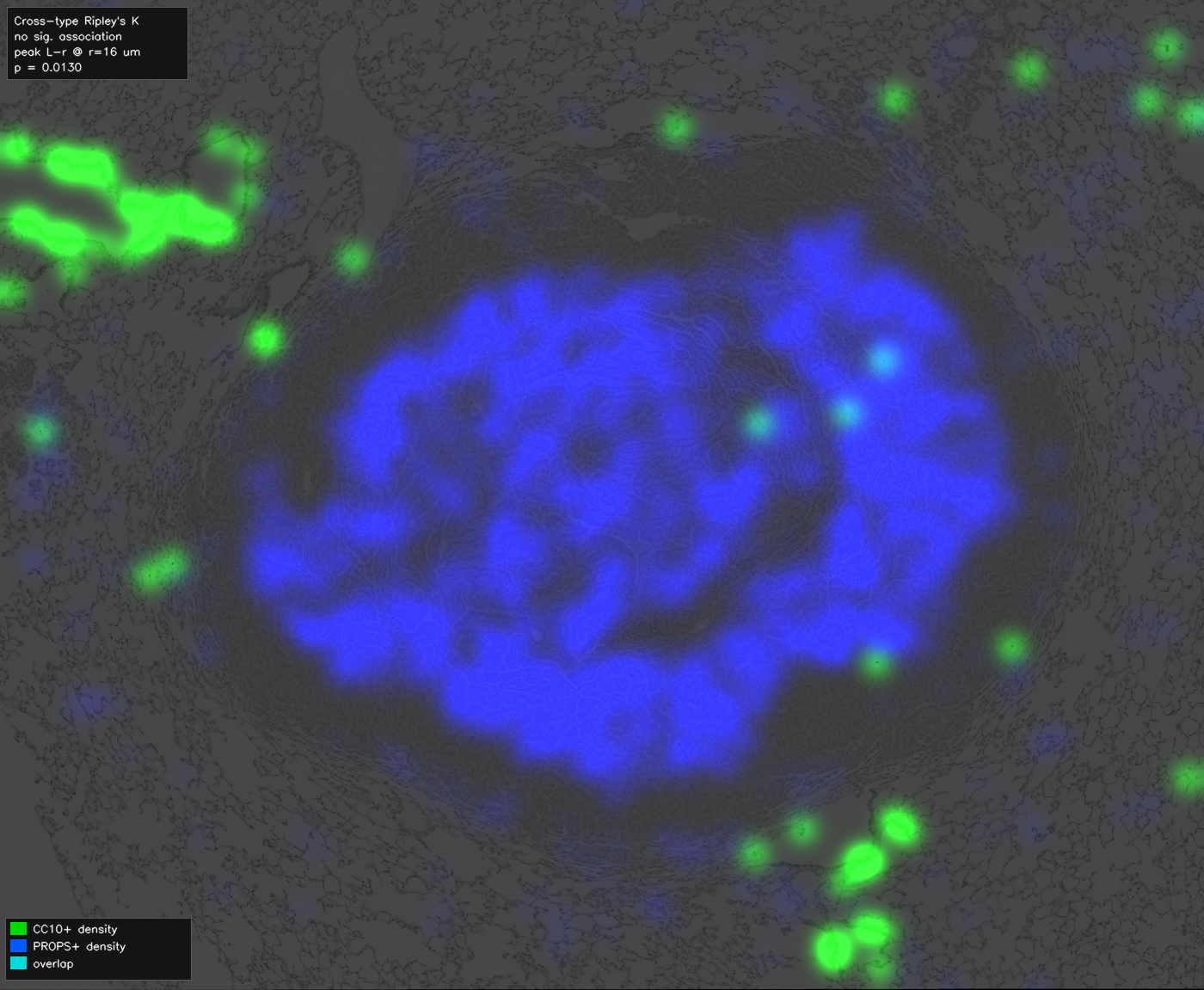
Density overlay
-
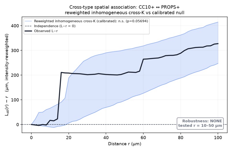
L−r null plot
-
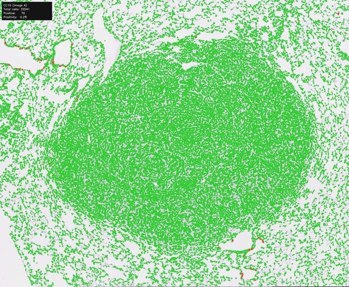
Spatial association: lung-lesion segmentation overlays
-
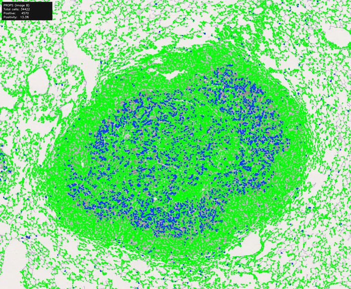
Spatial association: lung-lesion segmentation overlays
-
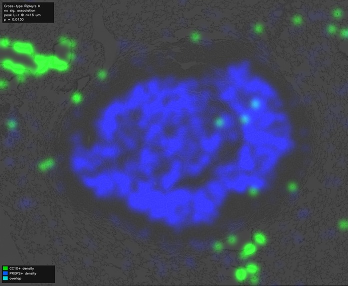
Density overlay
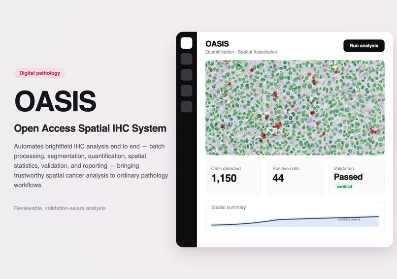
OASIS
Open Access Spatial IHC System
OASIS is a computational pathology platform that unifies the entire brightfield immunohistochemistry (IHC) workflow — from batch processing and segmentation to quantification, spatial analysis, validation, and reporting — into a single reproducible computational instrument. Every computational conclusion is explicitly validated before it reaches the researcher.
🚀 Try the Interactive Demo (recommended)
The best way to understand OASIS is to use it. Walk the complete workflow — from loading pathology images to reviewing a validation-aware report — in about two minutes, on preloaded example data. No installation, no setup, no uploads.
- 🔗 Interactive Demo: https://oasis-interactivedemo.vercel.app
- 💻 GitHub Showcase: https://github.com/smukilan9-ship-it/OASIS-showcase
Computational pathology has a software problem
Modern tissue analysis is rarely limited by a lack of algorithms. It is limited by fragmented workflows, disconnected software, expensive infrastructure, and the difficulty of determining which computational results can actually be trusted.
Researchers routinely move between independent tools for segmentation, quantification, registration, spatial statistics, visualization, spreadsheets, and reporting — manually stitching together an analysis pipeline while assuming every intermediate result is scientifically valid.
OASIS reimagines that entire process as a single computational instrument.
Unlike conventional digital pathology software that focuses on individual stages of analysis, OASIS unifies the complete workflow into one reproducible platform where every computational conclusion is accompanied by explicit validation before it reaches the researcher.
What it does
OASIS is a working desktop and command-line platform. It takes the images a pathology lab already produces and turns them into cell-level measurements, spatial maps, and validation-aware reports — through one guided workflow instead of a folder of disconnected tools.
OASIS is built around large-scale pathology workflows rather than single-image analysis. Entire cohorts can be processed automatically through one reproducible pipeline — 18 slides, 4,911 nuclei, 0 errors, in a single click — before users move into three complementary analysis paths:
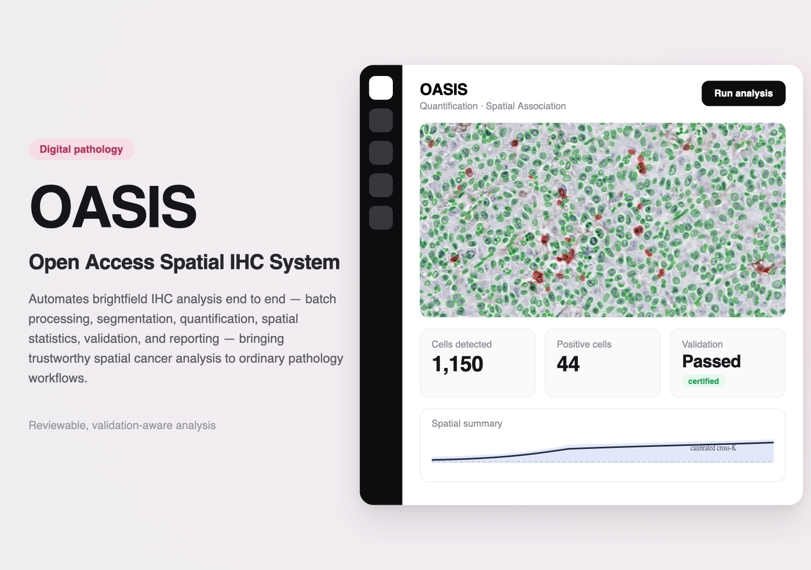
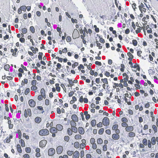
- Quantification — detects nuclei and counts marker-positive cells from ordinary brightfield IHC. In the demo: 1,150 nuclei, 44 DAB-positive (3.8%) on a tumour slide.
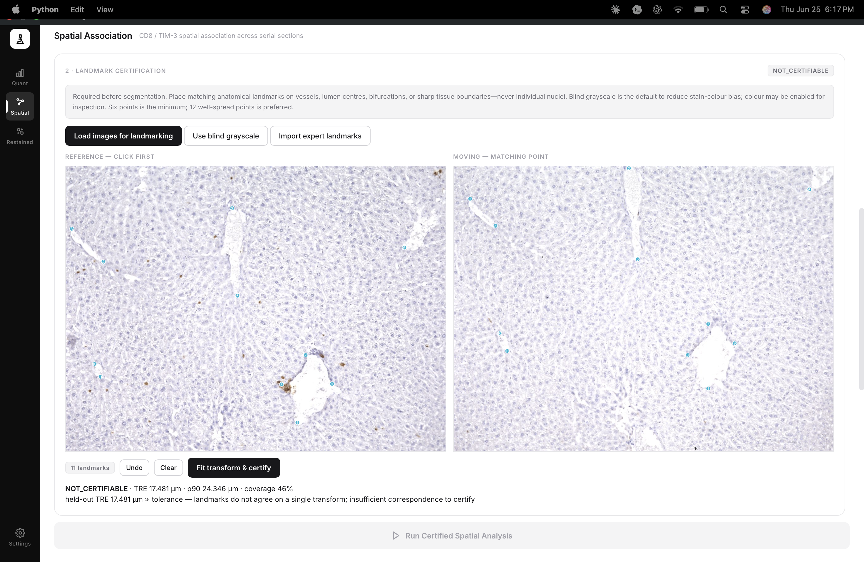
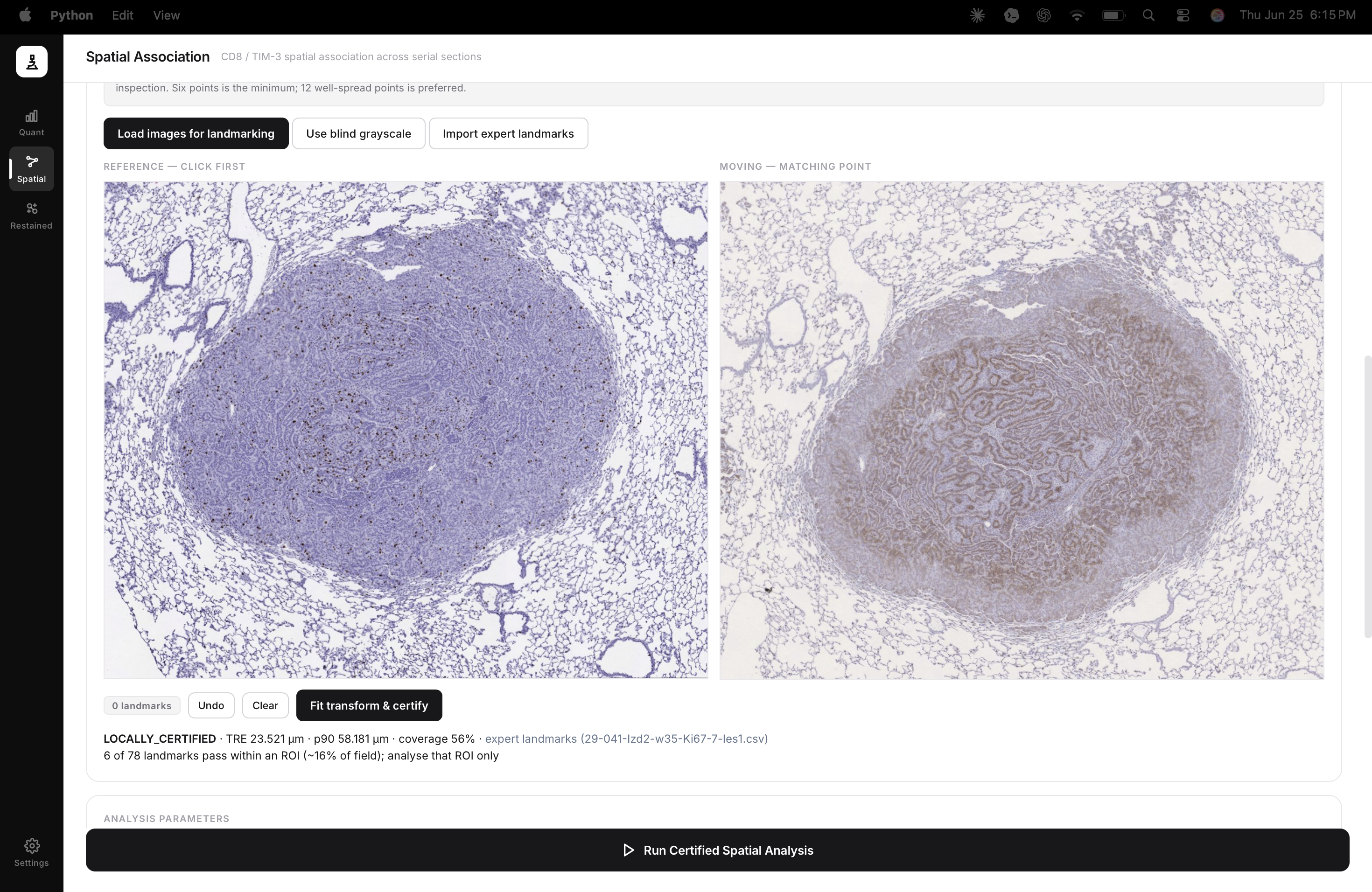
- Serial-section spatial association — compares two cell populations across adjacent sections as a population-level spatial relationship (never single-cell co-expression — they are different physical slices).
- Same-section restained co-expression — calls true single-cell co-expression only when the stains share one physical section and correspondence is certified. In the demo: CD8 vs FOXP3 — 28 CD8⁺, 11 FOXP3⁺, 0 double-positive across 439 cells.
The defining characteristic is not that OASIS automates analysis. It is that when registration cannot be certified, correspondence cannot be established, or the evidence is insufficient, the platform communicates uncertainty instead of producing an unsupported conclusion. Scientific restraint is a feature, not a limitation.
Why existing software falls short
Existing digital pathology software often excels at solving a single computational task — segmentation, registration, visualization, or quantification — but the researcher is still responsible for integrating those outputs into a coherent, defensible scientific workflow.
This leaves three persistent gaps:
- fragmented software ecosystems,
- limited reproducibility,
- and uncertainty about which computational conclusions are scientifically defensible.
OASIS is built to close all three at once.
Validation as a first-class feature
OASIS was designed around a simple principle: scientific software should validate conclusions, not merely compute them. Throughout development, that principle was tested with real data and hard numbers:
- Segmentation validated against independent expert annotations — micro-F1 0.776 against expert masks across 268 tiles from 8 patients (an external HNSCC comparison set), the harder external number rather than a flattering pilot.
- Spatial statistics independently verified — the cross-type estimator agrees with the R
spatstatreference to ~10⁻¹⁴. - A null model rejected after it failed its own test — an early significance model fired on 85–100% of pure-noise data; it was rebuilt to 3.2% false positives while retaining 100% / 99.2% power to detect planted attraction.
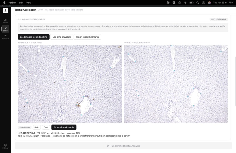
- The platform refusing a false positive — on a real certified serial pair, a naïve test reported a strong association (p = 0.001). After correcting for shared tissue preference, OASIS returned p = 0.32 and flagged it as a shared-tissue artifact rather than a biological finding. Same data. Two nulls. The design is what stopped the wrong answer from being believed.
These were not implementation details. They fundamentally shaped the platform's design philosophy.
Democratizing computational pathology
While advanced spatial-biology platforms keep expanding what can be measured biologically, OASIS expands who can participate in computational pathology.
Picture a cancer research laboratory with 15,000 archived brightfield IHC slides collected over a decade but no access to a multiplex imaging platform. Today, much of that data remains computationally underutilized. OASIS cannot create biological information that was never captured — but it can dramatically expand the amount of trustworthy computational analysis that laboratory can perform using the pathology infrastructure it already possesses.
By building on routine brightfield IHC — the infrastructure already present in hospitals and research laboratories worldwide — this free, open-access platform reduces computational barriers without lowering scientific standards.
How it was built
OASIS combines established digital-pathology tools with a deterministic, reproducible pipeline:
- QuPath + InstanSeg for nucleus segmentation,
- Python for orchestration, measurement, overlays, exports, and reporting,
- pywebview + HTML/JavaScript for the desktop interface,
- R
spatstatas an independent reference for validating the spatial-statistics engine.
Every stage of the workflow is reviewable, reproducible, and traceable.
Challenges
The hardest problem was never producing a graph — it was preventing the wrong graph from becoming a claim. Each challenge was an engineering decision about where to draw that line: enforcing the boundary between serial-section association and same-section co-expression in code; moving to landmark-certified registration after automated quality metrics proved unreliable; rebuilding the spatial null after stress testing exposed unacceptable false-positive rates; and unifying every stage into one coherent platform that communicates uncertainty rather than hiding it.
Accomplishments
- Built a complete desktop + command-line digital pathology platform on commodity brightfield IHC.
- Unified batch processing, segmentation, quantification, spatial analysis, validation, and reporting into one reproducible workflow.
- Automated entire pathology experiments rather than single images.
- Validated segmentation and spatial statistics against independent references, with the failures kept on the record.
- Engineered validation-aware gates that block or flag unsupported outputs instead of reporting them.
Rather than maximizing positive findings, OASIS is designed to maximize defensible scientific evidence.
Learnings
The future of computational pathology will not be defined by software that produces more results — it will be defined by software that produces more trustworthy ones. Lowering computational barriers should never require lowering scientific standards; OASIS was built around the opposite philosophy: accessibility and scientific rigor should reinforce one another. The project evolved from solving individual computational problems into designing an instrument that makes advanced tissue analysis more accessible while remaining transparent about its own limits.
What's next
Grounded next steps: more demonstration data to exercise the same-section co-expression path end to end, expert cell-level labels to validate marker thresholds, broader validation across additional tissue types, and continued refinement of the interactive product experience.
Built with
Python · QuPath · InstanSeg · SimpleITK · OpenCV · NumPy · SciPy · Shapely · matplotlib · pywebview · HTML/CSS/JavaScript · R · spatstat
Honest scope
OASIS is a research method-development platform — not a clinical diagnostic tool, and it makes no biological claims and does not replace advanced multiplex imaging. Serial sections support population-level spatial association only; same-section restaining supports true co-expression only when correspondence and thresholds are certified. The example datasets are used to demonstrate and validate the software.
The next era of computational pathology will not be defined solely by better imaging systems — but by computational instruments that make advanced tissue analysis reproducible, trustworthy, and widely accessible. OASIS is a working blueprint for that future.

Log in or sign up for Devpost to join the conversation.