-
-
Neura Health
Neura Health Simulator
Problem Statement
Medical imaging analysis is vital for early disease detection but remains a resource-intensive and time-consuming process. Doctors face enormous workloads, and diagnostic errors can have serious consequences. Deep learning models offer immense promise in assisting clinicians, but real-world deployment is complex, requiring significant computational resources, stable environments, and seamless integration into workflows.
We identified the urgent need for a lightweight, intuitive, and effective solution that could support healthcare professionals by enabling fast, accessible diagnostic insights from medical images. Our project aims to bridge this gap by offering an interactive simulator that mimics AI-driven analysis, setting the foundation for future deployment of fully trained models in clinical settings.
Why Our Solution Matters for Doctors
- Rapid Triage: Doctors can quickly upload chest X-rays, brain MRIs, or lung CT scans and receive immediate diagnostic feedback, aiding faster decision-making.
- Early Detection: By offering a preliminary assessment, it supports early intervention strategies, which are critical for conditions like pneumonia, brain tumors, and lung cancer.
- Ease of Use: A simple, visually appealing, web-based interface requires no specialized training or installation.
- Scalability: Future integration of real-time AI models will allow doctors to seamlessly upgrade diagnostic capabilities without changing workflows.
- Reduced Burnout: Tools that assist in initial screenings can alleviate cognitive load and improve overall efficiency.
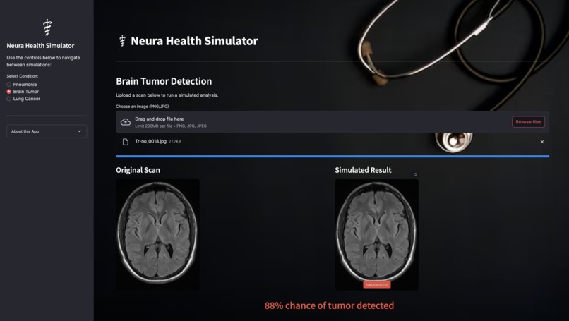
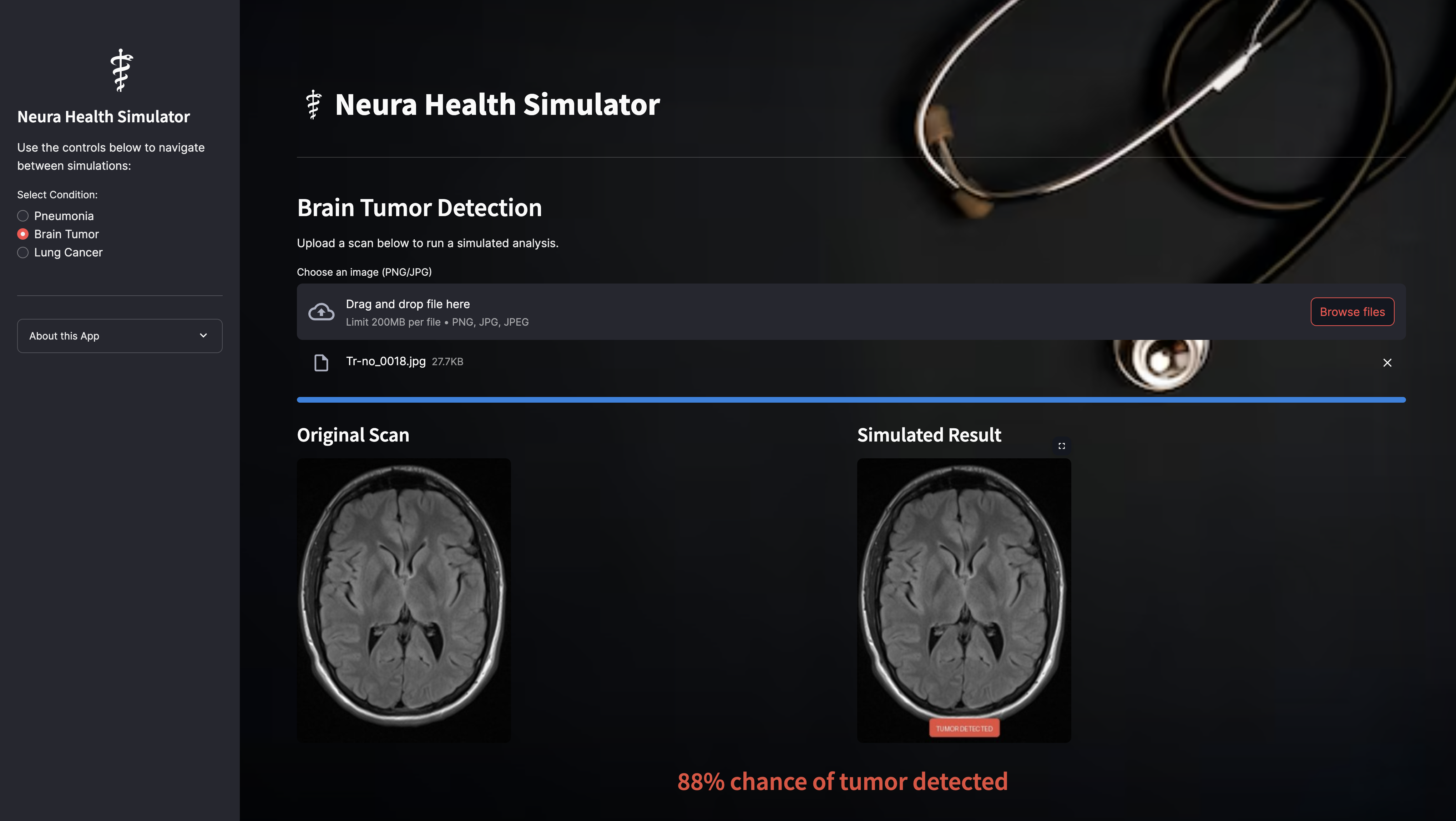
What It Does
- Condition Selection: Choose between Pneumonia, Brain Tumor, and Lung Cancer detection.
- File Upload: Uploads medical scans in PNG or JPG format.
- Visual Analysis: Animated analysis to simulate model processing.
- Result Display: Clearly labeled output (e.g., "NORMAL", "PNEUMONIA") directly overlaid on the scan.
- Professional UX: Full-screen background, modern fonts, intuitive button layouts, medical snake logo branding (⚕️).
Models We Worked On
Pneumonia Detection Model
- Architecture: DenseNet121 (transfer learning from ImageNet)
- Preprocessing: Resized to 224×224 pixels; pixel normalization [0, 1]
- Training:
- Binary Crossentropy Loss
- Adam Optimizer
- Accuracy and AUC Metrics
- Data augmentation (random flips, rotations)
- Libraries: TensorFlow 2.16, Keras, scikit-learn
Brain Tumor Detection Model
- Architecture: Custom CNN with multiple Conv2D and MaxPooling layers
- Preprocessing: MRI scans resized to 128×128 pixels
- Training:
- Binary Crossentropy Loss
- Adam Optimizer
- Accuracy, Precision, and Recall Metrics
- Libraries: TensorFlow, Keras, OpenCV
(Integration of Lung Cancer model is pending.)
Libraries Used
- Streamlit: Core framework for rapid UI prototyping
- Pillow (PIL): Image processing
- NumPy: Numerical computations
- TensorFlow/Keras: Deep learning model development
- Matplotlib: Training visualizations
- OpenCV: MRI image preprocessing
- Requests/Base64: Asset handling
How We Built It
- Customized CSS with background blur and overlay shadows
- Responsive design with Streamlit's
columnsfor side-by-side image comparison - Embedded base64 assets to eliminate dependency on external file servers
- Progress indicators and animated loaders to enhance user trust during analysis
Challenges We Ran Into
- Managing TensorFlow version conflicts for multi-model workflows
- Streamlit deprecation updates and maintaining compatibility
- Designing responsive, visually clean UI across devices
- Preparing backend APIs for seamless future model integration
Accomplishments We're Proud Of
- Delivering an intuitive, professional diagnostic tool prototype
- Building and training pneumonia and brain tumor detection models with real datasets
- Ensuring a seamless flow from condition selection to scan analysis to feedback display
What We Learned
- Thoughtful UX is just as critical as model accuracy in healthcare tools
- Backend flexibility and frontend polish can co-exist even in early prototypes
- Bridging AI potential to clinical reality requires user-centered design thinking
Future Plans
- Model Deployment: Integrate real trained DenseNet121 and CNN models
- Explainability: Grad-CAM visualization to highlight important diagnostic regions
- Secure Uploads: Ensuring HIPAA-compliant temporary image storage
- Cloud Hosting: Deployment on Streamlit Cloud or AWS
- Expand Scope: Adding skin cancer detection, diabetic retinopathy analysis, and more
This README was generated as part of the Neura Health Simulator project, focused on empowering doctors with AI-driven insights through seamless, interactive medical diagnostics.


Log in or sign up for Devpost to join the conversation.