-
-
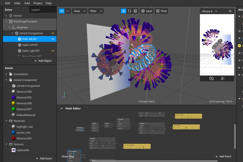
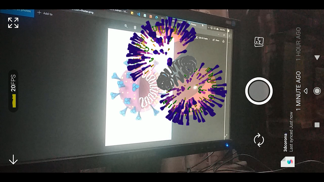
Testing with Spark-AR Player
-
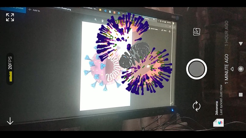
Spark-AR Studio
-
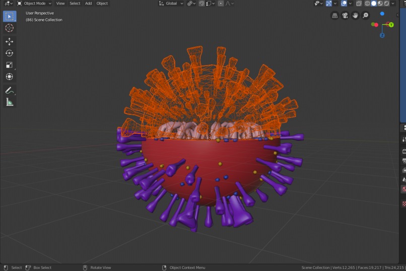
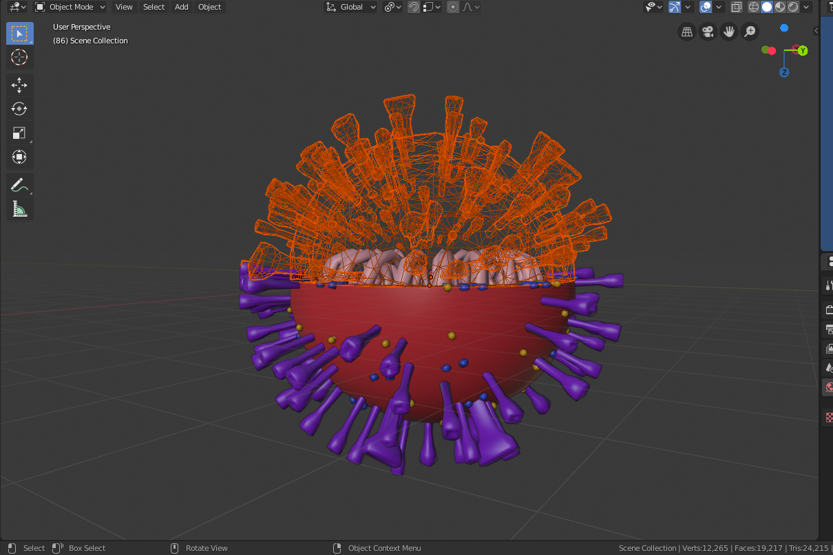
Blender - 3D Model of COVID-19
Inspiration
I always wanted to see microorganisms but a microscope is expensive and out of my reach. Later on, I got to know about Spark-AR and got this idea to make microbes on a bigger scale. He/She who faces the same problem as I, can have the experience of looking microbes under a microscope.
What it does
It shows different parts of a microbe, in this case, it's COVID-19. When tapping on the screen the microbe splits in half and shows the inner anatomy. It will only show up when the camera is placed on the COVID-19 poster made by our team.
How we built it
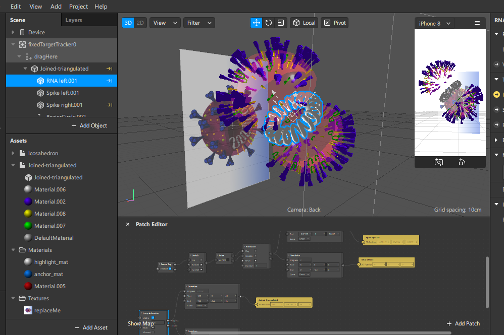
It was done in two parts, the 3D model of the microbe was made in Blender. Then the animations and functionality are done in patch editor in Spark-AR.
Challenges we ran into
While exporting the 3D model as .fbx format we could not export the transparent material of the orb that was done in Blender. But it was later added by the Material feature of Spark-AR.
Accomplishments that we're proud of
Because of Spark-AR, we gained the experience of working with AR and turned our imagination into reality.
What we learned
We learned how to take a digital 3D model and make the experience of observing it in the real-world using Spark-AR. While doing it we also learned a lot about the internal structure of the microbe.
What's next for Microbes Anatomy
We want to add more microbes such as bacteria, archaea, fungi, protozoa, algae, and viruses with detailed information. We also want to reach schools with no lab or equipment so that they can also experience and learn about the microbes.
Built With
- blender
- spark-ar


Log in or sign up for Devpost to join the conversation.