-
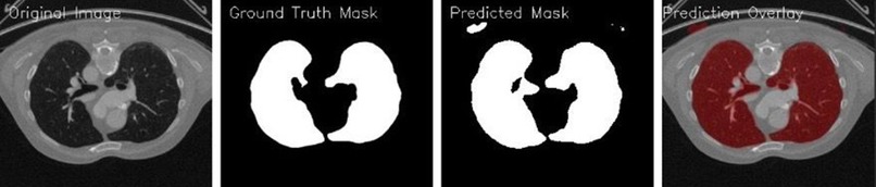
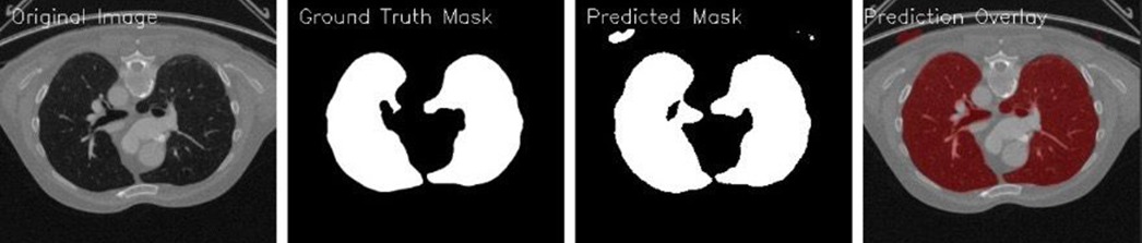
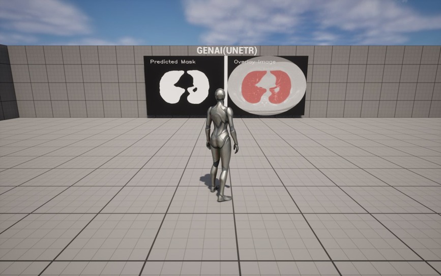
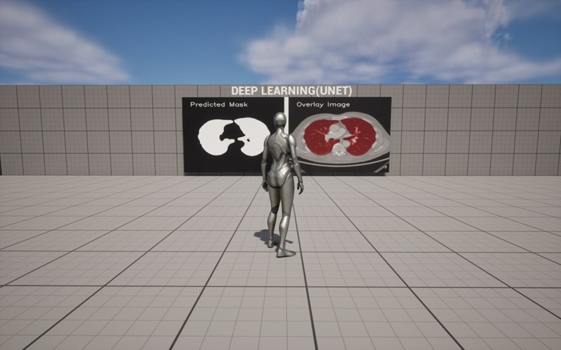
Prediction overlay
-
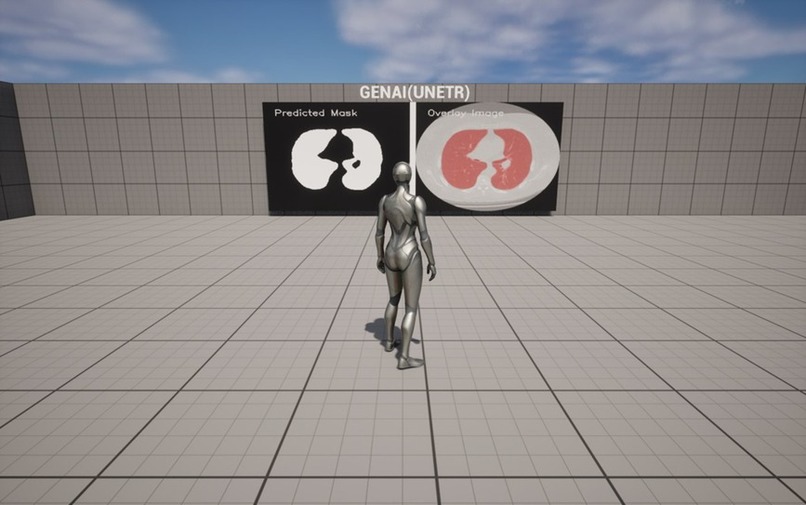
Prediction in GenAI- UNETR
-
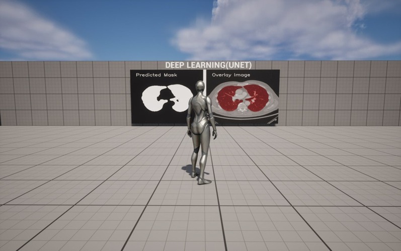
Prediction in DL- UNET
Inspiration
MedDiagnify was created to bridge the gap in current medical imaging tools. Despite advancements in imaging technologies, healthcare professionals still face challenges in accurately diagnosing complex conditions, such as cancers and orthopedic abnormalities, due to time constraints, limited spatial understanding, and the difficulty of identifying subtle anomalies in images. Our inspiration came from the need to enhance diagnostic efficiency, precision, and pre-surgical planning through AI-powered 3D medical imaging.
What it does
MedDiagnify is an AI-driven diagnostic and imaging tool designed to assist radiologists, pathologists, and surgeons in their medical image analysis. It offers:
- Advanced Cancer Detection: Uses AI to identify subtle anomalies in medical images, supporting early detection of cancer (lung, breast, colon).
- 3D Orthopedic Visualization: Generates 3D reconstructions from CT scans, giving surgeons a virtual, detailed view of bone structures for more effective pre-surgical planning.
- Automated Image Analysis: Scans large volumes of medical image data to highlight critical markers and potential complications, aiding faster and more accurate assessments.
How we built it
We leveraged cutting-edge Generative AI and Deep Learning techniques to train the app on extensive datasets of medical images. Using advanced convolutional neural networks (CNNs), we developed algorithms that could process, interpret, and generate high-quality 3D models and diagnostic insights from 2D scans. Additionally, cloud infrastructure and API integrations enable real-time analysis and easy collaboration across healthcare teams.
Challenges we ran into
- Data Quality and Availability: Gathering large and diverse medical image datasets with accurate annotations was a challenge. Ensuring that the data reflected various patient demographics and conditions was critical for training a robust AI model.
- Model Generalization: Ensuring that our AI models worked effectively across different imaging modalities (e.g., CT scans, X-rays, MRIs) required rigorous testing and optimization.
Accomplishments that we're proud of
- Accuracy Improvements: Our AI model consistently outperforms traditional image analysis techniques in identifying early-stage cancer and providing surgical planning insights.
- Real-time 3D Rendering: The tool’s 3D rendering feature allows surgeons to interact with bone structures in a dynamic, virtual environment, reducing guesswork in pre-surgical planning.
What we learned
- AI in Medicine is a Game-Changer: The integration of AI with medical imaging can dramatically reduce analysis time, improve diagnostic precision, and assist in critical decision-making processes.
- Collaboration is Key: Partnering closely with medical students, radiologists, and surgeons throughout the development process allowed us to fine-tune the tool and ensure it truly meets their needs.
- Data Quality Over Quantity: The quality of the data used to train our models is just as important, if not more, than the quantity. Focused, high-quality datasets yielded better results in model accuracy.
What's next for MedDiagnify
- Expand into Other Specialties: We plan to extend the platform’s capabilities to other medical fields, such as cardiology and neurology, to aid in the diagnosis and treatment of heart diseases and neurological conditions.
- Integrating with Hospital Systems: Further development will focus on integrating MedDiagnify with existing Electronic Health Record (EHR) systems to streamline workflows for clinicians and hospitals.
- Continuous AI Training and Updates: We will continue to train our AI models with new data and feedback from clinicians to improve accuracy, adapt to emerging conditions, and ensure that the system stays on the cutting edge of medical imaging technology.

Log in or sign up for Devpost to join the conversation.