-
-
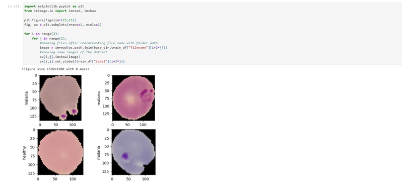
Image Samples
-
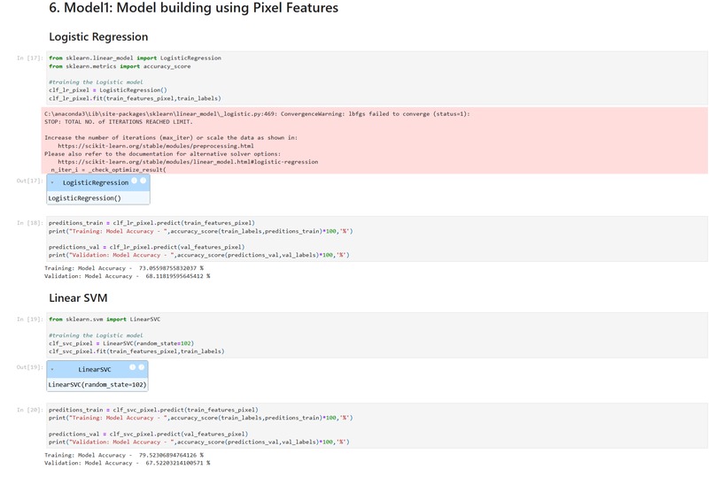
Pixel Features
-
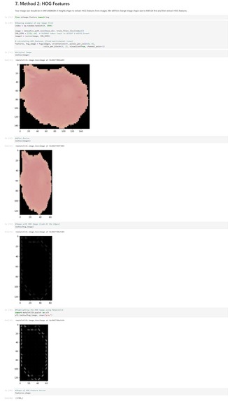
Hog Feature Image
-
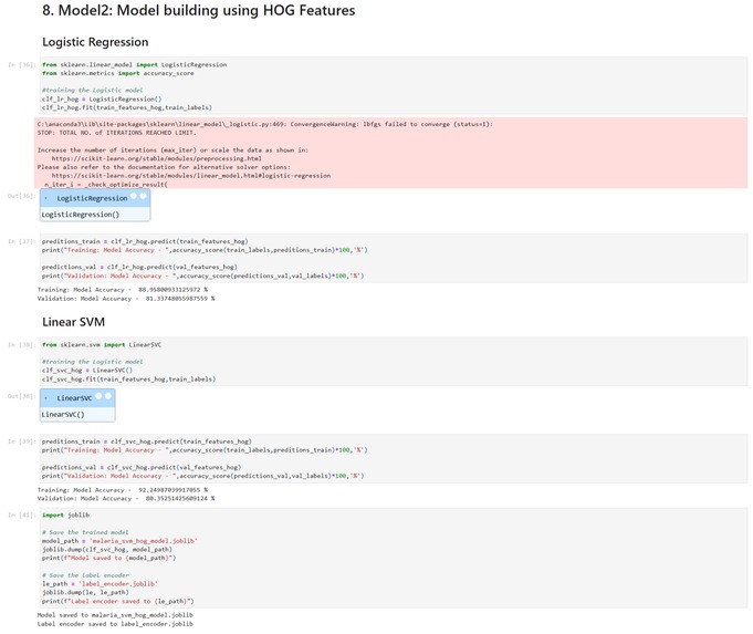
Hog Feature
-
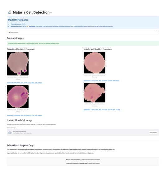
Streamlit app
-
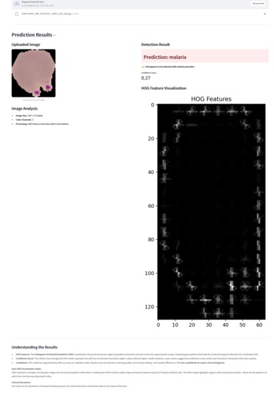
Malaria cell Prediction Result
-
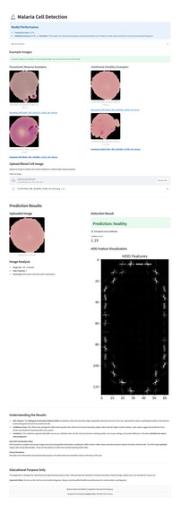
Healthy Cell Prediction Result


Inspiration
Manual malaria diagnosis from blood smear images is time-consuming and requires expertise. We aimed to automate this process using machine learning for faster and more accessible detection.
What it does
Detects malaria in cell images using a trained Linear SVM model with HOG features. Provides a Streamlit app for users to upload images and receive predictions and confidence scores.
How we built it
- Explored and analyzed the dataset
- Preprocessed images and encoded labels
- Extracted features using raw pixels and HOG
- Trained and evaluated Logistic Regression and Linear SVM models
- Saved the best model and label encoder with joblib
- Developed a Streamlit app for deployment and visualization
Challenges we ran into
- Preprocessing and resizing images for feature extraction
- Selecting the best feature extraction and model combination
- Integrating model inference with the Streamlit interface
Accomplishments that we're proud of
- Automated malaria cell detection workflow
- Achieved over 81% validation accuracy with SVM and HOG features
- Built a user-friendly web app for predictions and visualization
What we learned
- The impact of feature extraction on image classification
- Model evaluation and selection for real-world data
- Deploying machine learning models with Streamlit
What's next for Malaria Cell Detection
- Improve model accuracy with more data or advanced techniques
- Add support for additional image formats
- Enhance the Streamlit app UI and user experience
- Generate more advanced and detailed reports for predictions
- Add export options for results (PDF, CSV, etc.)
- Make prediction results and explanations easy to understand without medical jargon
Built With
- github
- huggingface-spaces
- joblib
- jupyter-notebook
- numpy
- pandas
- python
- scikit-images
- scikit-learn
- streamlit