


Inspiration
Lung cancer remains one of the most fatal diseases due to its often late detection. This inspired us to build a system that could assist radiologists in early and accurate detection of pulmonary nodules—a critical indicator of lung cancer. The potential to save lives through earlier diagnosis using artificial intelligence was a strong motivator for our team.
What it does
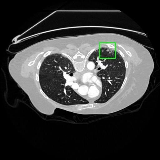
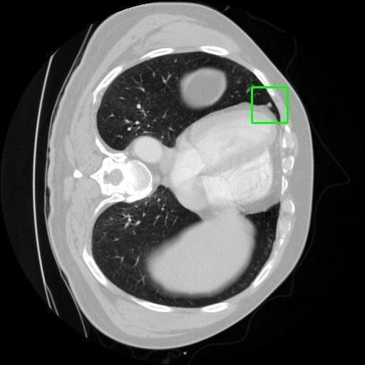
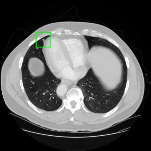
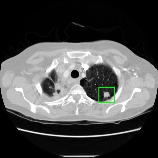
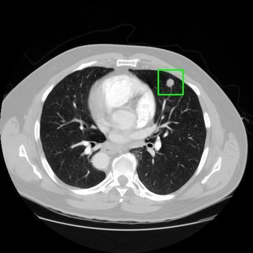
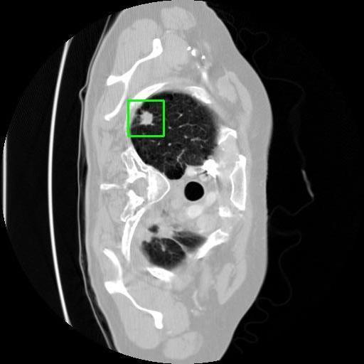
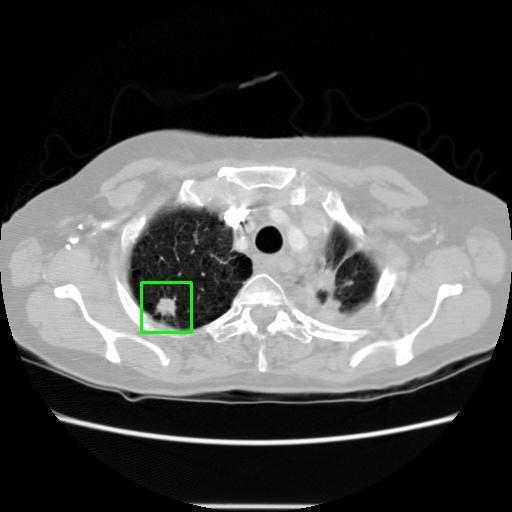
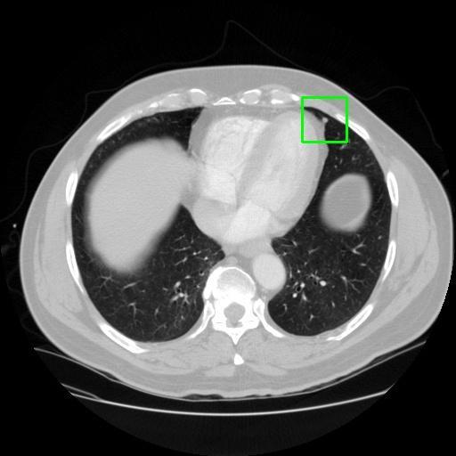
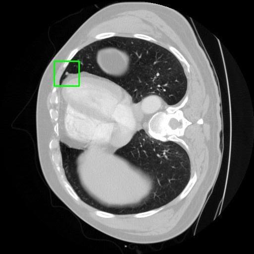
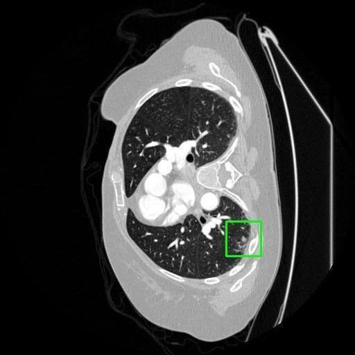
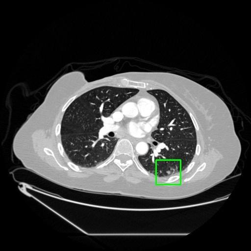
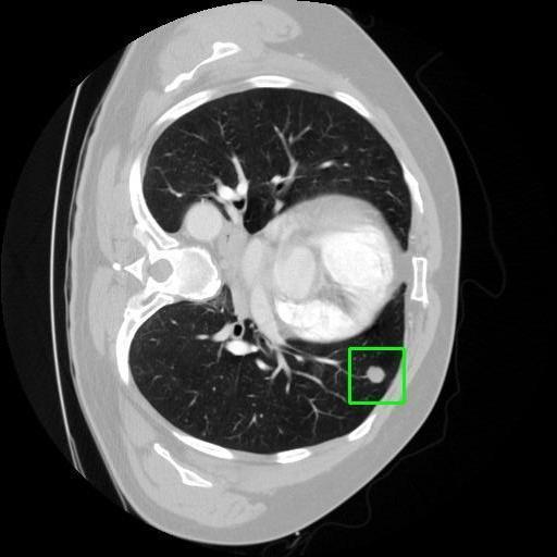
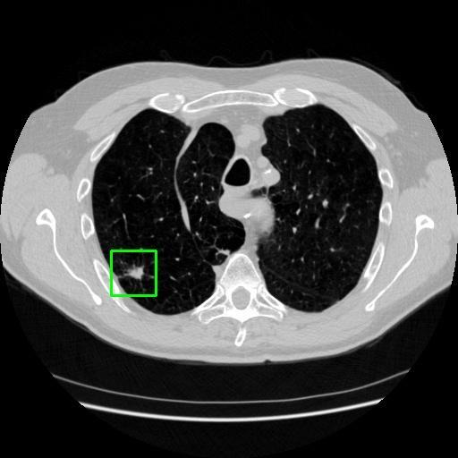
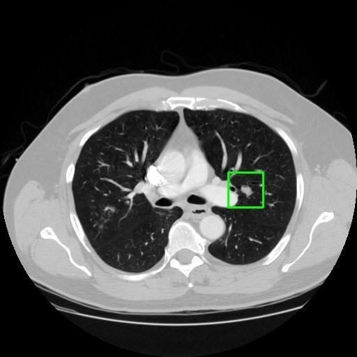
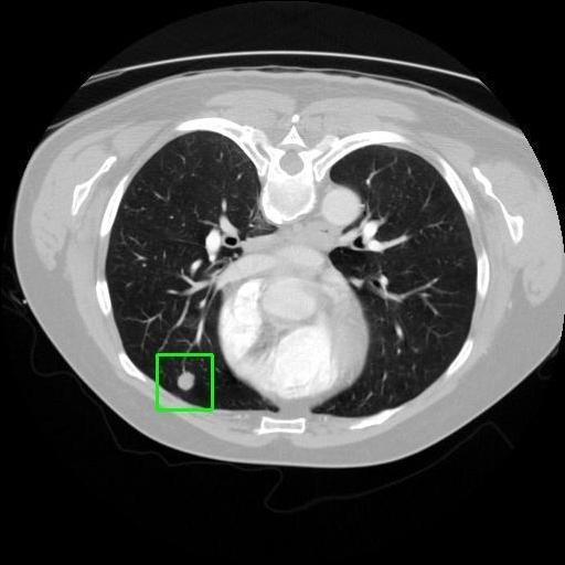
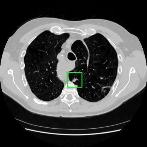
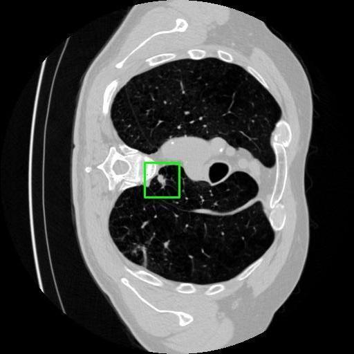
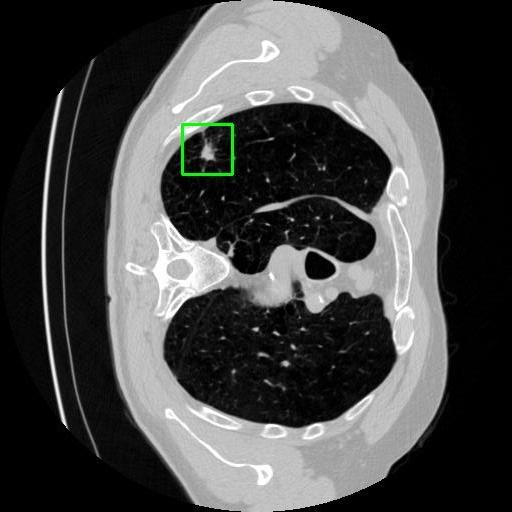
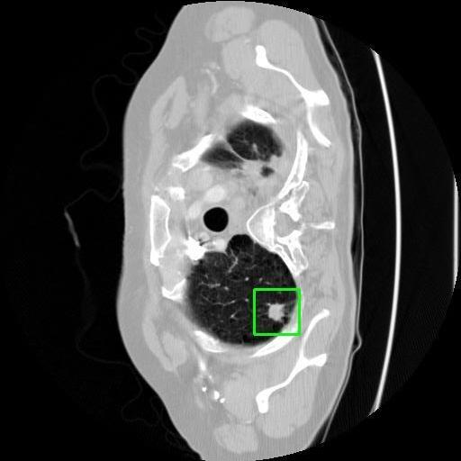
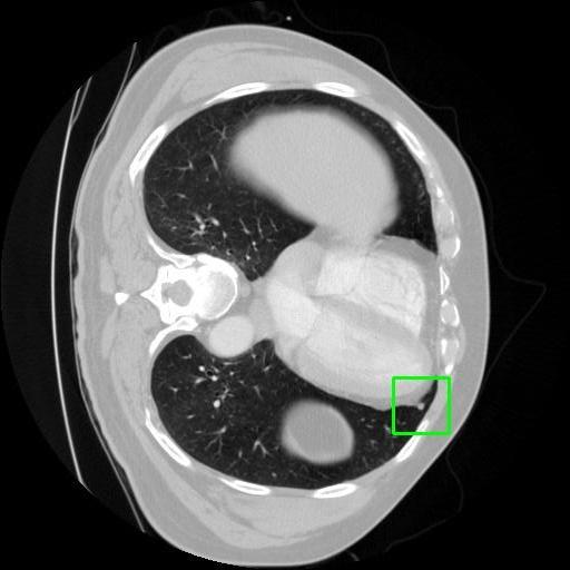
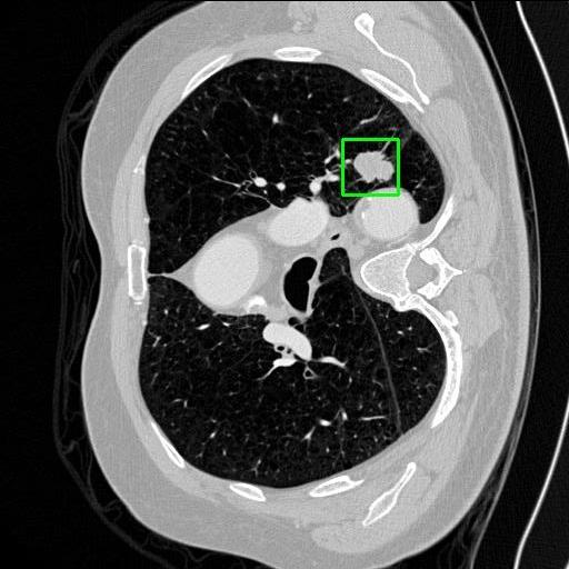
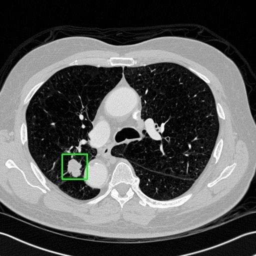
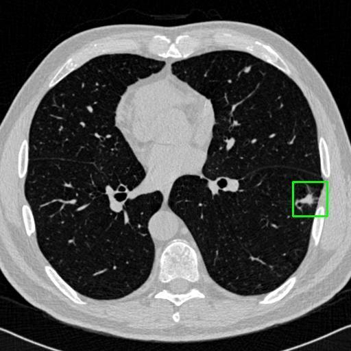
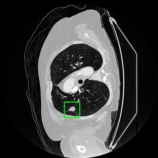
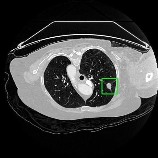
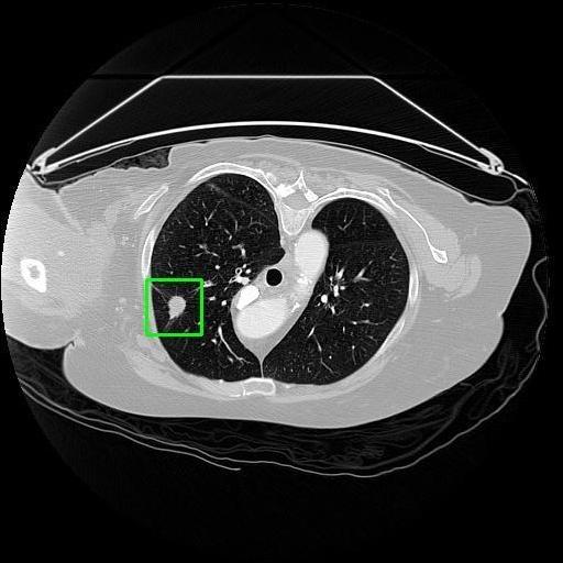
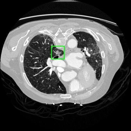
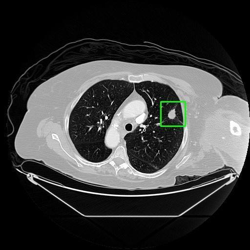
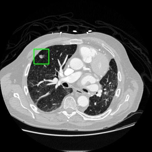
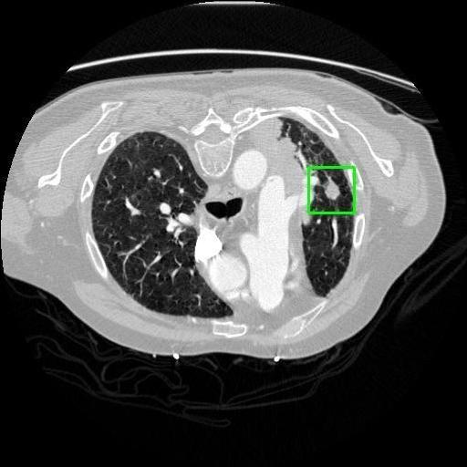
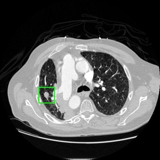
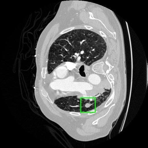
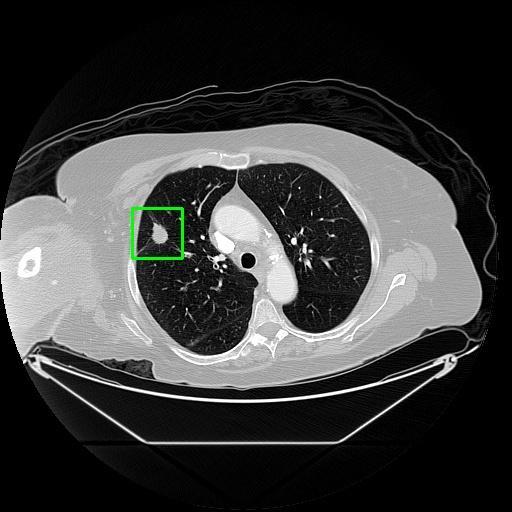
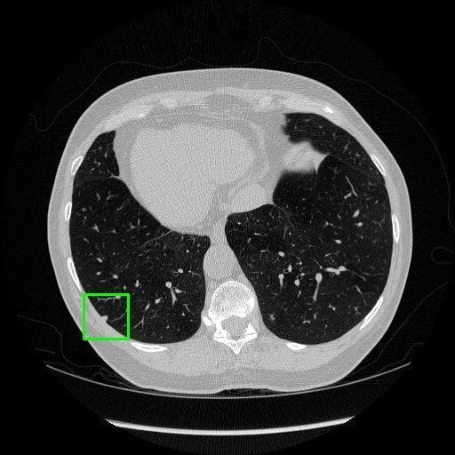
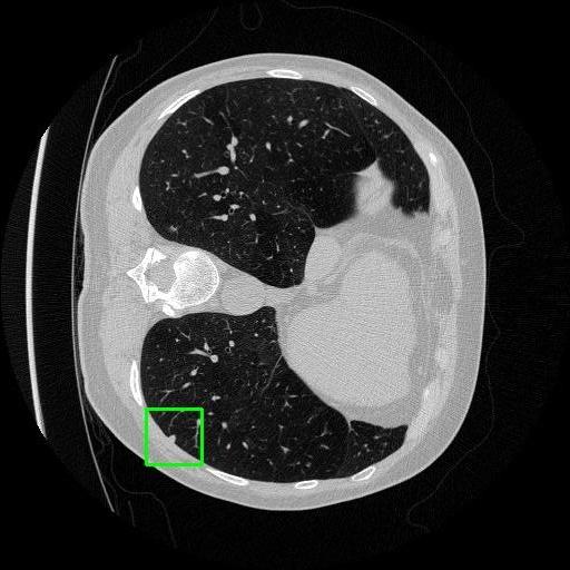
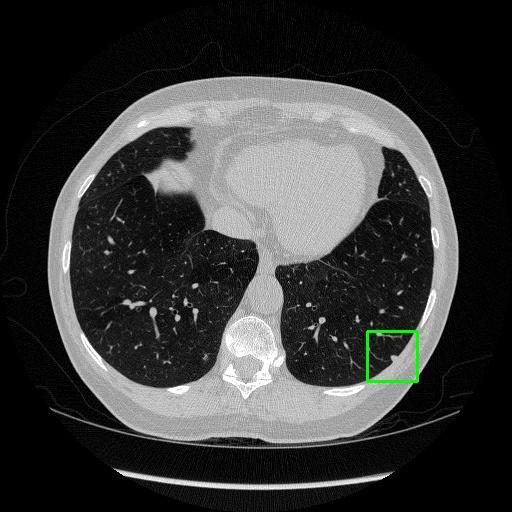
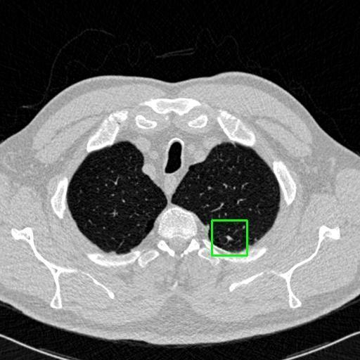
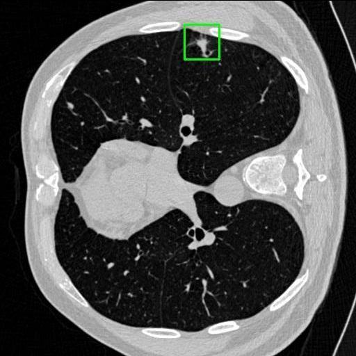
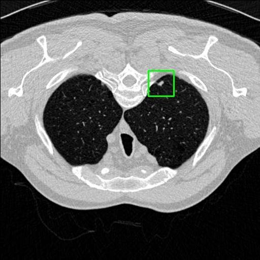
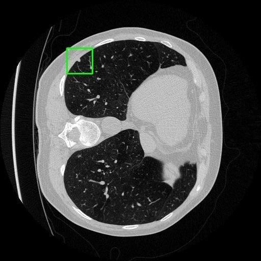
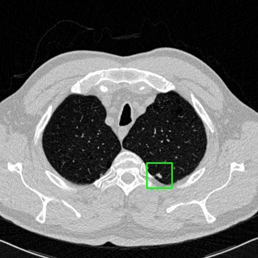
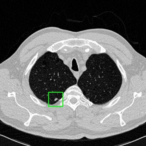
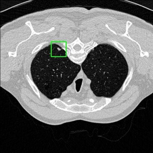
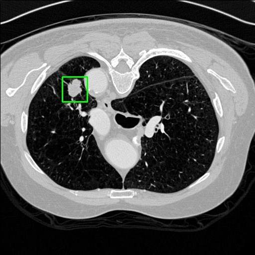
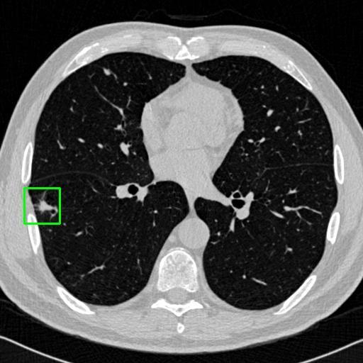
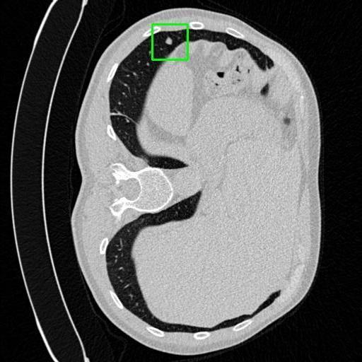
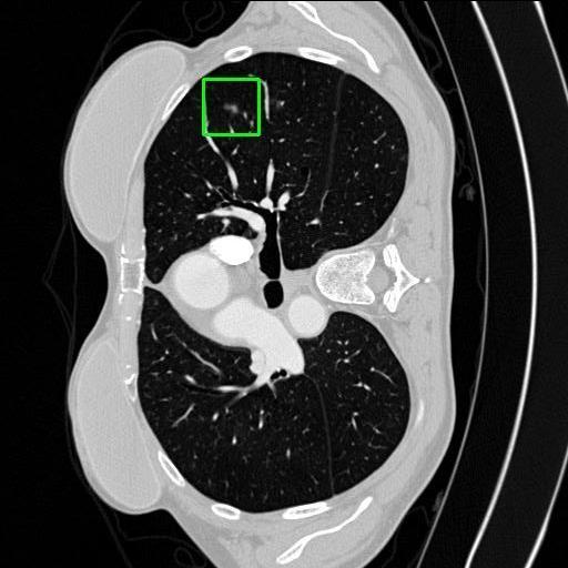
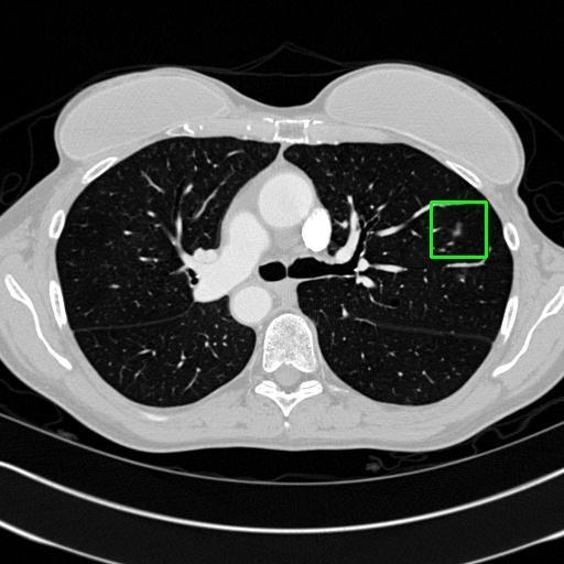
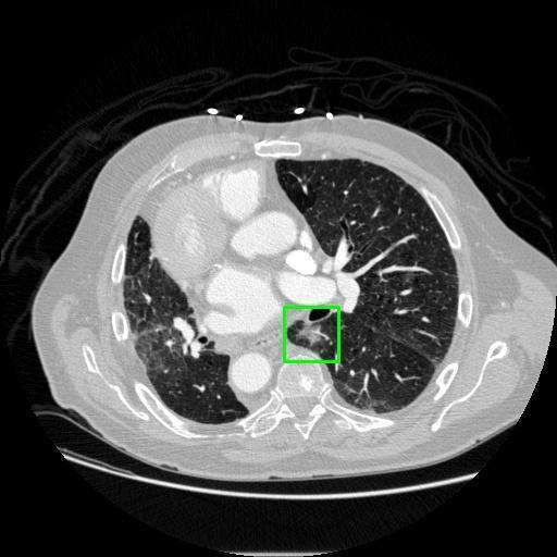
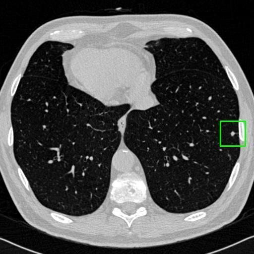
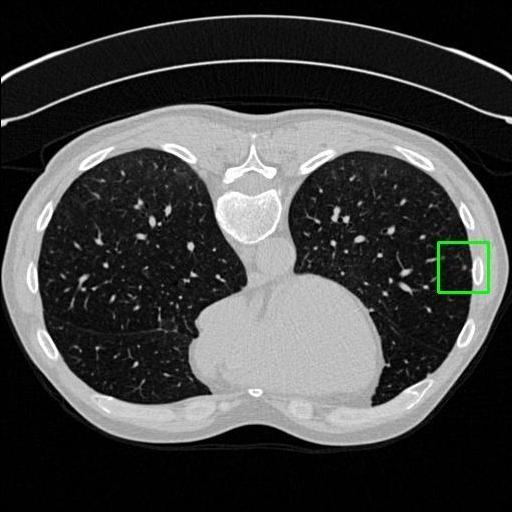
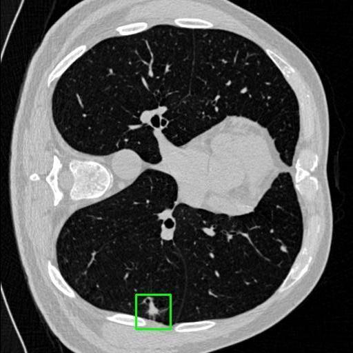
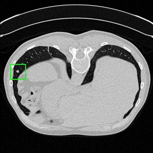
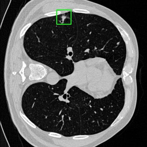
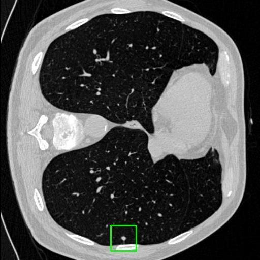
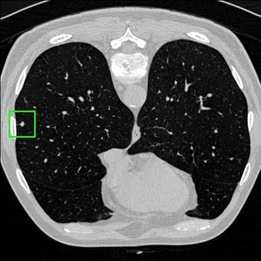
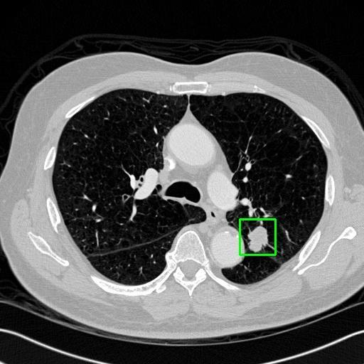
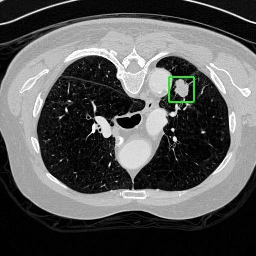
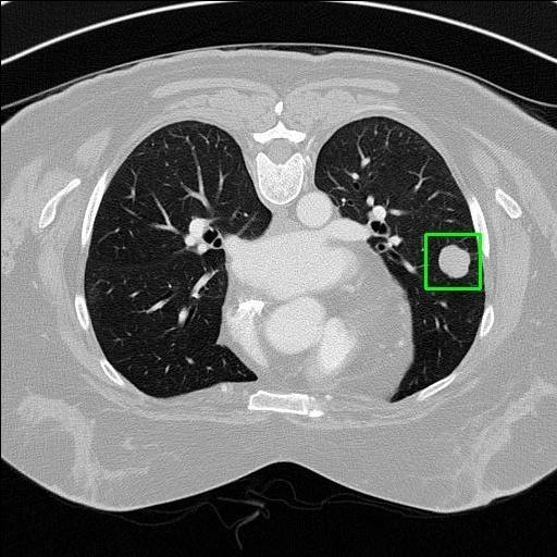
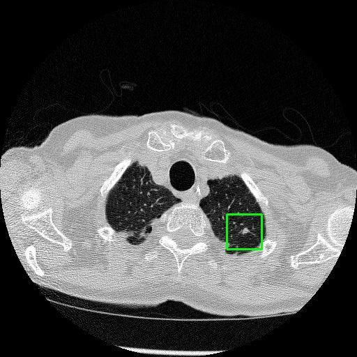
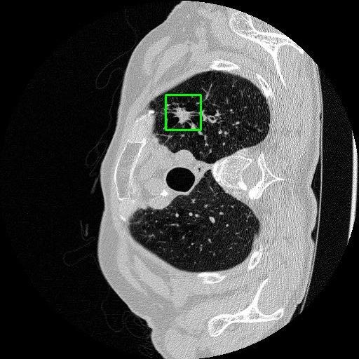
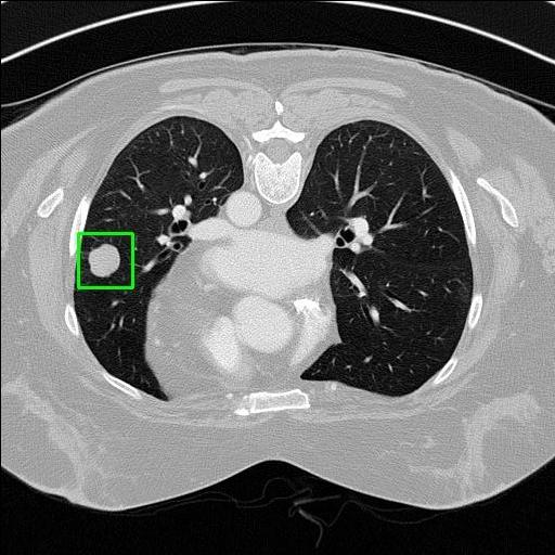
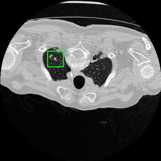
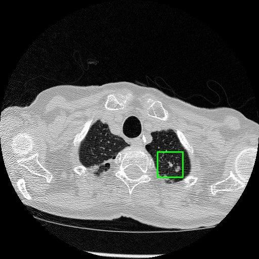
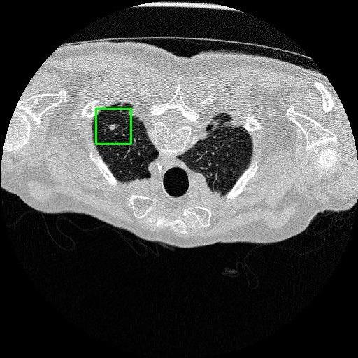
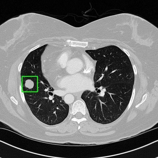
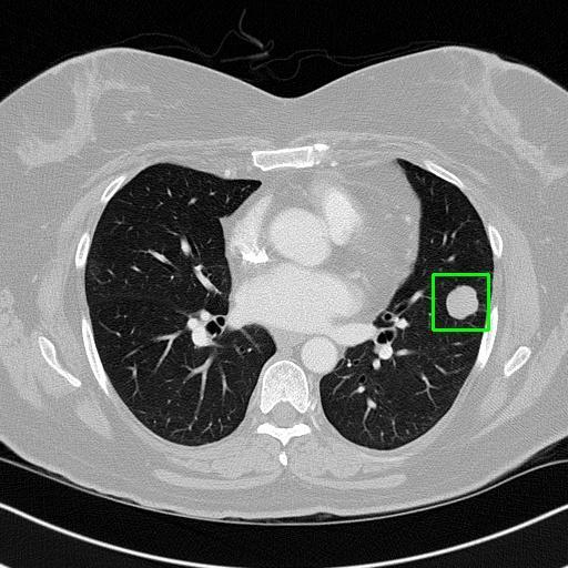
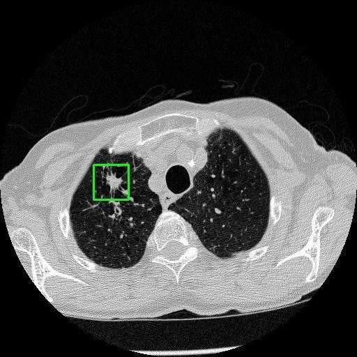
We developed an AI-based system using deep learning models for analyzing lung CT scans to detect and classify pulmonary nodules. We chose the YOLOv11 architecture for its enhanced object detection capability and adapted it specifically for medical imaging, incorporating pixel-level precision and severity classification.
The primary goals were:
To detect nodules in CT scans.
To classify them as benign or malignant.
To label the severity (null, moderate, severe) for further clinical action.
How we built it
Tools & Technologies: YOLOv11: For object detection and severity classification.
Python & PyTorch: For training and implementation.
Roboflow: For dataset curation and preprocessing.
LIDC-IDRI dataset and other open medical image sources.
Key Features: Real-time detection of nodules.
Classification into three severity levels with colored bounding boxes.
Data augmentation techniques to improve generalization: flipping, rotation, scaling, contrast adjustments, and noise injection.
Architecture Highlights: SPPF and C2PSA Blocks for multiscale feature attention.
C3K2 Block for lightweight but powerful feature aggregation.
CBS layers for normalization and efficient activation.
Final Detect Layer to output bounding boxes, objectness scores, and class predictions.
Challenges we ran into
Annotating the medical dataset was time-consuming and required high domain knowledge.
Medical image characteristics (non-RGB, soft tissue contrast) made general-purpose models less effective without fine-tuning.
Distinguishing between similar-looking benign and malignant nodules posed classification difficulty.
Maintaining performance on small, complex features like micro-nodules tested the limits of traditional object detectors.
Accomplishments that we're proud of
Successfully built and deployed an AI model (YOLOv11) capable of detecting lung nodules in CT scans with high accuracy and real-time performance.
Achieved ~0.90 mAP@0.5 on validation data, showing strong object detection capabilities even on complex medical images.
Designed a severity classification system that categorizes nodules into null, moderate, and severe using colored bounding boxes, assisting in rapid clinical decision-making.
Implemented advanced data augmentation techniques to improve the generalization of the model and simulate real-world variability in medical imaging.
Developed a user-friendly interface (in progress or planned) for uploading CT images and visualizing predictions for non-technical medical professionals.
Gained deeper insights into the challenges of medical image analysis, particularly the need for 3D context, pixel-level precision, and interpretability.
Demonstrated the potential of AI-assisted diagnosis in real-world healthcare by reducing radiologist workload and increasing diagnostic speed.
What we learned
Deep learning architectures like U-Net and CNNs (ResNet, DenseNet) are often more suitable than general object detectors for medical image tasks.
High-quality annotated datasets are crucial—bad data can ruin even the best models.
Real-world medical image processing needs 3D context awareness which 2D models like YOLO must adapt for.
Continuous model validation and tuning (over 50 epochs) led to improved precision, recall, and mAP values: Box loss dropped from 0.71582 to 0.48289 Classification loss dropped from 0.50189 to 0.44476 Precision increased to 0.82, Recall to 0.89, mAP@0.5 to ~0.90
What's next for LUNG CANCER DETECTION AND ANALAYSIS WITH AI
3D Model Integration: Transitioning from 2D slice-based analysis to 3D convolutional models to capture full volumetric context of CT scans for more accurate diagnosis. Web-Based Deployment: Launching a secure web portal where radiologists and clinicians can upload scans, receive instant AI-based assessments, and download reports. Expanded Dataset Training: Incorporating more diverse datasets—including PET scans and low-dose CTs—for better generalization across patient demographics and conditions.

Log in or sign up for Devpost to join the conversation.