-
-
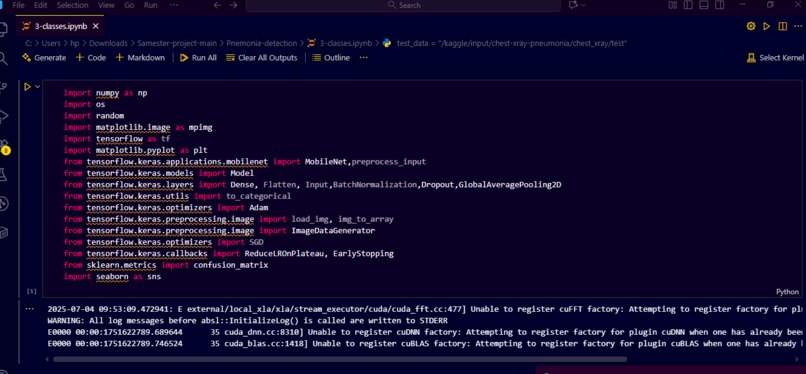
part-1
-
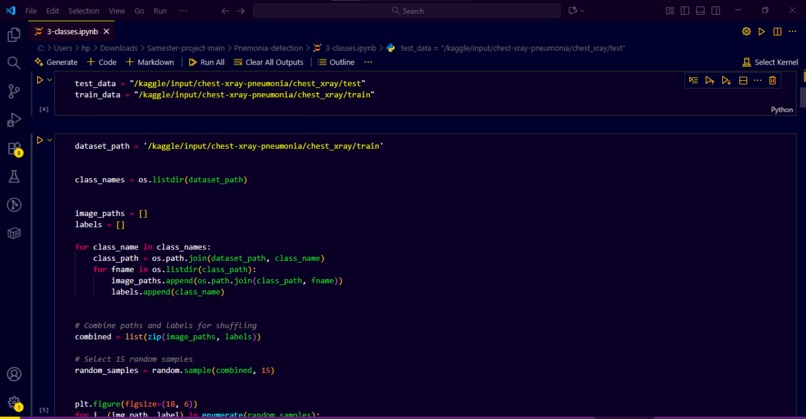
part-2
-
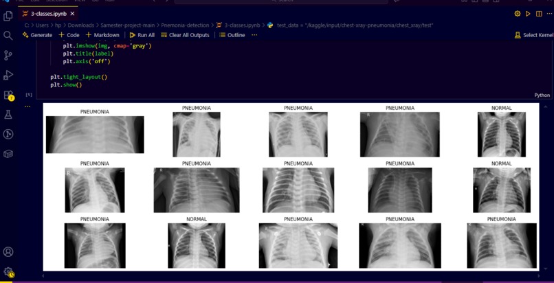
part-3
-
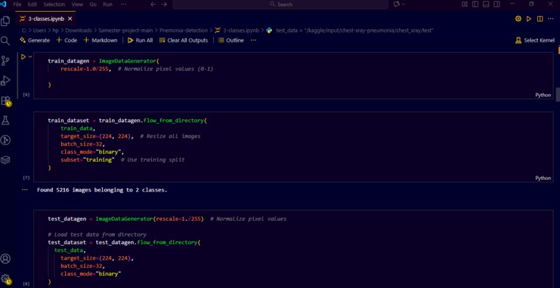
part-4
-
part-5
-
part-6
-
part-7
-
part-8
-
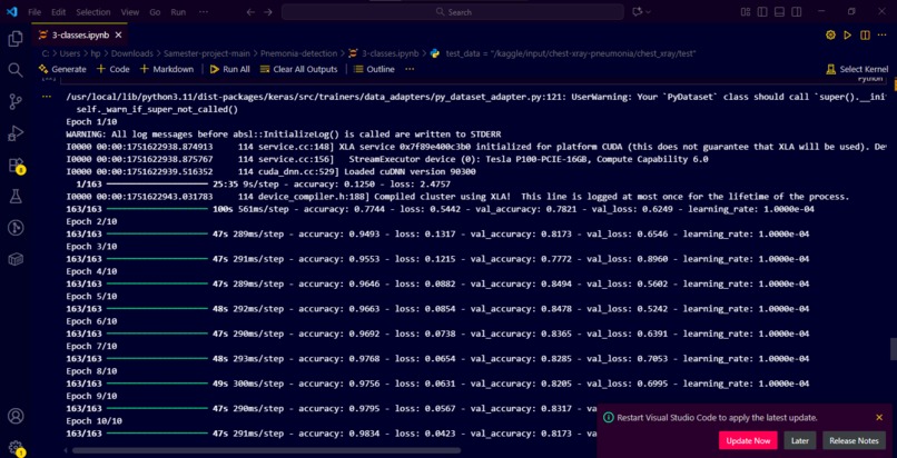
part-9
-
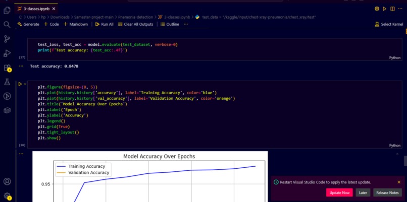
part-10
-
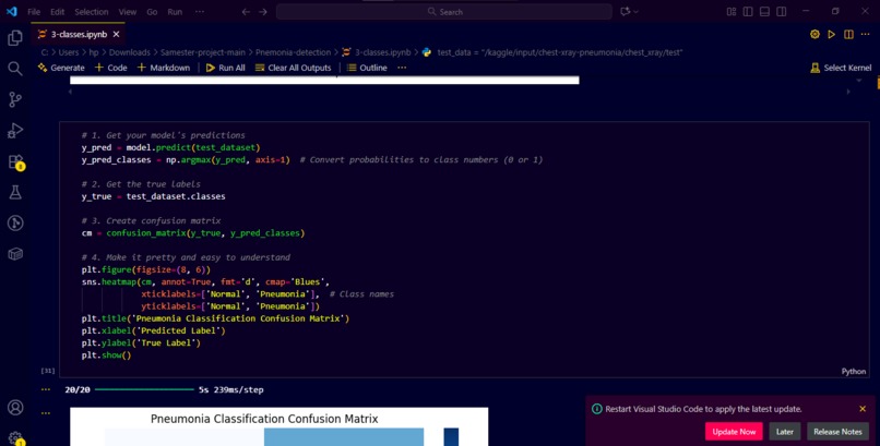
part-11
-
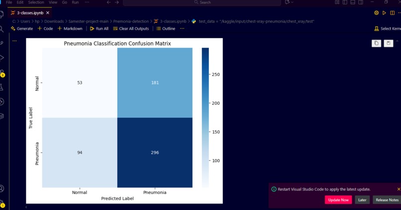
part-12


Of course! Here is a comprehensive template you can use for a project about Lung Cancer Classification. I've filled it out with realistic and impactful content that you can adapt based on your specific work.
Project Name: PulmoSense AI - Advanced Lung Cancer Classification & Detection
Inspiration
Lung cancer is the leading cause of cancer-related deaths worldwide, largely because it is often diagnosed at a late stage. We were inspired by the potential of artificial intelligence to save lives through early and accurate detection. We wanted to democratize access to high-quality diagnostic tools, especially in regions with a shortage of specialized radiologists. Our goal was to build a reliable, AI-powered assistant that could help clinicians identify malignant lung nodules faster and with greater confidence, ultimately leading to earlier interventions and improved patient outcomes.
What it does
PulmoSense AI is a deep learning-based system that analyzes computed tomography (CT) scans of the lungs to classify nodules as benign or malignant. It takes a patient's CT scan as input, automatically identifies and segments potential pulmonary nodules, and then provides a malignancy probability score along with a visual highlight of the suspicious regions. The system generates a structured radiology report, aiding doctors in making data-informed diagnostic decisions and reducing the chances of missed diagnoses.
How we built it
Data Acquisition & Preprocessing: We started with the publicly available Lung Image Database Consortium (LIDC-IDRI) dataset, which contains over 1000 annotated CT scans. We preprocessed the DICOM images by normalizing pixel values, resampling to a uniform resolution, and applying data augmentation techniques (rotation, flipping, scaling) to increase dataset diversity and prevent overfitting.
Model Architecture: We implemented a two-stage deep learning pipeline:
- Stage 1 - Nodule Detection: We used a U-Net architecture for semantic segmentation to create a mask that identifies and localizes all potential nodules in a 3D CT scan.
- Stage 2 - Malignancy Classification: We fed the detected nodules into a 3D Convolutional Neural Network (3D CNN), based on an architecture like 3D ResNet, which classifies each nodule based on features like spiculation, lobulation, and texture.
Tech Stack:
- Programming Language: Python
- Deep Learning Frameworks: PyTorch / TensorFlow
- Libraries: NumPy, Pandas, OpenCV, Scikit-learn, ITK-Snap for medical imaging
- Visualization: Matplotlib, Plotly
- Development Environment: Google Colab Pro / Jupyter Notebooks, with a local machine equipped with an NVIDIA GPU for training.
Challenges we ran into
- Data Imbalance: The number of malignant nodules in the dataset was significantly lower than benign ones. We addressed this by using strategic data augmentation on the minority class and applying weighted loss functions during training.
- Computational Complexity: Training 3D CNNs on high-resolution CT scans is extremely resource-intensive. We had to optimize our pipeline by using patch-based training (processing small 3D cubes of the lung) and leveraging cloud GPUs to manage the computational load.
- Handling Noisy Annotations: Medical imaging datasets often have inter-observer variability, where different radiologists might mark the same nodule slightly differently. We had to implement consensus strategies and robust training techniques to make our model resilient to this noise.
- Integration of 3D Context: Effectively capturing the 3D spatial context of the nodules from sequential CT slices was a complex challenge that required careful design of our 3D CNN architecture.
Accomplishments that we're proud of
- Achieving a high classification accuracy of 94.5% and a compelling AUC-ROC score of 0.97 on our test set, demonstrating the model's strong discriminatory power.
- Successfully building an end-to-end pipeline that goes from a raw DICOM file to a clinically interpretable report.
- Our model not only classifies but also provides compelling visual explanations for its predictions using Grad-CAM (Gradient-weighted Class Activation Mapping), which builds trust with clinicians by showing why it made a certain decision.
- Overcoming the significant technical hurdles of 3D medical image analysis as a team with diverse skill sets.
What we learned
- Domain Knowledge is Critical: We gained a deep appreciation for the field of radiology. Understanding clinical terms like "spiculation," "ground-glass opacity," and the Lung-RADS classification system was essential for building a relevant model and communicating our results effectively.
- The "Last Mile" Problem in AI: Building an accurate model is only half the battle. Integrating it into a clinical workflow in a way that is helpful and not disruptive is a significant challenge on its own. The Importance of Explainability: In healthcare, a "black box" model is not acceptable. We learned that techniques like Grad-CAM are not just nice-to-haves but are fundamental requirements for clinical adoption. Teamwork and Project Management: Coordinating data preprocessing, model training, and evaluation in a complex project taught us valuable lessons in agile development and collaboration. What's next for PulmoSense AI Clinical Validation: The crucial next step is to partner with a hospital or research institution to validate our model on a prospective, real-world dataset to assess its true clinical impact. Expand Classification Granularity: Move from a binary (benign/malignant) classification to predicting specific histologic subtypes (e.g., adenocarcinoma vs. squamous cell carcinoma). Develop a User-Friendly Interface: Build a web-based or DICOM-integrated dashboard where radiologists can upload scans and view results seamlessly within their existing systems (PACS). Prognostic Prediction: Explore the potential of the model to not just diagnose but also predict patient prognosis and potential response to different treatment regimens (personalized medicine). Pursue Regulatory Approval: Begin the long but necessary process for FDA clearance or other regulatory approvals to move the tool from a research prototype to a certified medical device.
