Inspiration
Cardiovascular disease remains the leading cause of mortality globally. While conventional statistical calculators can output a raw risk percentage from patient lab values, a flat number like 78% risk fails to give clinicians or patients an intuitive spatial understanding of where pathology is developing inside the coronary anatomy or which physiological signals triggered a specific artery alert.
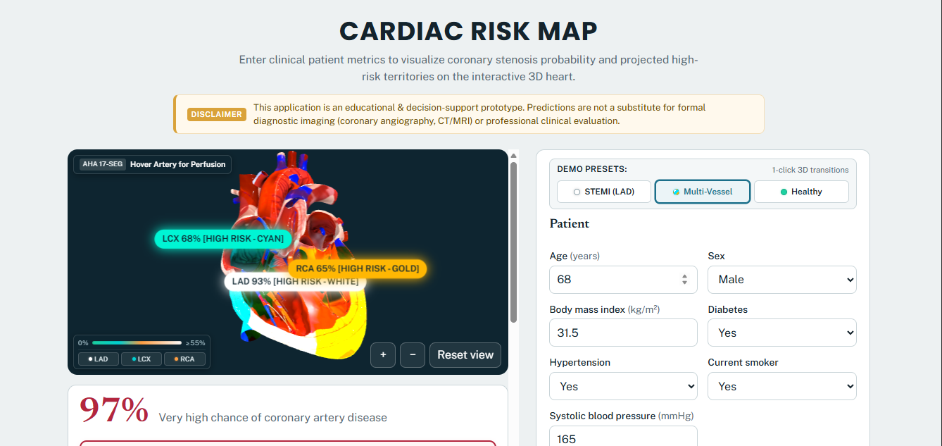
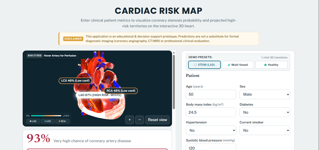
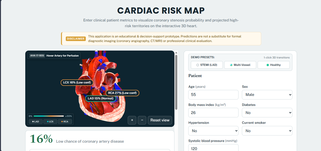
We built Cardiac Risk Map (GitHub Repository) to bridge the gap between 1D tabular clinical data and 3D human anatomy—combining four leak-free machine learning classification pipelines, real-time per-vessel SHAP (shap.TreeExplainer) interpretability, and a 60 FPS Three.js / WebGL beating human heart mapped to the American Heart Association (AHA) 17-segment myocardial perfusion territories.
Clinical Safety Disclaimer: This application is an educational and clinical decision-support prototype. Predictions are not a substitute for formal diagnostic imaging (coronary angiography, cardiac CT/MRI) or professional clinical evaluation.
What It Does
- Multi-Vessel Stenosis & Overall CAD Prediction: Predicts both Overall Coronary Artery Disease (
Cath) and vessel-specific stenosis probabilities (≥ 50% luminal narrowing) across the three major epicardial arteries:- LAD (Left Anterior Descending): Supplies the anterior wall and anterior interventricular septum.
- LCX (Left Circumflex): Supplies the anterolateral and posterolateral left ventricular walls.
- RCA (Right Coronary Artery): Supplies the right ventricle, inferior wall, and basal inferoseptal segments.
- Real-Time 3D WebGL Beating Heart Mapping: Dynamically shades the 3D myocardium (
beating-heart.glb) using GPU vertex color buffers based on predicted vessel probabilities (0%Normal →25%Guarded →≥ 55%High-Risk / 3D White Overdrive), complete with an interactive AHA 17-Segment Perfusion HUD, 3D Risk & Territory Scale legend, orbit/zoom camera controls, and clickable vessel inspection. - Per-Vessel Explainable AI (SHAP): Allows users to toggle the Feature Contributions (SHAP) panel across Overall CAD, LAD, LCX, and RCA to inspect exact risk-raising (red) and risk-lowering (blue/green) patient biomarkers in real time.
- 1-Click Clinical Presets for Rapid Evaluation: Includes three pre-configured clinical profiles (High-Risk Anterior STEMI (LAD), Multi-Vessel CAD, and Healthy Baseline) for instant live testing.
How We Built It
1. Zero Target Leakage & Feature Engineering (src/config.py, src/data.py)
Trained on the Z-Alizadeh Sani Cardiology Dataset (N = 303 patients from Tehran Heart Center), our feature matrix X ingests 50+ non-invasive predictors spanning four clinical domains:
- Demographics & History:
Age,Sex,BMI,DM(Diabetes),HTN(Hypertension),Current Smoker,FH(Family History),DLP(Dyslipidemia),CRF,CVA,CHF. - Symptoms & Vitals:
Typical Chest Pain,Atypical,Nonanginal,Exertional CP,Dyspnea,BP(Blood Pressure),PR(Pulse Rate),Edema,Lung Rales. - 12-Lead Resting ECG:
Rhythm,Q Wave,ST Elevation,ST Depression,Tinversion(T-Wave Inversion),LVH,Poor R Progression. - Echocardiography & Laboratory Markers:
EF-TTE(Ejection Fraction),Region with RWMA(Regional Wall Motion Abnormality),VHD,FBS,Cr,TG,LDL,HDL,HB,K,Na,WBC,PLT.
Because Cath, LAD, LCX, and RCA all originate from invasive coronary angiography, all four target columns are unconditionally excluded from X across every model pipeline to guarantee zero target leakage. Furthermore, Region with RWMA is used strictly as a clinical feature by the classifier rather than a direct 3D coordinate.
2. Multi-Target ML Pipeline & 5-Fold Stratified Cross-Validation (src/train.py)
For each target (Cath, LAD, LCX, RCA), we constructed an independent scikit-learn Pipeline combining inside-fold StandardScaler() normalization with a balanced ensemble classifier:
Pipeline([
    ("scaler", StandardScaler()),
    ("clf", RandomForestClassifier(
        n_estimators=300,
        max_depth=6,
        min_samples_leaf=2,
        class_weight="balanced",
        random_state=42
    ))
])
Evaluated via 5-Fold Stratified Cross-Validation (StratifiedKFold(n_splits=5, shuffle=True, random_state=42)), our deployed pipelines achieve:
| Target Vessel / Disease | Accuracy | Majority Baseline | ROC-AUC | Sensitivity (Recall) | Precision | F1-Score | Clinical Confidence Tier |
|---|---|---|---|---|---|---|---|
Overall CAD (Cath) |
86.5% | 71.3% | 0.930 | 90.3% | 90.7% | 90.5% | Moderate (Single-Center Cohort) |
| LAD Artery (Anterior) | 77.2% | 58.4% | 0.845 | 80.8% | 80.6% | 80.6% | Moderate (Strong Anterior ECG Signal) |
| LCX Artery (Lateral) | 63.7% | 60.7% | 0.727 | 58.8% | 54.7% | 56.5% | Low / Guarded (Posterior Subtlety) |
| RCA Artery (Inferior) | 65.7% | 62.4% | 0.711 | 57.9% | 55.3% | 56.2% | Low / Guarded (Inferior Overlap) |
3. Algorithmic Benchmarking & Probability Calibration (src/benchmark.py)
In docs/BENCHMARK_REPORT.md, we benchmarked our Balanced Random Forest against XGBoost, Gradient Boosting, and post-hoc probability calibration (CalibratedClassifierCV with Isotonic and Sigmoid scaling) evaluated via the Brier score (Brier = (1/N) Σ (pᵢ - yᵢ)²):
| Target Vessel | Random Forest ROC-AUC | XGBoost ROC-AUC | Gradient Boosting ROC-AUC | Uncalibrated Brier | Isotonic Calibrated Brier |
|---|---|---|---|---|---|
Overall CAD (Cath) |
0.930 ± 0.04 | 0.918 ± 0.05 | 0.912 ± 0.05 | 0.1030 | 0.0932 |
| LAD Stenosis | 0.845 ± 0.06 | 0.832 ± 0.07 | 0.825 ± 0.07 | 0.1684 | 0.1591 |
| LCX Stenosis | 0.728 ± 0.10 | 0.710 ± 0.11 | 0.696 ± 0.11 | 0.2215 | 0.2140 |
| RCA Stenosis | 0.704 ± 0.07 | 0.697 ± 0.08 | 0.678 ± 0.09 | 0.2268 | 0.2195 |
4. Sub-50ms Cached SHAP Engine & 60 FPS Three.js WebGL Pipeline (src/predict.py, static/heart3d.js)
To explain individual predictions, we compute exact additive feature attributions (f(x) = φ₀ + Σ φⱼ(x)) using shap.TreeExplainer, where φ₀ is the baseline model expectation and φⱼ(x) is the exact contribution of clinical feature j toward the predicted stenosis probability. By pre-instantiating all four TreeExplainer objects in memory at server boot (src/predict.py), our Flask /predict endpoint returns all four probabilities and four full SHAP vectors in under 50 ms. On the client (static/heart3d.js), Three.js maps those probabilities onto the 3D heart's GPU attributes.color vertex buffers at 60 FPS without requiring a dedicated GPU.
Challenges We Ran Into
- Anterior (
LAD) vs. Posterolateral (LCX) & Inferior (RCA) Signal Asymmetry: Standard 12-lead resting ECG places precordial leads (V1–V6) directly over the anterior wall supplied by the LAD (0.845ROC-AUC), whereas the posterolateral wall supplied by the LCX (0.727ROC-AUC) lacks direct posterior leads (V7–V9) in routine exams. Rather than masking this clinical limitation, we added explicit Low / Guarded Confidence indicators on LCX and RCA cards and documented the single-center (N = 303) scope transparently. - Real-Time SHAP Latency: Constructing
TreeExplainerper HTTP request caused a>400 msdelay during interactive slider/preset updates. Moving explainer initialization to worker startup reduced inference + attribution latency to<50 ms. - Smooth 3D Perfusion Shading Without Heavy Shaders: Instead of expensive multi-pass fragment shaders that lag on integrated laptop GPUs, we implemented spatial vertex weight interpolation directly on the Three.js mesh geometry buffers.
Accomplishments That We're Proud Of
- Zero Data Leakage & Rigorous Validation: Achieved
0.930ROC-AUC (90.5%F1) on Overall CAD and0.845ROC-AUC (80.6%F1) on LAD stenosis under strict 5-Fold Stratified Cross-Validation with zero angiography feature leakage. - True Per-Artery Explainability: Clinicians can click any individual vessel (
LAD,LCX,RCA) to see both its 3D AHA 17-segment myocardial territory illumination and its dedicated SHAP biomarker breakdown. - Production Cloud Deployment: Deployed end-to-end on Render (cardiac-risk-prediction-k2jh.onrender.com) with 1-click judge demo presets and comprehensive technical documentation.
What We Learned & Team Contributions
- Shivam Chauhan (Team Lead — System Architecture, Backend API, ML/SHAP Engine & 3D WebGL Pipeline): Architected the Flask REST inference backend (
app.py,src/predict.py), boot-timeshap.TreeExplainercaching, leak-free 5-fold Stratified CV training and benchmarking suite (src/config.py,src/train.py,src/benchmark.py), and the 60 FPS Three.js GPU vertex shading engine (static/heart3d.js,static/app.js). - Haseen Babu (Clinical Data Mapping, Preset Curation & UI QA): Mapped physiological reference bounds and unit labels across the 50+ input controls (
templates/index.html), configured the 1-click clinical test case presets, and conducted cross-viewport QA testing.
What's Next for Cardiac Risk Map
- External Multi-Center Validation: Expanding training beyond the single-center Tehran Heart Center cohort (
N = 303) to multi-institutional registries to elevate clinical confidence from Moderate to High. - Sub-Branch Coronary Resolution: Extending the AHA 17-segment vertex map to include Diagonal (
D1/D2), Obtuse Marginal (OM), and Posterior Descending (PDA) sub-branches.
Built With
- 3d-visualization
- cardiology
- css3
- flask
- gunicorn
- healthcare-ai
- html5
- javascript
- machine-learning
- medical-ai
- numpy
- pandas
- python
- scikit-learn
- three.js
- webgl

Log in or sign up for Devpost to join the conversation.