

About the Project: RetinaMyopiaAI
Inspiration:
The inspiration for this project came from the growing prevalence of myopia worldwide and the need for early, accurate diagnosis. Many patients experience progressive vision loss due to delayed detection. I wanted to leverage AI and retinal imaging to create a tool that can assist ophthalmologists and improve patient outcomes.
What I Learned:
Through this project, I gained hands-on experience in:
- Medical image processing, including retinal fundus image enhancement and segmentation.
- Machine learning and deep learning, particularly CNNs (Convolutional Neural Networks) for image classification.
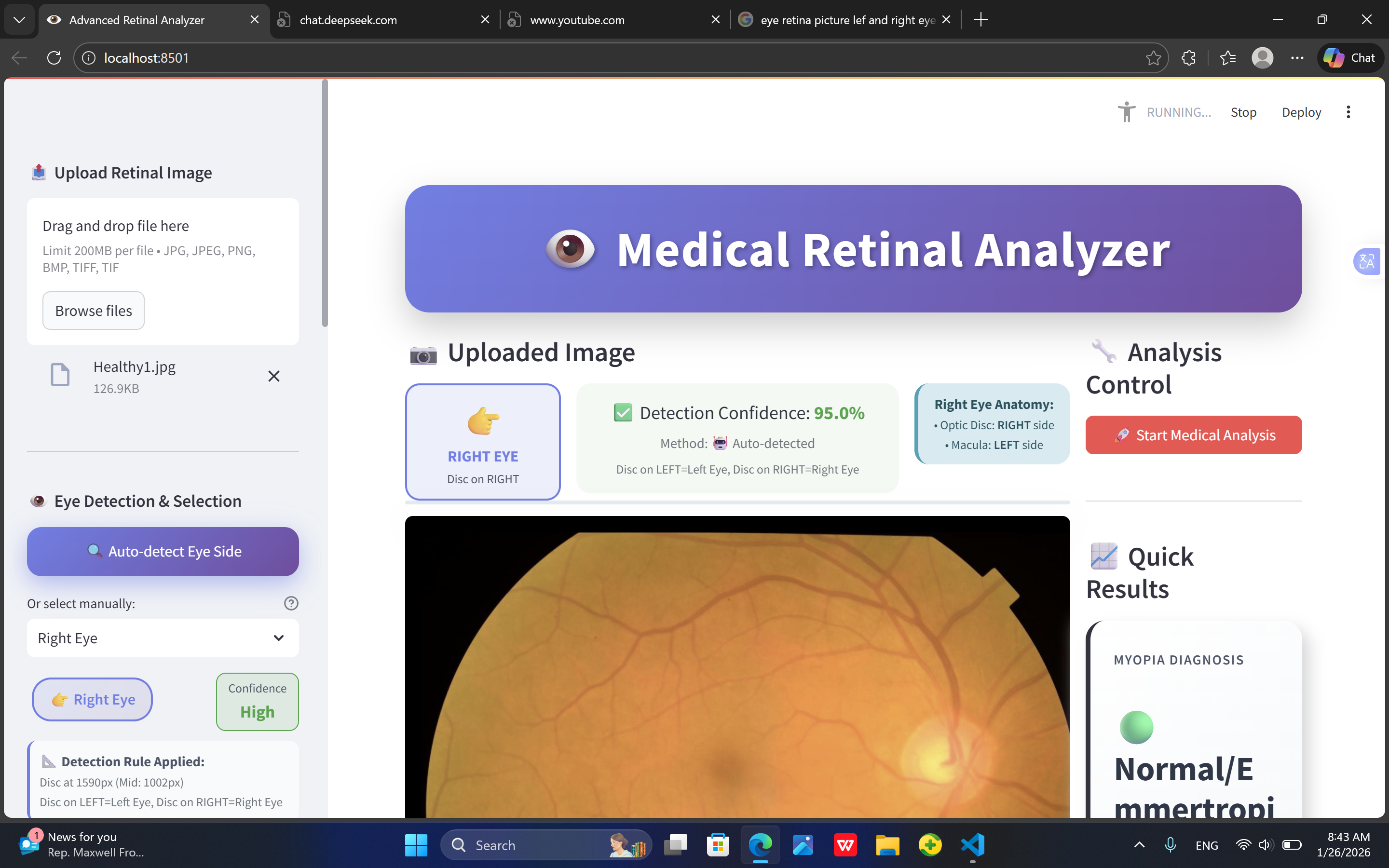
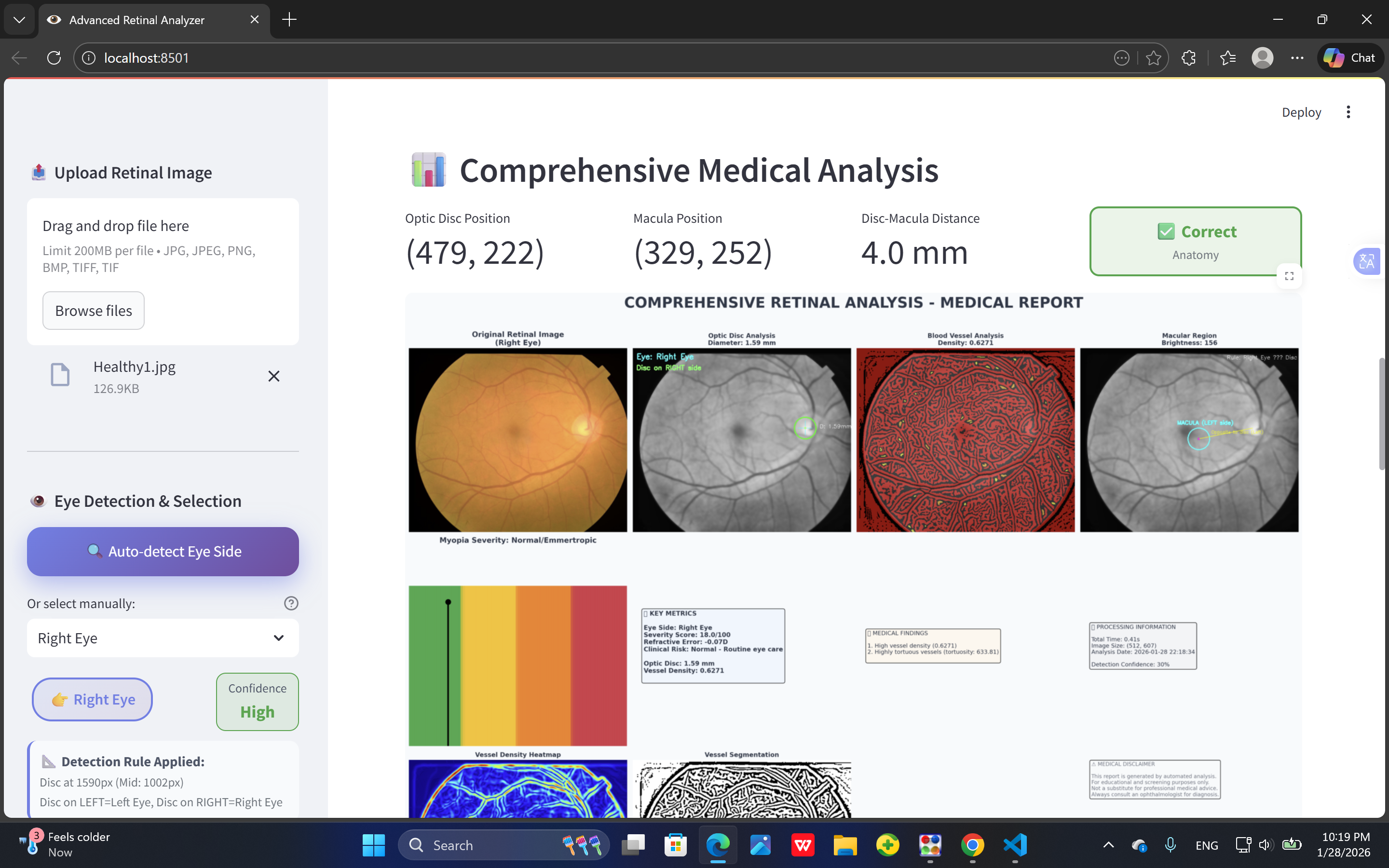
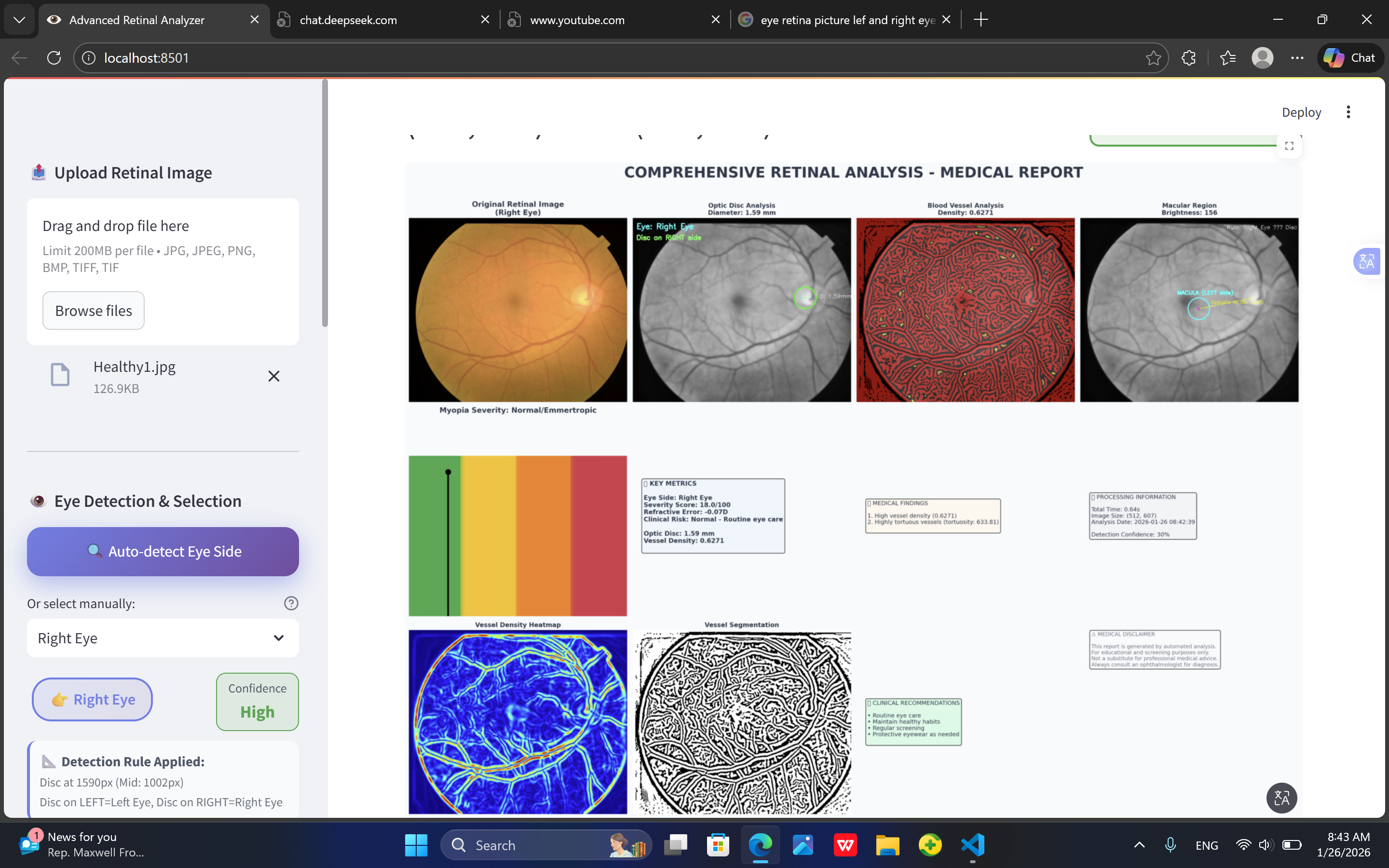
- Clinical relevance of retinal biomarkers, such as optic disc size, cup-to-disc ratio, and peripapillary atrophy.
- Data annotation and preprocessing, ensuring images are normalized and labeled for training.
How I Built It:
The project was implemented in Python using libraries such as OpenCV, scikit-image, and PyTorch.
- Data Collection: Retinal fundus images were collected from public datasets.
- Preprocessing: Images were resized, normalized, and enhanced to highlight key retinal structures.
- Model Development: A CNN-based classifier was trained to detect myopia severity levels (mild, moderate, high).
- Evaluation: Model performance was measured using accuracy, precision, recall, and F1-score.
Challenges Faced:
- Image quality variability: Different fundus cameras produced images of varying resolution and brightness, requiring extensive preprocessing.
- Limited labeled data: Annotated myopia images were scarce, necessitating data augmentation and careful training.
- Class imbalance: Severe myopia images were less frequent, which required weighted loss functions and oversampling techniques.
Impact:
RetinaMyopiaAI demonstrates how AI can assist in early detection of myopia using fundus images. This project combines medical knowledge, computer vision, and machine learning to create a practical tool for ophthalmology.
Example of a simple retinal measurement (using LaTeX):
The optic disc radius ( r ) can be converted from pixels to millimeters:
[ r_{mm} = r_{px} \times 0.025 ]
Where ( r_{px} ) is the radius measured in pixels. This helps quantify structural changes in myopic eyes.

Log in or sign up for Devpost to join the conversation.