-

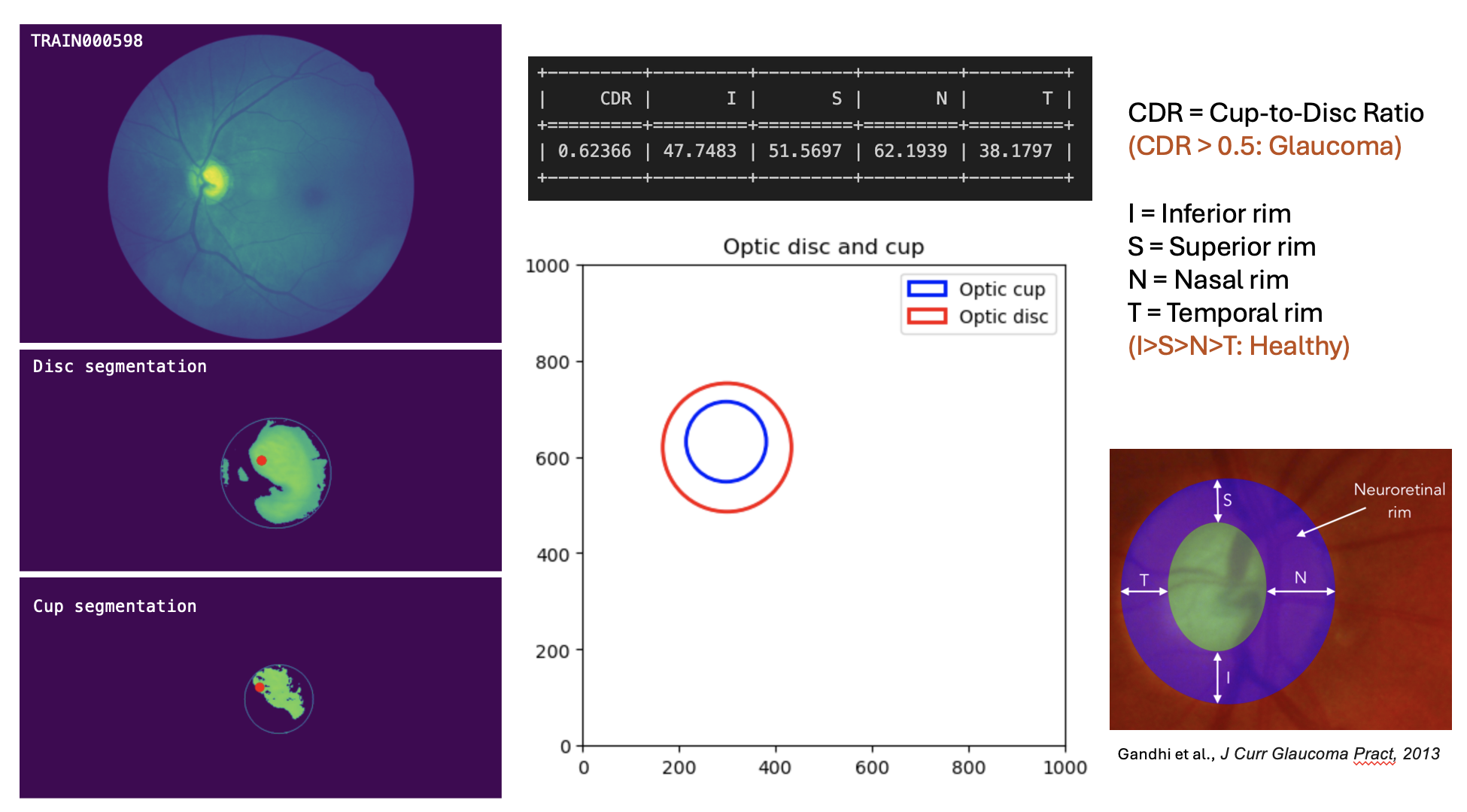
Image analysis pipeline
-

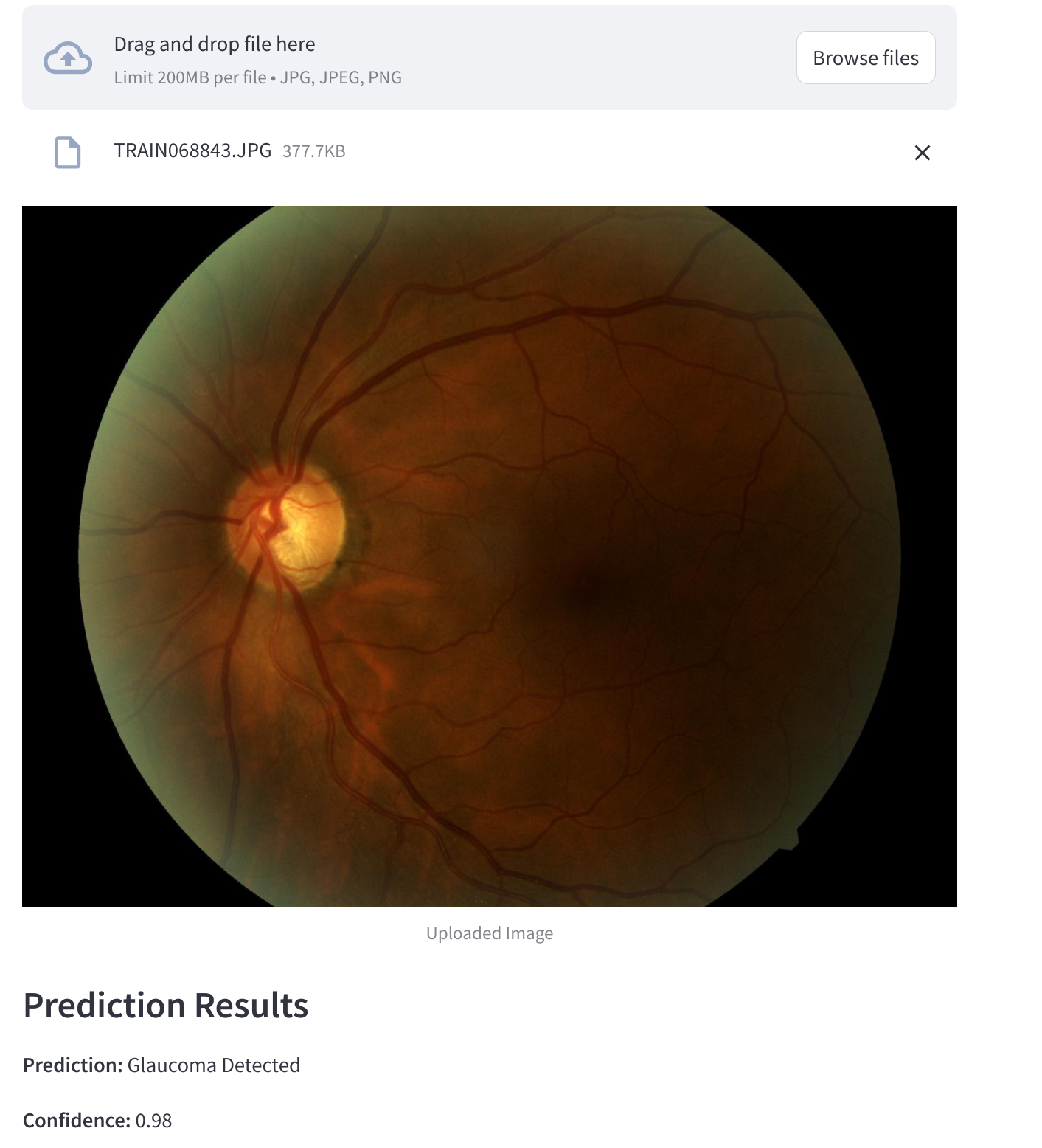
Example color fundus photo and model output
Inspiration
Glaucoma is one of the leading causes of irreversible blindness globally, often referred to as the "silent thief of sight." It affects over 3 million adults over the age of 40 in the United States alone, and the World Health Organization estimates that it accounts for 12% of global blindness. The insidious nature of glaucoma lies in its asymptomatic progression; many individuals remain unaware of their condition until significant vision loss has occurred. Early detection is paramount in managing glaucoma effectively, as timely intervention can slow or halt the progression of the disease, preserving the patient's quality of life.
Our team at Dr. Castillo's Dynamic Medical Image and Computing Lab is driven by the desire to leverage technology to tackle pressing healthcare challenges. We recognized that one of the barriers to early glaucoma detection is the reliance on specialized equipment and expert interpretation, which may not be readily available in all healthcare settings, especially in underserved communities. By developing an algorithm that can analyze standard color fundus photographs—a more accessible imaging modality—we aim to democratize glaucoma screening and assist clinicians in making timely diagnoses.
Our inspiration stems from the potential to make a tangible impact on public health by reducing preventable blindness through technology. By integrating traditional image analysis techniques with advanced machine learning models, we aspire to create a tool that not only detects glaucoma with high sensitivity and specificity but also provides clinically relevant markers to justify its decisions. This explainability is crucial for gaining trust in AI-assisted diagnostics and for aiding clinicians in understanding the underlying pathology. Our ultimate goal is to contribute to a future where no one has to suffer from avoidable vision loss due to late detection of glaucoma.
What it does
InsEYEte is an advanced diagnostic tool designed to detect glaucoma from color fundus photographs. Given a standard retinal image, our algorithm not only determines the presence of glaucoma but also identifies clinically relevant defects and markers to justify its decision. These markers include optic nerve cupping, retinal nerve fiber layer thinning, and vascular abnormalities—all critical indicators of glaucoma progression. By providing both a diagnosis and the underlying reasons, InsEYEte offers a comprehensive assessment that can assist clinicians in making informed decisions.
How we built it
We began by developing a GPU-accelerated preprocessing pipeline to handle high-resolution retinal images efficiently. This pipeline enhances image contrast and focuses on the region of interest around the optic disk, which is crucial for detecting glaucoma-related changes. We utilized traditional image analysis techniques with libraries like SciPy and OpenCV to isolate key markers such as optic nerve cupping and nasalization of vessel trunks.
To capture more complex features, we fine-tuned a ConvNext encoder—a state-of-the-art convolutional neural network—on our dataset. The latent space derived from this encoder encapsulates high-level abstractions of the image data. By concatenating these latent features with those obtained from traditional image analysis, we created a hybrid model that leverages the strengths of both approaches. Training was conducted on our lab's NVIDIA DGX station equipped with an A100 80GB GPU, allowing us to process large datasets and complex models effectively.
Challenges we ran into
One of the primary challenges was handling the variability in image quality and size. Retinal images can differ significantly due to factors like camera equipment, lighting conditions, and patient movement. This variability made it difficult to maintain consistent detection of the optic disk using simple solutions. We had to implement more sophisticated image processing techniques to ensure our algorithm could robustly identify regions of interest across diverse images.
Another challenge was balancing the need for high-resolution images to capture detailed markers with the computational limitations of processing large images. We had to optimize our pipeline to preserve essential details while ensuring that the processing time remained practical for clinical use.
Accomplishments that we're proud of
We are proud to have achieved promising preliminary results, demonstrating a sensitivity of 0.75 on a held-out test dataset at a specificity of 0.95. This indicates that our model is effective at correctly identifying patients with glaucoma while minimizing false positives. Additionally, the integration of traditional image analysis with deep learning provides not only accurate predictions but also explainable outputs, which is crucial for clinical acceptance.
What we learned
This project was a significant learning experience for our team. Transitioning from working with CT and echocardiogram datasets to retinal images required us to adapt our skills to a new imaging modality. Team members with machine learning backgrounds gained insights into traditional computational methods, while those experienced in image analysis delved into advanced deep learning techniques. This interdisciplinary collaboration highlighted the value of combining different methodologies to tackle complex problems effectively.
What's next for InsEYEte
Looking ahead, we plan to refine our preprocessing pipeline to enhance its robustness against varying image quality issues, such as low contrast or blurriness. We aim to expand our image analysis algorithms to extract additional clinically relevant features, like retinal nerve fiber layer thickness and hemorrhages, to improve diagnostic accuracy. Furthermore, we intend to validate InsEYEte on larger and more diverse datasets to enhance its generalizability. Ultimately, our goal is to develop a reliable, explainable tool that can be seamlessly integrated into clinical workflows to aid in the early detection and management of glaucoma.
Built With
- pytorch
- streamlit

Log in or sign up for Devpost to join the conversation.