-
methodology
About the Project
Inspiration
Lung cancer diagnosis often depends on the detailed examination of tissue samples, where histopathological images reveal important cellular and structural characteristics. However, manually examining a large number of images can be difficult, time-consuming, and subject to variation. We were motivated to explore how artificial intelligence could assist in analyzing these images and provide a reliable automated classification approach.
What We Built
We developed “Histopathological Image-Based Lung Cancer Classification Using Feature Fusion and Metaheuristic Optimization”, an AI-based framework for classifying lung cancer from histopathological images.
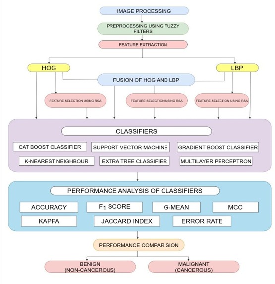
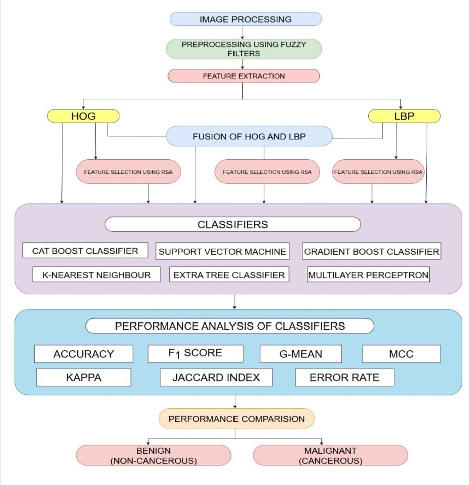
The proposed system combines different feature representations to capture diverse visual patterns present in lung tissue. These features are integrated using feature fusion, followed by metaheuristic optimization to identify the most useful and relevant information for classification. This helps create a more effective feature representation for the final prediction.
Workflow:
Input Images → Image Preprocessing → Feature Extraction → Feature Fusion → Feature Optimization → Classification → Final Prediction
How We Built It
We designed the project as a sequential image-processing and machine-learning pipeline. Initially, histopathological images are processed to make them suitable for further analysis. Relevant visual characteristics are then extracted from the images.
Instead of depending on a single feature representation, we combine multiple feature sets to preserve complementary information. The fused feature space may contain redundant or less useful information, so a metaheuristic optimization technique is used to select an effective set of features.
The optimized features are subsequently provided to the classification model, which generates the final lung cancer prediction.
What We Learned
Developing this project helped us understand the practical application of AI in medical image analysis. We gained hands-on knowledge in:
- Histopathological image processing
- Image feature extraction
- Multi-feature fusion
- Metaheuristic optimization
- Machine learning classification
- Performance evaluation
- Designing an end-to-end medical AI pipeline
A key learning from the project was that better feature representation and intelligent feature selection can significantly influence classification performance, rather than relying only on the classifier itself.
Challenges We Faced
Histopathological images contain complex patterns involving cells, tissues, textures, and structural variations. Handling these variations and extracting meaningful information was one of the major challenges.
We also faced the problem of feature redundancy after combining multiple feature representations. A large feature space can increase computational requirements and may introduce unnecessary information into the classification process.
To address this, we incorporated metaheuristic optimization to search for a more effective feature subset. Finding a suitable balance between feature quality, computational cost, and classification performance was an important part of our development process.
Impact
Our project explores how feature fusion and optimization techniques can strengthen AI-based analysis of histopathological lung images. The resulting framework can serve as an AI-assisted classification system to help researchers and medical professionals analyze tissue images more efficiently.
While the system is not intended to replace clinical diagnosis, it demonstrates the potential of combining advanced computational techniques to support faster and more consistent medical image analysis.
Built With
- aritificialintelligence
- cnn
- computervision
- deeplearning
- featureextraction
- featurefusion
- imageanalysis
- imageclassification
- machine-learning
- numpy
- pandas
- python

Log in or sign up for Devpost to join the conversation.