-
-

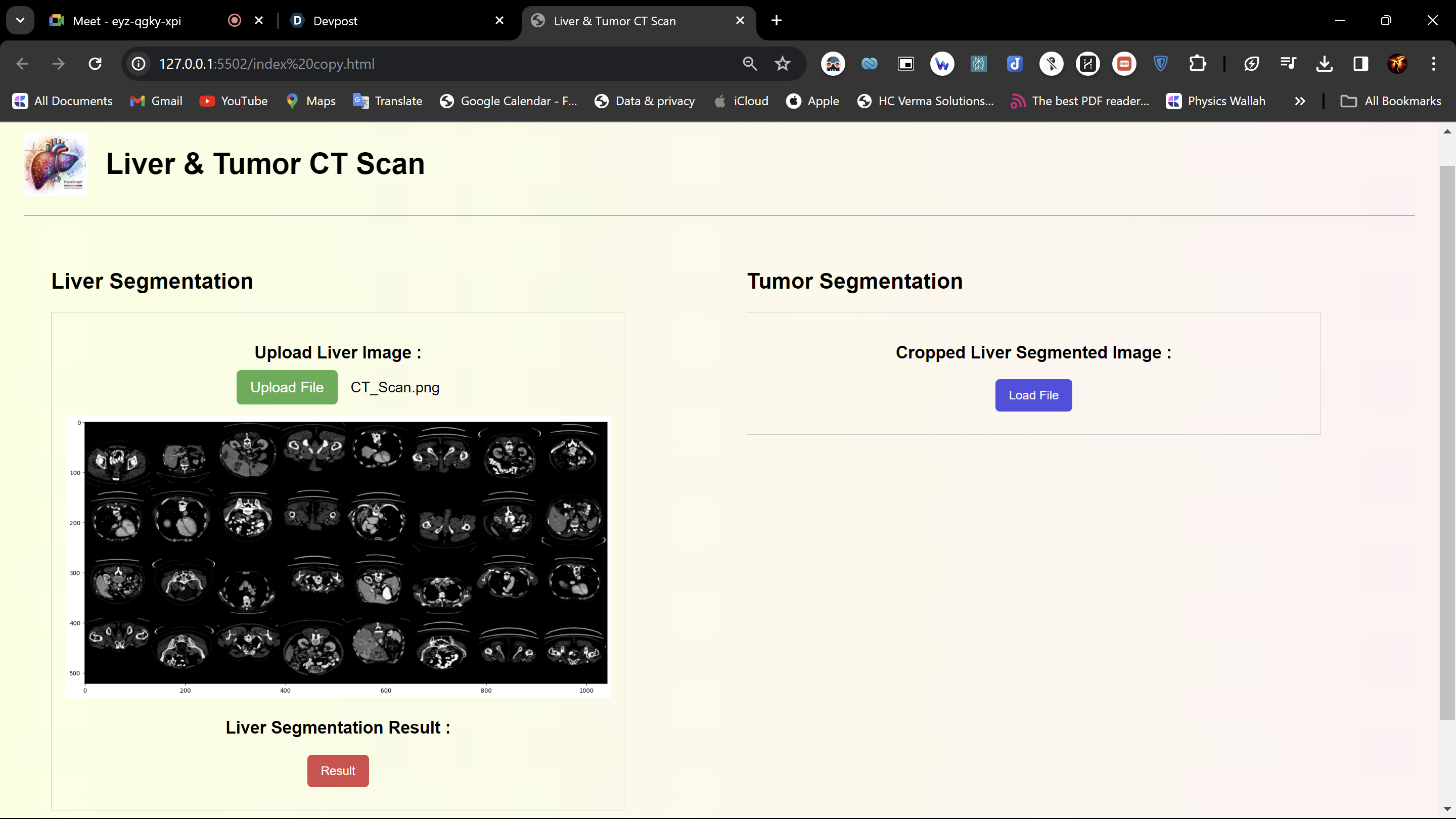
Uploading the image of the CT Scan for Liver Segmentation
-
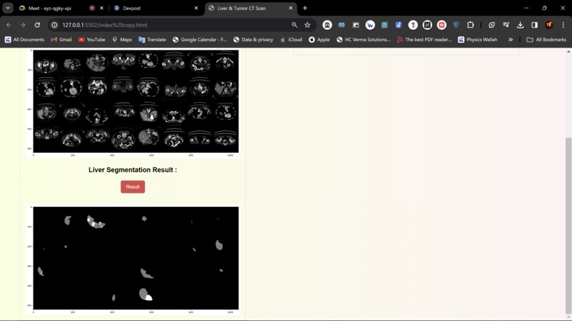
Liver Segmented by processing of CT Scan Image
-
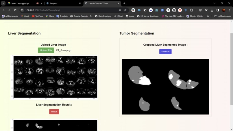
Cropped Tumor & Liver Segmented Image


Inspiration :
Imagine this: doctors battling liver diseases like tumors need a faster, more reliable way to pinpoint the exact location of the problem. That's where our project comes in. Livers can be tricky to manage, and sometimes things like special dyes used for imaging can make it even harder to see what's going on. Our goal is to develop a super-smart tool that can automatically analyze scans and identify the liver, no matter what it looks like. This would be a game-changer for doctors, allowing them to diagnose and treat patients with greater accuracy and speed.
What it does :
The Advanced Liver Segmentation Project is all about developing smart tools that can accurately outline the liver in medical images, especially in CT scans. This project is tackling the tricky parts of liver segmentation, like dealing with different liver shapes, handling contrast agents, and making sure the segmentation is spot-on for diagnosing and planning treatments for liver issues such as tumors. By using fancy deep learning tricks and advanced image processing techniques, the project aims to make liver segmentation more efficient and precise. This is super important for doctors and medical teams in real-world situations, helping them diagnose and treat liver diseases more effectively.
How we built it:
The Advanced Liver Segmentation Project was all about bringing together smart tech, medical know-how, and a ton of CT scans with liver outlines marked up to teach a computer how to spot livers like a pro.We did some fancy stuff to the images first, like cleaning up noise, making sure the colors were just right, and enhancing the contrast to make the pictures clearer for the computer to learn from ,then used these cool things called convolutional neural networks (CNNs) to teach the computer how to pick out liver bits accurately from the images & also borrowed some smarts from other pre-trained models to help the computer get really good at spotting livers specifically. To make sure the computer was doing a great job, we tested it on different sets of images to see how well it could find livers, how sensitive it was to spotting them, and how specific it was in its detections. We tweaked and fine-tuned the model to make it work even better and be super reliable. In a nutshell, the Advanced Liver Segmentation Project was a mix of tech wizardry, medical expertise, and a whole lot of testing to create a top-notch liver-spotting tool for medical imaging tasks.
Challenges we ran into:
Addressing the variability in liver morphology proved to be quite a challenge for segmentation models due to the diverse shapes and sizes of livers in medical images. Robust algorithms were needed to accurately capture these differences in liver morphologies present in the dataset. Dealing with the presence of contrast media in imaging techniques posed another significant challenge for liver segmentation. The introduction of artifacts and inconsistencies by contrast-enhanced imaging required the development of methods to handle these variations effectively and ensure precise segmentation even in the presence of contrast media. One critical challenge was ensuring that the segmentation model could generalize well to unseen data and different imaging modalities. Fine-tuning the model parameters and optimizing the architecture to enhance generalization performance demanded extensive experimentation and validation to achieve reliable results.
Accomplishments that we're proud of:
The project was all about making liver segmentation more efficient and accurate, dealing with the complexities of liver morphology variations, challenges posed by contrast media, and the critical need for precise segmentation in diagnosing and planning treatment for hepatic diseases like liver tumors.
To achieve this, the project successfully created automated segmentation methods using cutting-edge deep learning algorithms and advanced image processing techniques. These methods were specifically designed to accurately segment the liver in medical imaging, with a special focus on CT scans. By developing these innovative approaches, the project aimed to streamline and enhance the process of liver segmentation, ultimately improving the quality of diagnosis and treatment planning for various liver-related conditions.
What we learned:
Impact of Data Quality on Model Performance: The quality of annotated data directly influences the performance of segmentation models. Ensuring high-quality, diverse datasets is crucial for training accurate and generalizable deep learning models for liver segmentation. Importance of Automation: Automating liver segmentation tasks can significantly improve efficiency, consistency, and accuracy compared to manual segmentation methods. The project highlighted the benefits of automation in streamlining medical imaging processes.
What's next for HepaScope: Advanced Liver Segmentation Project:
Improving the segmentation of small liver tumors remains a challenging task, despite the advancements in some methods. It would be beneficial to explore techniques that can effectively segment a wide range of liver tumors.
Hierarchical Convolutional Deep Neural Networks (CDNNs) have shown promise in accurately segmenting liver tumors from coarse to fine levels and analyzing tumor burden. Further research into these models could potentially enhance segmentation outcomes.
Integrating real-time assessment of image quality for liver T2WI images could be a valuable addition to the segmentation process. By incorporating techniques for automated evaluation of image quality, it is possible to enhance image quality and mitigate the impact of suboptimal images on the segmentation process.


Log in or sign up for Devpost to join the conversation.