-
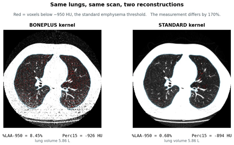
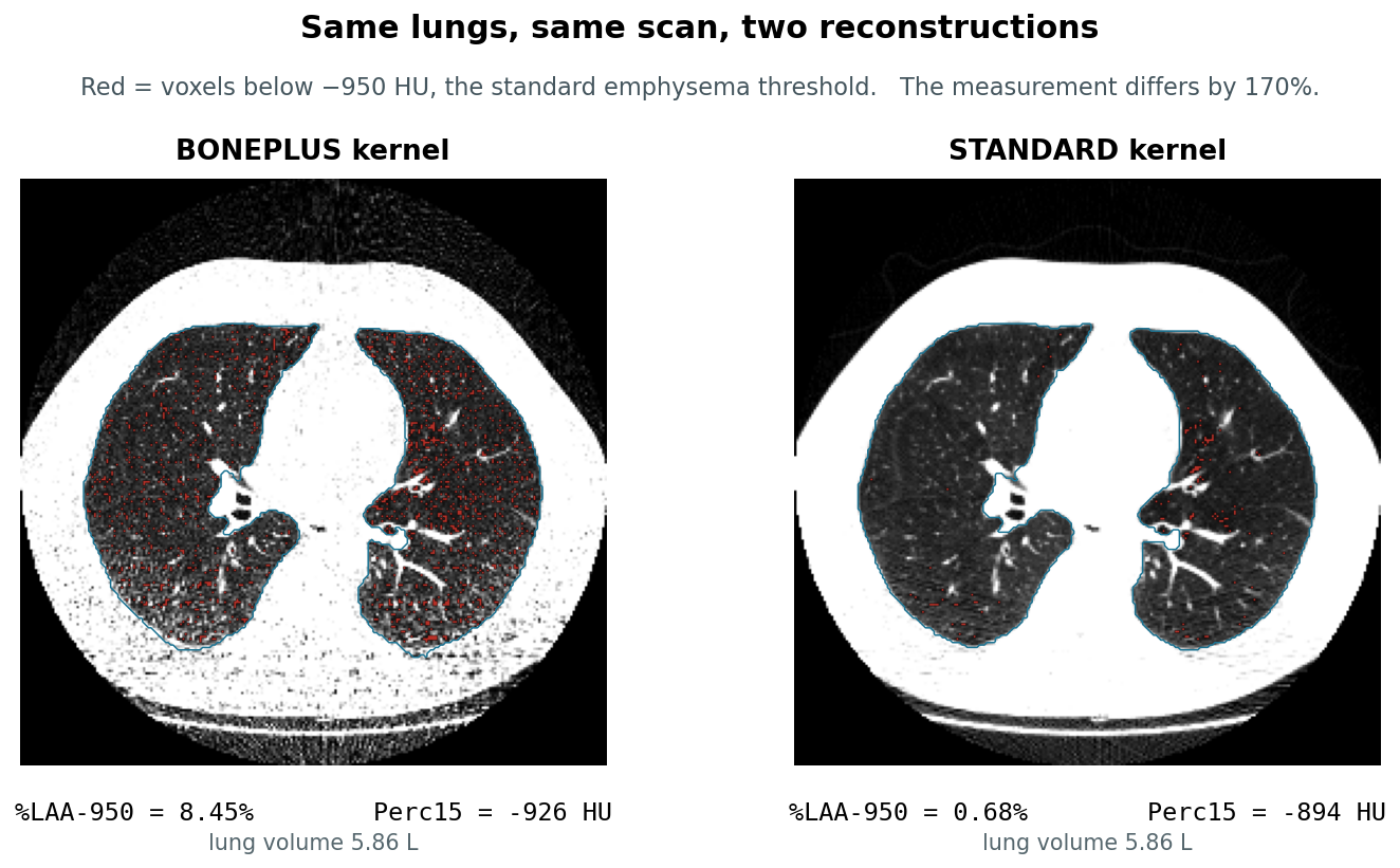
Same scan, two reconstructions. 8.45% vs 0.68% emphysema, identical 5.86 L lung. Left has emphysema, right does not.
-
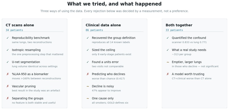
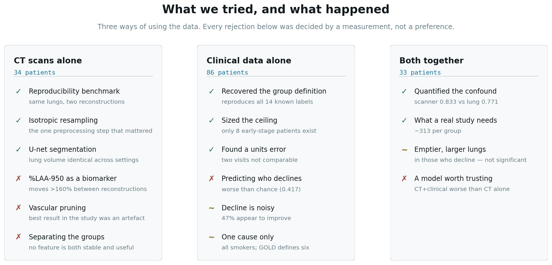
What we tried across CT alone, clinical data alone, and both combined. Every rejection was decided by a measurement.
-
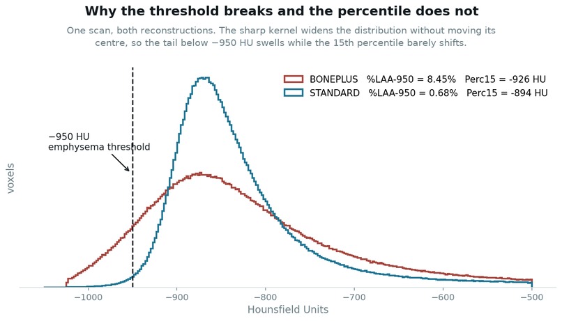
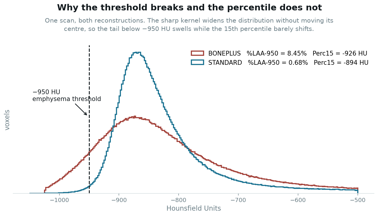
The sharp kernel widens the density distribution without moving its centre, so a fixed threshold counts noise as disease.
-
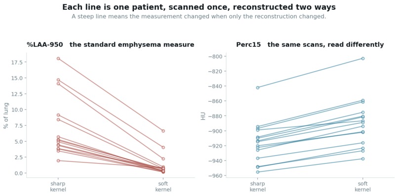
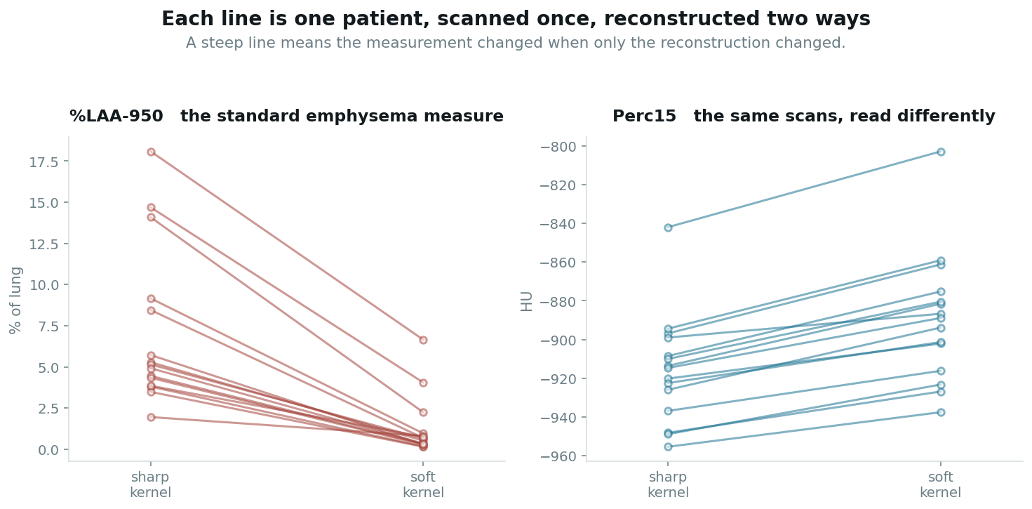
All 15 patients, both reconstructions. %LAA-950 reorders them — median shift 164%. Perc15 stays parallel at 2.9%.
Inspiration
COPD is almost always caught late, once spirometry crosses FEV₁/FVC < 0.7. But damage exists in the lung years earlier. The challenge was to find it on CT, in people whose breathing tests are still normal.
What we found
Not a biomarker. Something we think matters more.
The same lungs, one scan, two reconstructions:
| Sharp kernel | Soft kernel | |
|---|---|---|
| %LAA-950 | 8.45% | 0.68% |
| Lung volume | 5.86 L | 5.86 L |
On the left that patient has emphysema; on the right they do not. Across 15 paired patients the median shift is 164%. Perc15, read from the same scans, shifts 2.9%.
A sharp kernel adds noise, widening the density distribution without moving its centre. %LAA-950 counts voxels past a fixed line, so it reads that spread as disease:
%LAA-950 = the percentage of lung voxels below −950 HU.
We then scored every feature against the clinical label. Acquisition parameters beat every imaging feature — slice count AUC 0.833, tube voltage 0.750, against 0.771 for the best measurement of the lung. A model trained without protocol control learns the scanner.
How we built it
Python, SimpleITK, lungmask (R231 U-net), Frangi vesselness, scikit-learn.
One scan chosen per patient by explicit rules — reject expiratory, prefer soft kernel and 120 kVp, verify the plane from the direction cosines, never pair across studies. Every decision logged.
The key idea: each patient's scan is stored at several reconstructions. The patient did not change between them, so any difference is the machine. That makes a paired experiment possible with no labels at all.
Challenges
Our first segmentation deleted the lungs. It discarded air touching the volume border — but the trachea reaches the top slice, so lungs, airway and outside air are one component. One patient came out at 0.036 L against a true 6.2 L, and nothing in the table looked wrong.
Every new batch broke something. An angiogram treated as non-contrast. Two reconstructions paired across studies three years apart. A coronal reformat chosen over a real acquisition. Each would have produced a plausible, wrong number.
Our most impressive result was an artefact. Vascular pruning reached AUC 0.875 — the largest effect in the project — from the one feature that had already failed the robustness test. A proper vessel segmentation erased it.
What we learned
Measure your measurement first. Of 26 features, only 8 survived a change of kernel.
A negative result with a number beats a positive one without. Detecting the effect we see at 80% power needs roughly 313 patients per group. This cohort has 8 pre-COPD patients; we scanned all of them.
Clinical data told us nothing — age, pack-years, baseline FEV₁, BMI and sex predicted decline worse than chance (AUC 0.417). CT reached 0.613. The case for imaging is not that it works well; it is that nothing else works at all.
What's next
Report Perc15, not %LAA-950 — same scans, ρ = 0.961 against 0.664. Free improvement for anyone doing lung densitometry.
A cohort large enough to settle this is necessarily multi-centre, and that is exactly where reconstruction kernels get mixed. Protocol standardisation is a prerequisite, not a detail.
Still missing: expiratory scans, more early-damage patients, a third breathing test, and aetiologies beyond smoking — GOLD defines six, this cohort has one.
All data de-identified. Raw imaging deleted after analysis; no patient identifiers in the repository or any figure.
Built With
- biomarkers
- computer-vision
- copd
- ct-scan
- data-science
- databases
- healthcare
- image-segmentation
- medical-imaging
- numpy
- public-health
- pulmonology
- pydicom
- python
- pytorch
- quantitative-imaging
- radiology
- reproducibility
- scikit-image
- scikit-learn
- scipy
- simpleitk

Log in or sign up for Devpost to join the conversation.