-
-
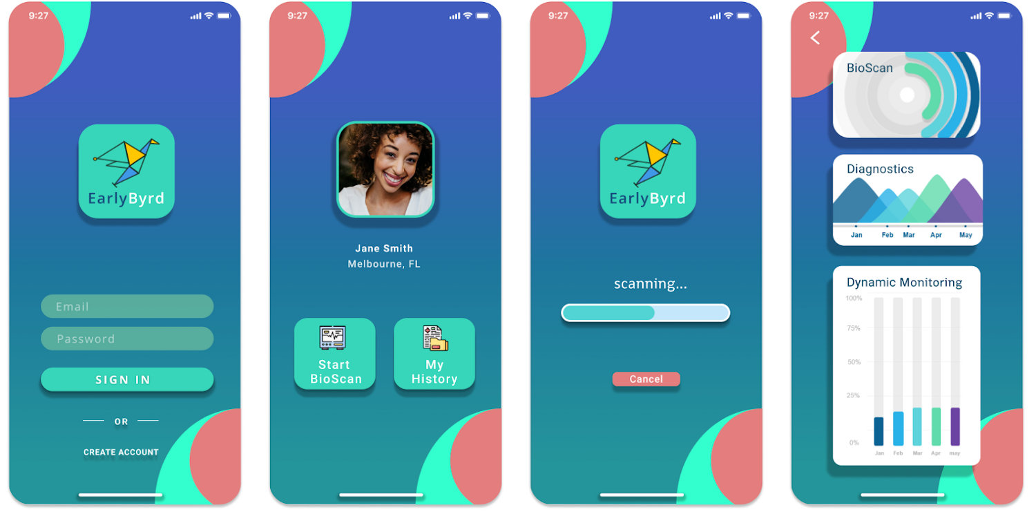
MobileApp_Prototype
-
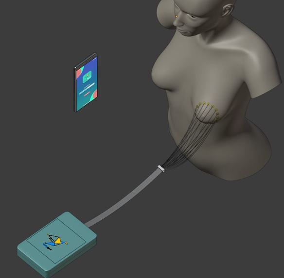
Bio-Scan
-

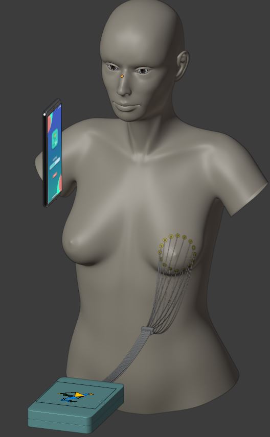
Front view
-
Electrode_Attachment_closeup
-
Top view


Purpose and Motivation
The pandemic has shed a light on the deficiencies in healthcare, particularly for developing nations. As someone that was born and grew up in Kenya, I noticed that there is a stark contrast between the healthcare tech available to citizens of developing nations as compared to citizens from a country like the United States. Focusing on cancer statistics, I found that although developing nations bear about 80% of the global cancer burden, only about 5% of global resources are devoted to cancer prevention and treatment. For example, in Kenya, 70–80% of cancer cases are diagnosed in late stages, and 78.5% of the victims do not survive. In the book Radical by Kate Pickert, Black women are “diagnosed with the disease more often than other races but are 40% more likely to die of it due to lack of access to high-quality treatments and other factors.” To combat this social challenge, we took a detailed look at the current breast cancer screening technology and found that Ultrasound has a high rate of false positives as it cannot distinguish between benign and malignant tumors. Furthermore, mammograms are unsafe for younger women due to the radiation exposure and studies have shown that repeated use of mammograms can contribute to cancer mortality. Our idea is novel because after talking to a radiologist in Kenya, we found out that nobody there is using EIT technology, which has been introduced recently in the biomedical imaging world in USA. We then did further research and realized that we could create a modernized, radiation-free device that works on the principle of bio-impedance that would only cost up to 200 USD to build.
How this application works
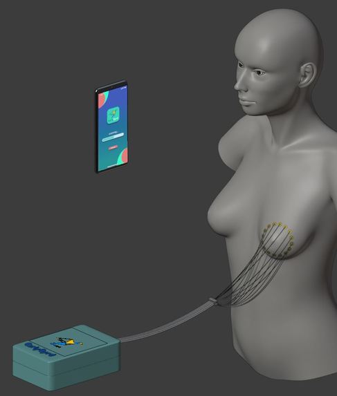
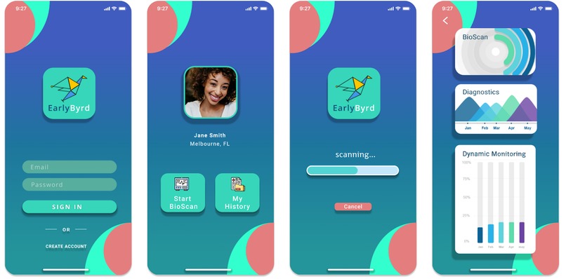
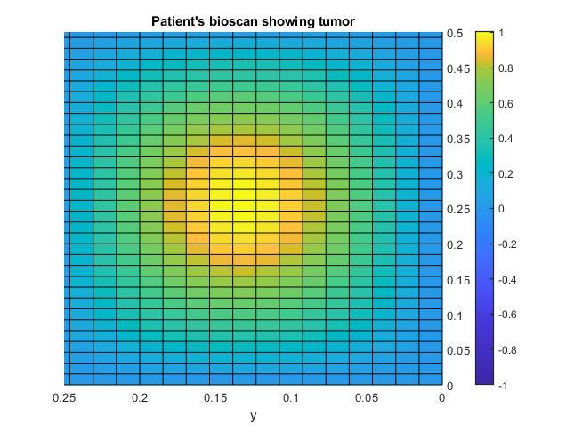
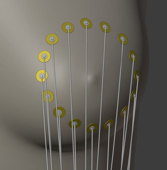
We want to use our skills and experience to develop a low-cost, non-invasive, radiation free biomedical device for early breast cancer detection. The device works on the principle of bio-impedance in order to detect cancerous tumor. It has already been shown that cancerous cells have significantly different conductivity properties as compared to healthy cells. To be more specific, cancerous cells are more conductive than healthy cells, which means they allow passage of more current. We are using precisely this property in order to record surface electrical activity from electrodes placed on breast tissue. Low current will be applied at a frequency of 50KHz as it has been shown from research that accurate readings of impedance can be obtained at this value. The electrodes record data in a pair-drive fashion, where adjacent electrodes will be passing current in turn while all other electrodes will be recording the voltage. After all impedance readings have been obtained, the data will be imported to a mobile app. For now, we are sending the data to a computer because our app isn't ready yet. Then an elegant reconstruction algorithm is implemented to create a 2D cross-sectional view of the breast to show if a tumor is present. Our mobile app will pair with the device and store the results of the bio-scans. Our mobile app idea will allow medical staff to easily access the results anywhere and at anytime. It also allows dynamic monitoring of patient's history. This is something that current technology such as Ultrasound, X-ray mammography and MRI/CT scans do not allow as repeated radiation exposure contributes to cancer mortality.
How we built it
We are currently in the process of obtaining the hardware components for the device. We have also carefully selected all the hardware components after reading a ton of research papers in the field of Electrical Impedance Tomography. Our prototype will be built using multiple electronic components including multiplexers, micro-controller, impedance analyzer, battery pack, electrodes, and wifi module. So far we have ordered the ESP32 micro-controller and the Impedance Analyzer and have written Arduino code in order to log impedance/ voltage data. We are doing a trade-off study to help us decide whether we should buy reusable, dry electrodes which are comfortable for the patient but very expensive, or non-reusable, wet electrodes which are uncomfortable for the patient when removed from their skin, but are cheap. We have already developed a 3D CAD model of the device in Creo (for the device) and Blender (for the patient) demonstrating how a human subject would interact with it. We have also built a UI of our mobile app using Figma. It shows the basic functionality of the app and what features are available. For the back-end of the app, we have written MATLAB and C code that will contain the reconstruction algorithm that uses the GMRES method with multigrid preconditioning to numerically solve a PDE in order to gather a complete picture of the conductivity map of a patient's breast.
How to use this application
The electrodes would be attached to the patient's breast and current would be applied to the tissue beneath. The impedance data will be recorded and sent to our mobile app. The mobile app will seamlessly connect to our device in order to perform scans with a few taps and in a matter of seconds. The application will run our reconstruction and inference algorithms in real-time and significantly aid doctors in performing diagnosis. The EarlyByrd mobile app will also store patients’ history. As such, it will serve as a great tool to track the progress of treatment.
Challenges we ran into
The team consists of engineers with a background in Mechanical, Aerospace and Applied Mathematics. As a result, we do not have much experience in the Medical field, so being able to read relevant research papers and understand the underlying biology proved to be a time-consuming and challenging task. We are having a tough time deciding what kind of electrodes we should proceed with so that it isn't too costly but at the same time we want to get accurate readings and ensure that the patient has a comfortable experience. Furthermore, both of us have no prior experience with building mobile apps, so this is the first time we have really pushed ourselves to work on this idea and build something that could be useful to millions of people. We are learning how to use Flutter and Dart to create our mobile app and have spent hours working on the UI/UX design for the app and website.
Accomplishments that I'm proud of
- We have completed the high-fidelity prototype design for our mobile app using Figma and Flutter.
- We have created a 3D CAD model of our device using Blender and Creo Parametric to demonstrate how a human subject would interact with it.
- We have completed coding a reconstruction algorithm in C and MATLAB that uses the GMRES method to generate a complete picture of the impedance activity in the breast tissue.
Go-to-Market Evaluation (How will this product be made scalable and accessible to the public?)
Our device is very cheap to manufacture (only $200) in comparison with current screening technology which can cost anywhere between $10,000 - $300,000. This means that the device is affordable to hospitals in under-resourced regions. It is portable and therefore can be easily shipped out. The mobile app is accessible to anyone with a smart phone and the results generated can be easily understood by medical staff. Smart phone penetration in sub-Saharan Africa has seen remarkable growth in recent years and will continue to grow according to Forbes. We would use smart phone adoption in the region to maximize our impact.


Log in or sign up for Devpost to join the conversation.