-
-
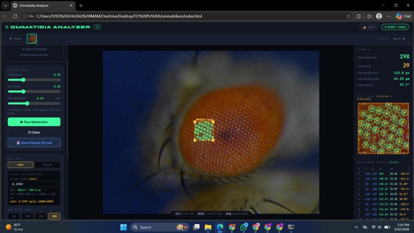
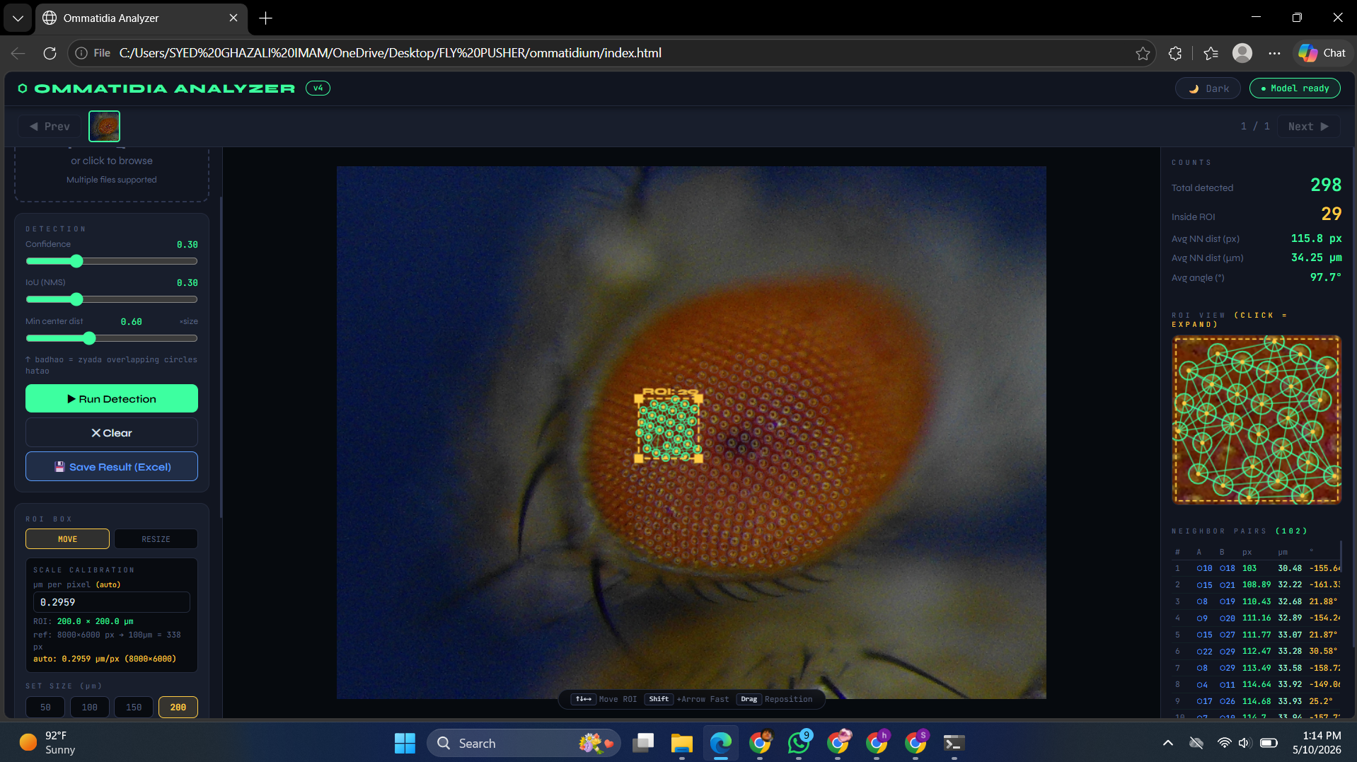
ommatidia counting and neighboring distance
-
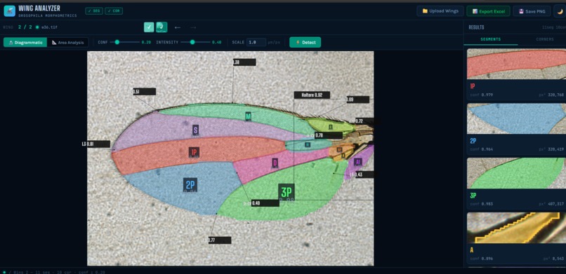
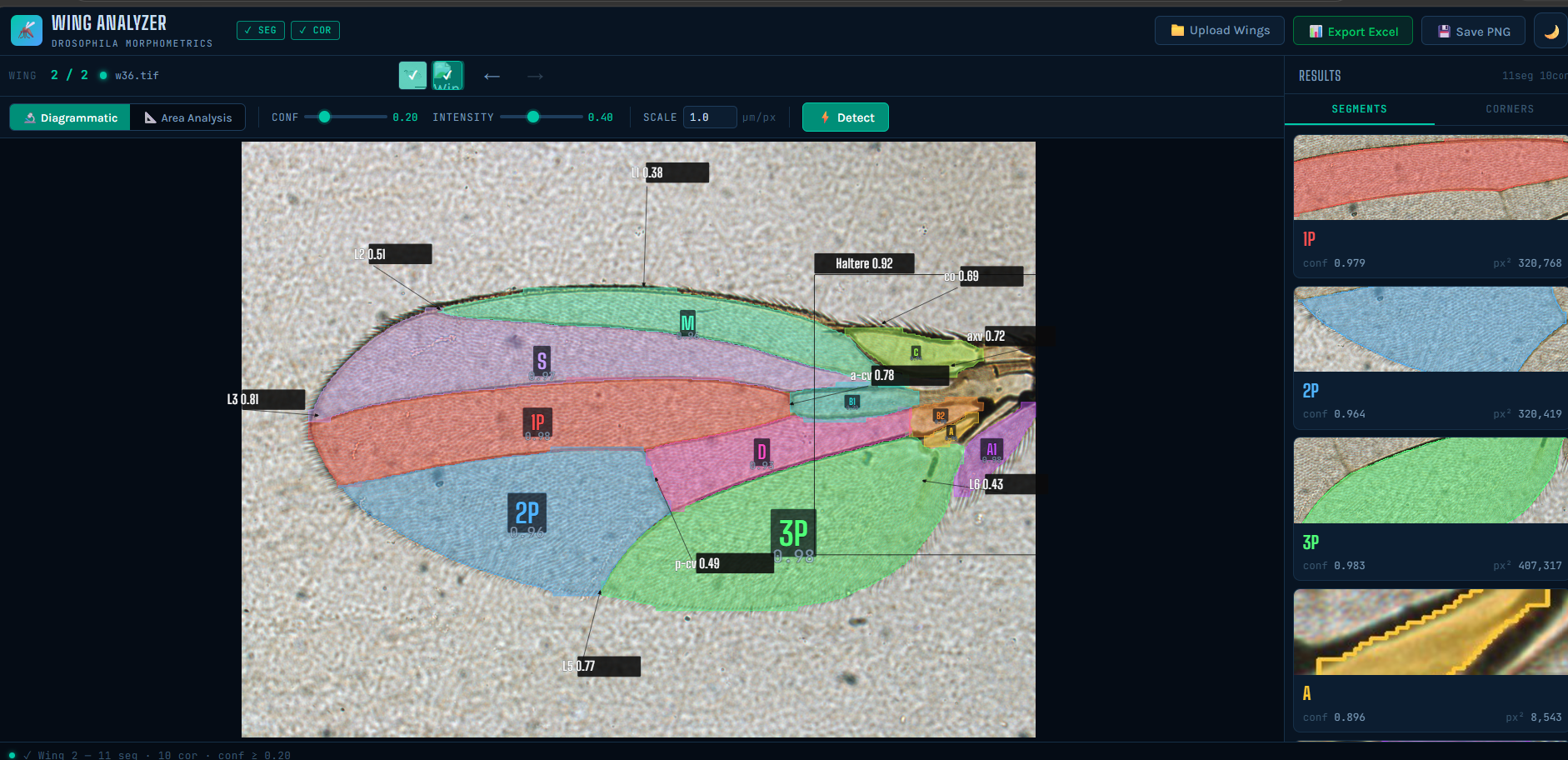
wing corners detection and area
-
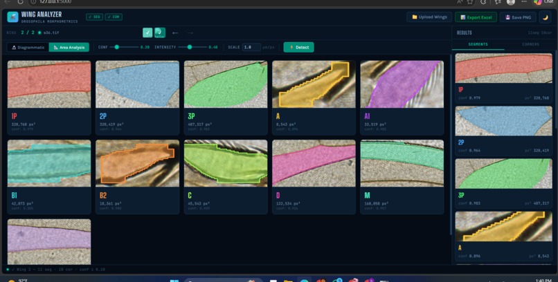
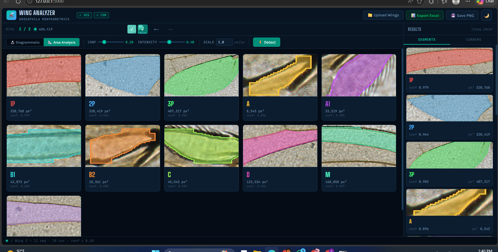
individual wing's segment area
-
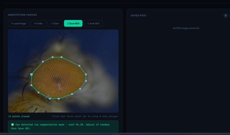
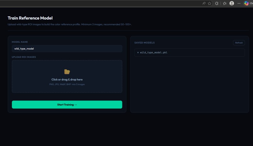
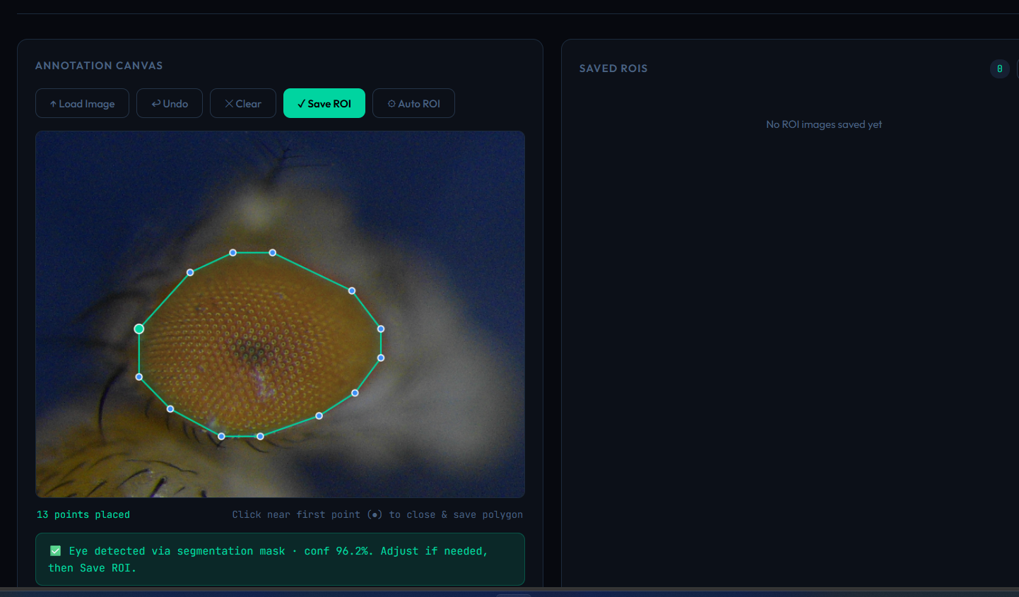
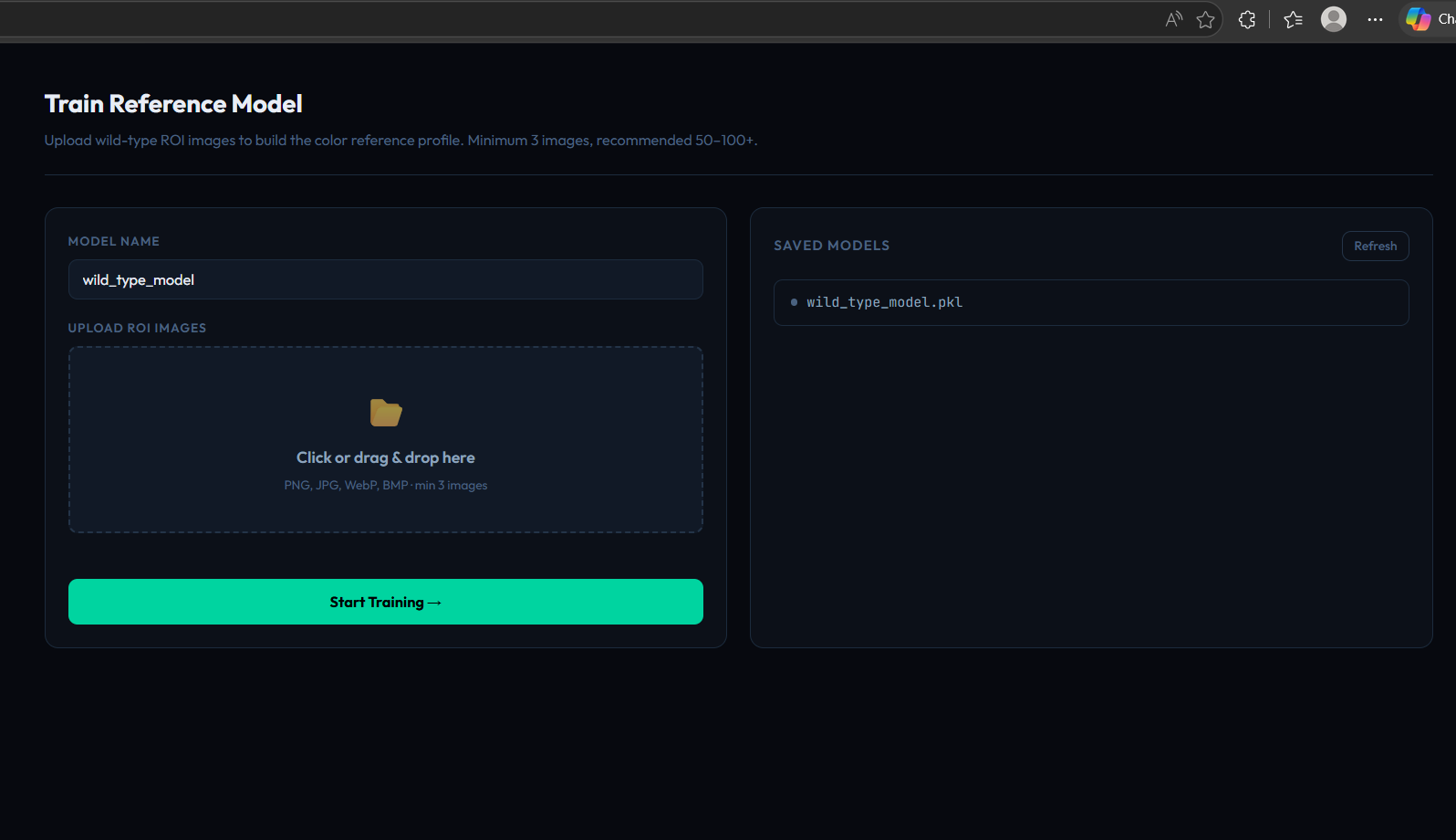
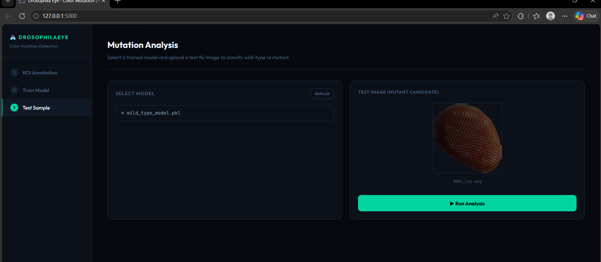
model for detecting eye part for colour analysis
-
-
-
-
-
-
-
-
-
-
-
-
Inspiration
Drosophila research relies heavily on precise morphological analysis, but many phenotyping tasks are still performed manually. Researchers may spend hours examining fly eyes, wings, and pigmentation, making the process time-consuming, repetitive, and susceptible to human error.
We were inspired to build a tool that could bring AI-assisted automation to Drosophila laboratories. Our goal was to make morphological analysis faster, more consistent, and easier to reproduce while allowing researchers to focus more on biological interpretation rather than repetitive measurements.
What it does
Drosophila Species Morphometrics is an AI-powered phenotyping platform designed to automate the analysis of key morphological characteristics of Drosophila.
The platform includes multiple analysis modules:
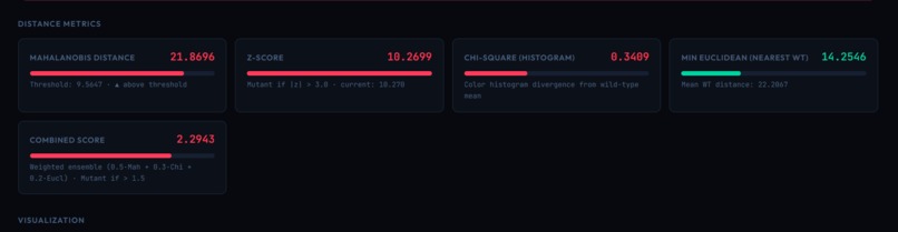
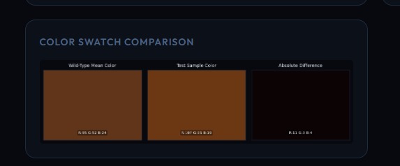
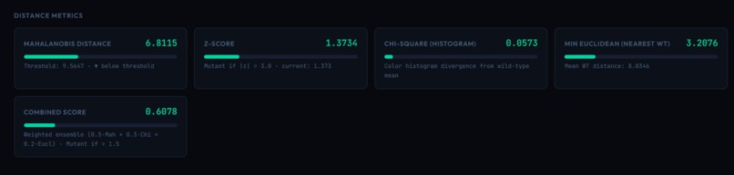
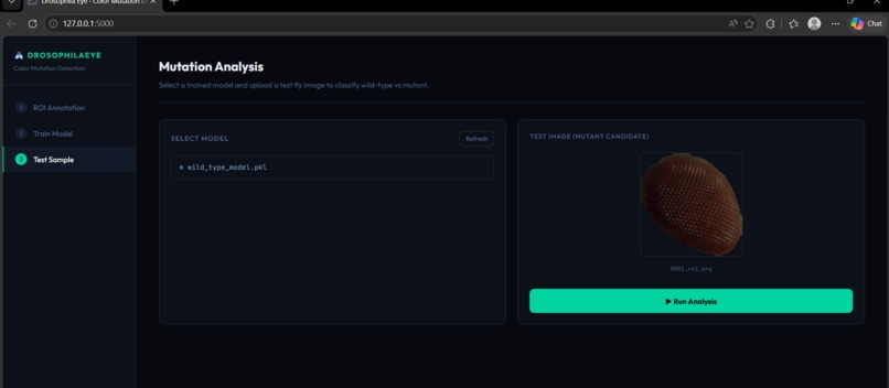
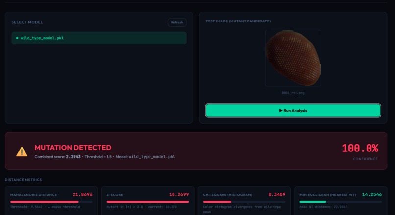
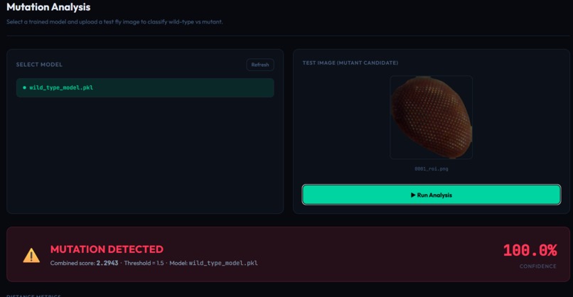
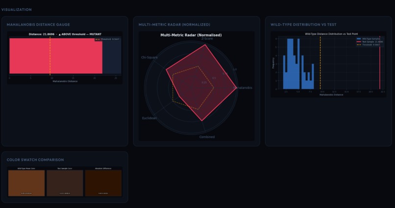
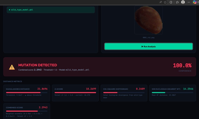
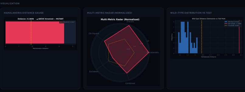
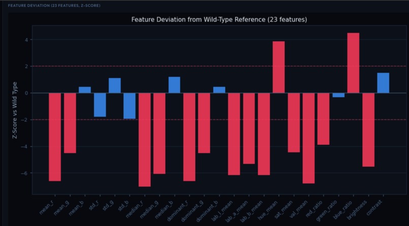
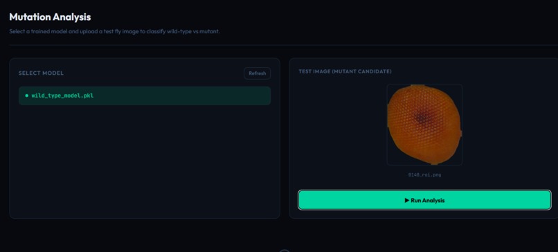
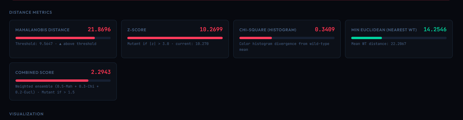
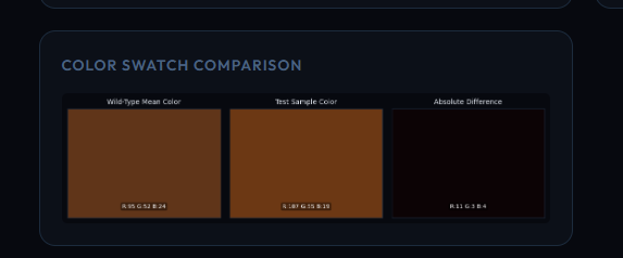
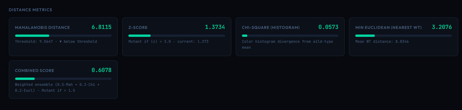
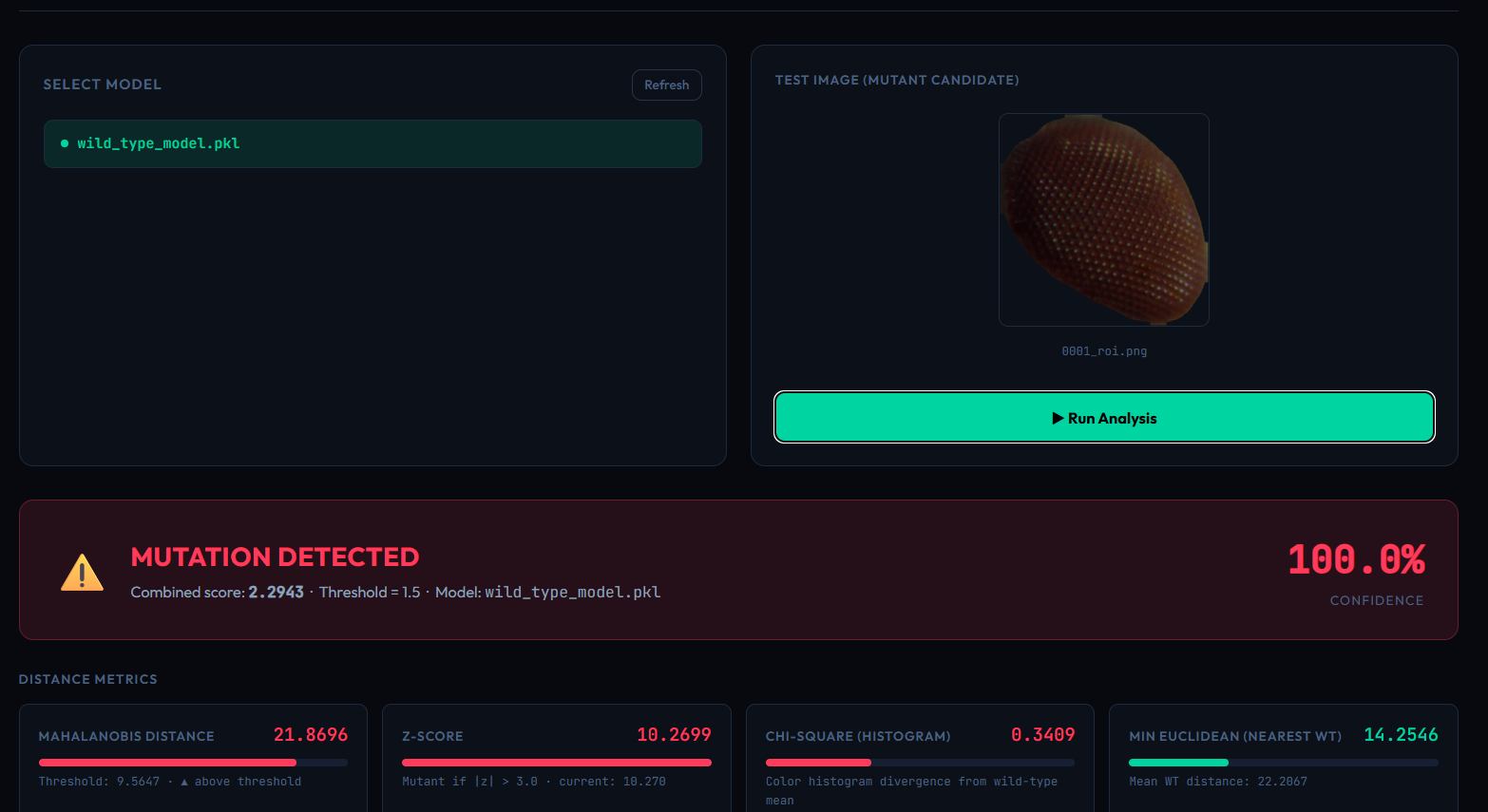
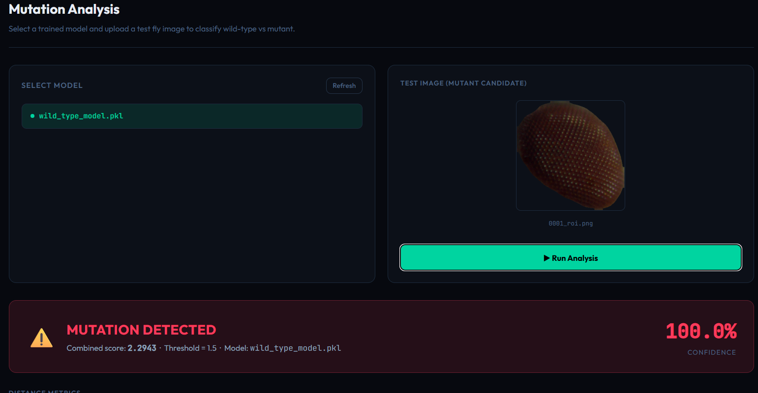
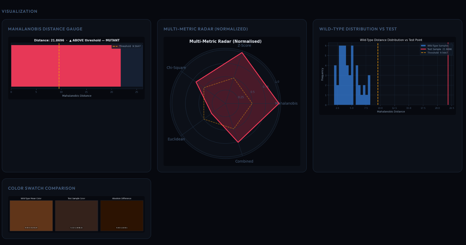
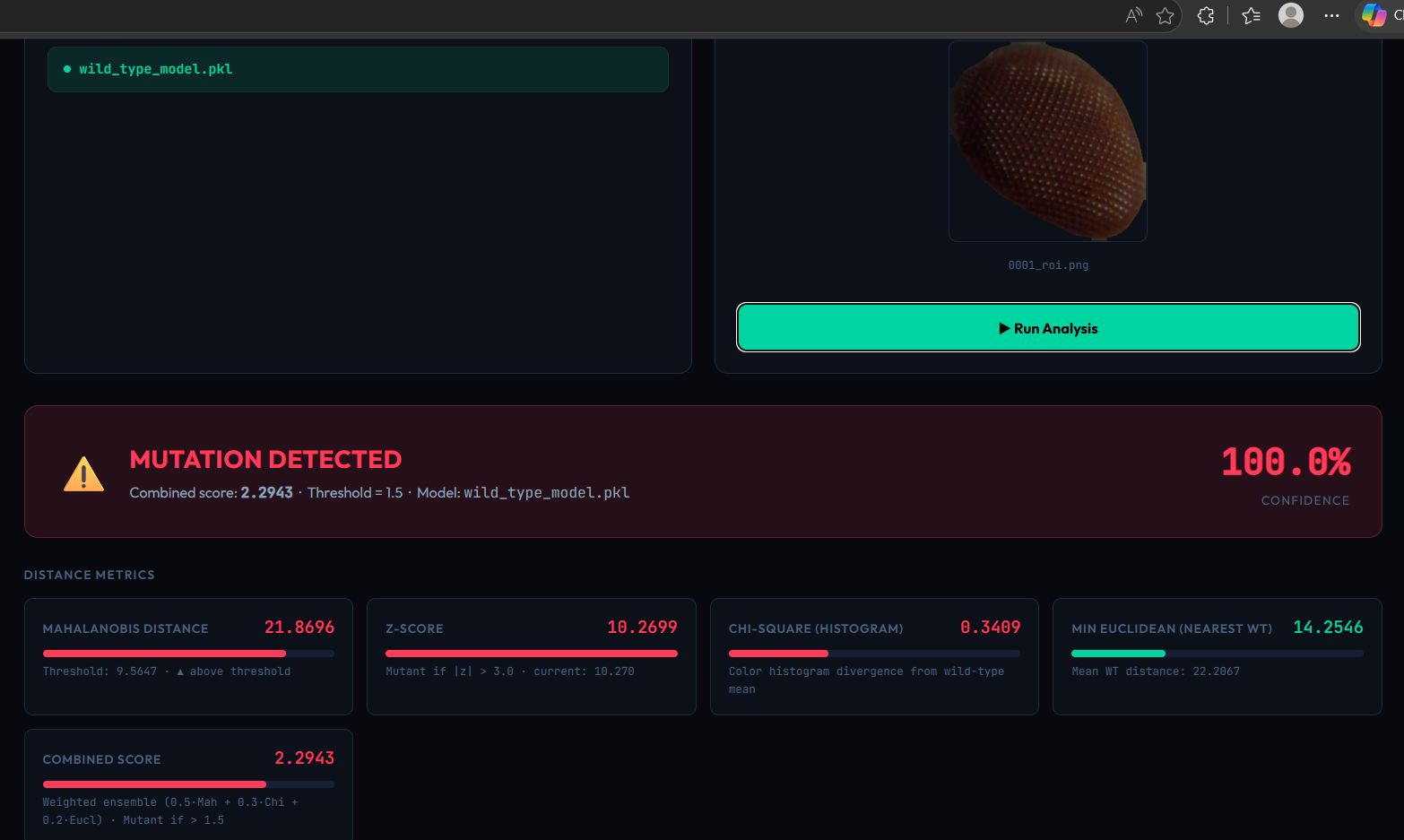
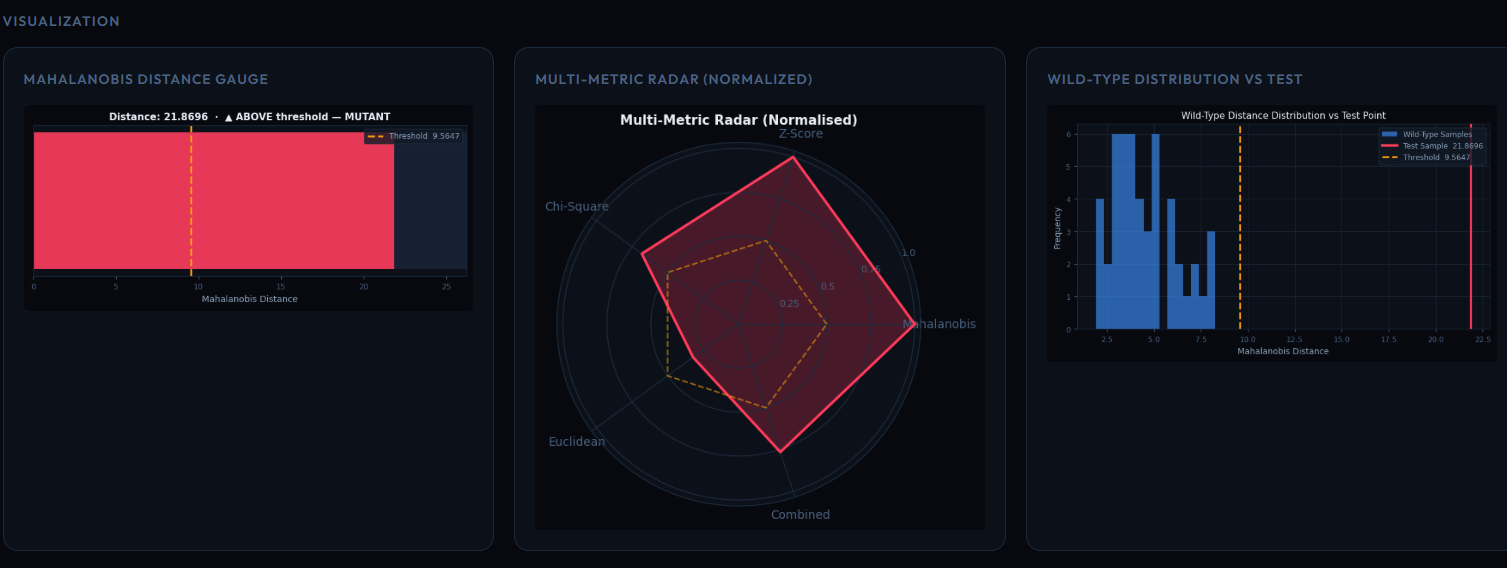
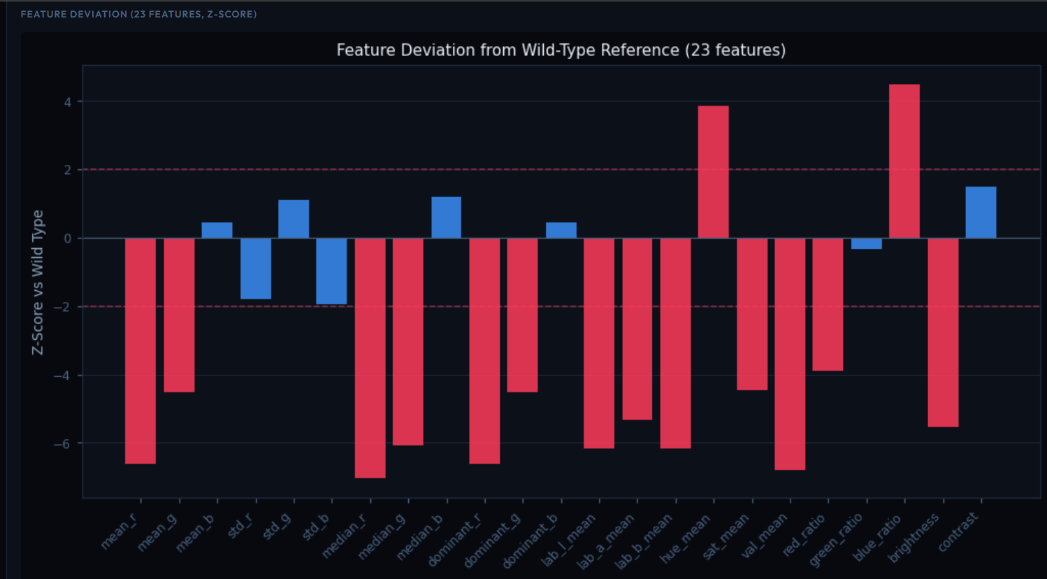
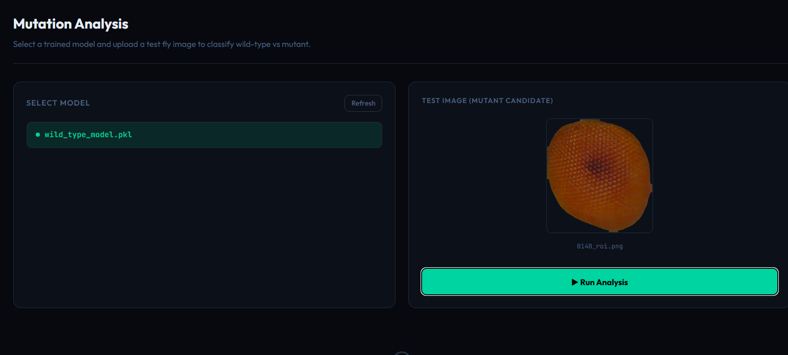
Ommatidia Analyzer — detects and analyzes ommatidia in Drosophila eyes using computer vision. Color Mutation Analyzer — compares eye coloration against a wild-type reference profile and identifies deviations using statistical analysis, including Mahalanobis distance. Wing Analyzer— uses computer vision models to analyze Drosophila wing morphology.
Instead of requiring researchers to manually inspect and measure images, the platform converts images into quantitative morphological information that can be used for downstream research and comparison.
How we built it
We combined computer vision, deep learning, statistical analysis, and a web-based interface to create the platform.
For object detection and morphological analysis, we experimented with modern detection architectures and developed customized pipelines for Drosophila-specific features. The Ommatidia Analyzer incorporates a custom YOLO-based model and distance-based Non-Maximum Suppression to improve detection in densely packed eye structures.
For color analysis, we established wild-type reference profiles and used statistical distance measures to quantify how strongly a sample deviates from the reference distribution.
The platform also includes visualization components that allow researchers to interpret the extracted measurements rather than receiving only raw model predictions.
We iteratively trained, evaluated, and refined our models using annotated Drosophila datasets, experimenting with different architectures and dataset splits to improve reliability.
Challenges we ran into
One of the biggest challenges was the limited availability of high-quality, consistently annotated Drosophila datasets. Biological images can vary significantly in lighting, orientation, image quality, and morphology, making generalization difficult.
Detecting small and closely packed structures such as ommatidia was particularly challenging because individual structures can overlap or appear extremely close to one another. This required us to go beyond standard object-detection pipelines and develop additional post-processing strategies.
We also had to balance model accuracy, computational requirements, and practical usability. A model that performs well experimentally is not necessarily useful in a laboratory if it is difficult to operate or interpret.
Accomplishments that we're proud of
We are proud to have transformed a traditionally manual biological analysis workflow into an AI-assisted morphometrics platform.
We successfully developed multiple specialized modules rather than building a single generic classifier. The platform can analyze eye structures, pigmentation, and wing morphology while producing quantitative and visual outputs.
We are particularly proud of combining deep learning with statistical phenotyping, allowing the system to not only detect biological features but also quantify deviations from a wild-type reference.
The project also demonstrated that computer vision can be adapted to specialized biological research problems where conventional off-the-shelf models are not sufficient.
What we learned
This project taught us that applying AI to biology requires much more than simply training a model. Data quality, annotation consistency, biological context, evaluation methodology, and interpretability are equally important.
We learned how challenging it can be to work with small, specialized datasets and how domain-specific preprocessing and post-processing can significantly affect model performance.
We also learned the importance of designing AI systems around the researcher's workflow, rather than treating the machine-learning model as the entire product.
Most importantly, the project showed us how computer vision and statistical methods can work together to turn biological images into meaningful quantitative measurements.
What's next for Drosophila Species Morphometrics
Our next goal is to expand the platform into a more comprehensive AI-assisted Drosophila phenotyping system.
We plan to:
Expand the dataset with more Drosophila species, strains, mutations, sexes, and developmental stages. Improve model robustness across different microscopes, imaging conditions, and laboratories. Add additional morphological features and automated measurements. Improve calibration and validation against expert biological measurements. Develop stronger statistical analysis and longitudinal comparison capabilities. Make the platform easier for researchers to integrate into their existing laboratory workflows. Explore additional AI approaches for discovering relationships between morphological phenotypes and genetic or environmental factors.
Ultimately, we want Drosophila Species Morphometrics to evolve from an image-analysis tool into a reliable AI-assisted research platform for automated Drosophila phenotyping and quantitative morphology

Log in or sign up for Devpost to join the conversation.