Inspiration
The bioimage processing community has, since its inception, worked on extracting quantitative information from microscopy images of cultured cell
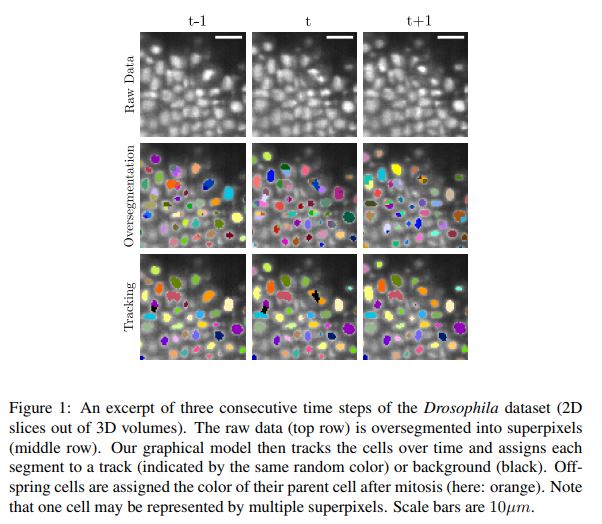
biology is developing a great hunger for robust and accurate automated cell tracking (Meijering et al. 2009, Kanade et al. 2011, Meijering et al. 2012, Gonzalez et al. 2013, Ma ´ ska et al. 2014)
Cell migration and proliferation are two important processes in normal tissue development and disease.
To visualize these processes, optical microscopy remains the most appropriate imaging modality. Some imaging techniques, such as phase contrast (PhC) or differential interference contrast (DIC) microscopy make cells visible without the need of exogenous markers. Fluorescence microscopy on the other hand requires internalized, transgenic, or transfected fluorescent reporters to specifically label cell components such as nuclei, cytoplasm, or membranes
What it does
a prototype to a future workflow for analyzing fluorescence microscopy.
How I built it
Currently working on a one-time process on an opensource platform (KNIME).
Challenges I ran into
Finding help
Accomplishments that I'm proud of
Learning!
What I learned
Image identification processes
What's next for Deep Cell
Working on a prototype with some help.
Built With
- ilastik
- knime
- matlab
Log in or sign up for Devpost to join the conversation.