-
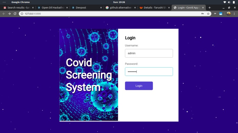
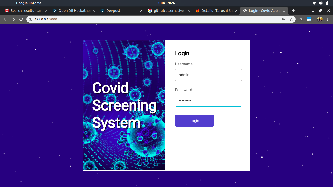
Login Screen
-
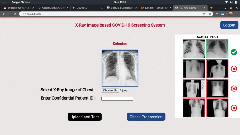
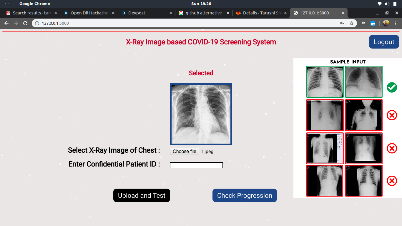
Landing Page
-
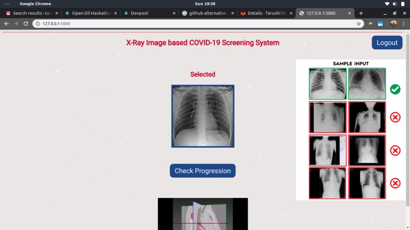
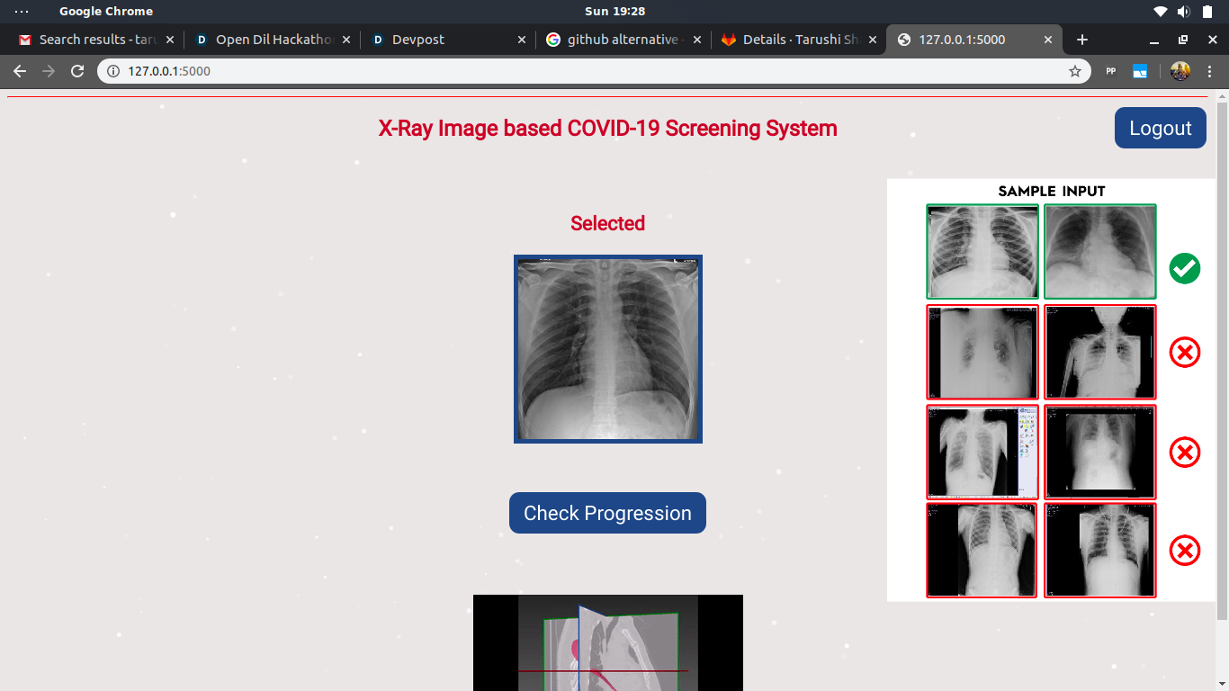
Loading Page
-
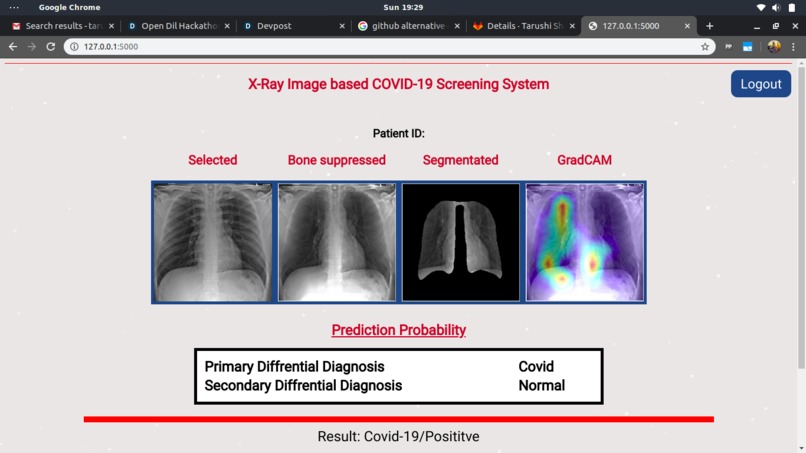
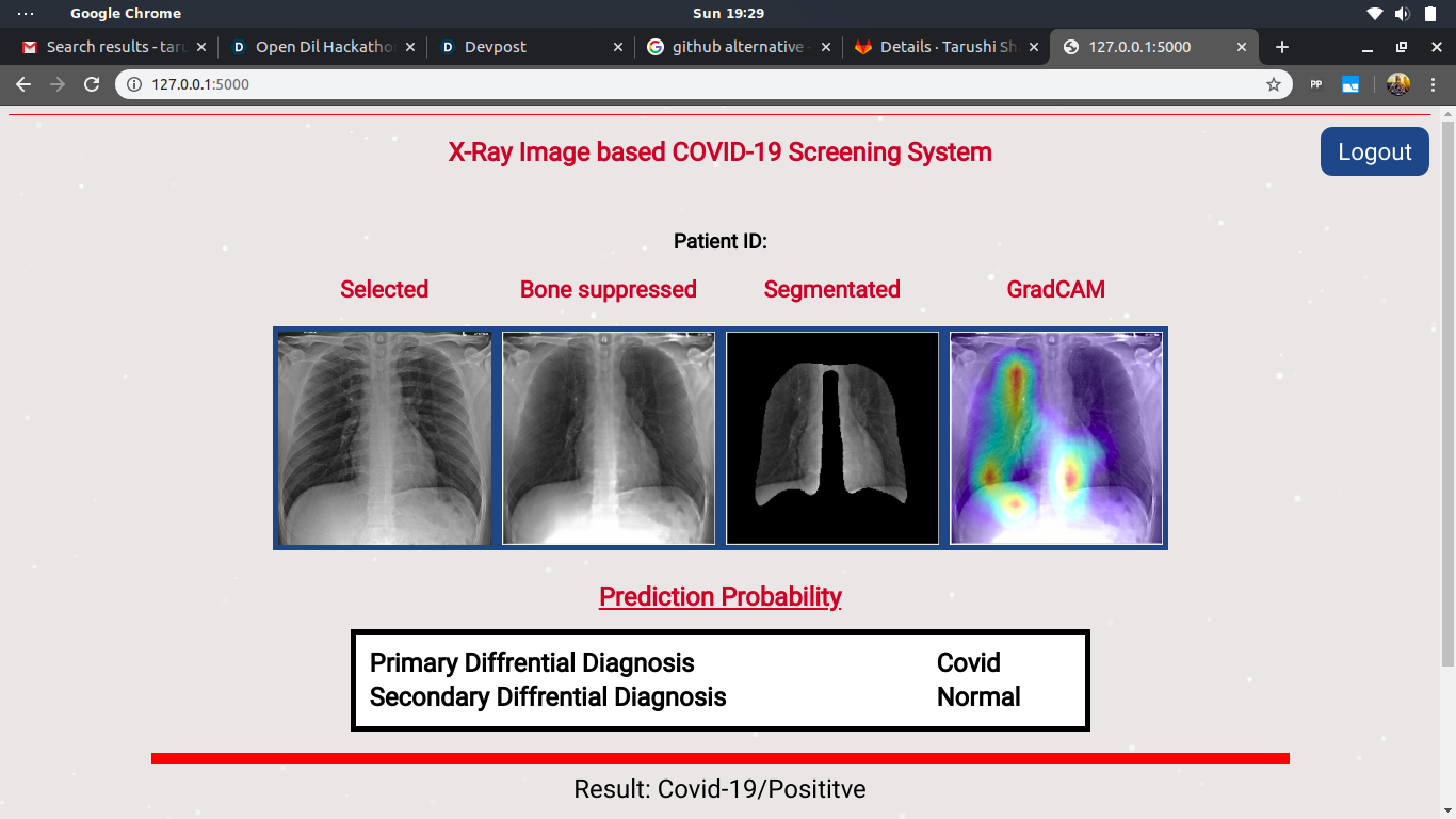
Final Output Page
Chest-Radiograpghy-Disease-Prediction
Inspiration
Amidst the recent worldwide outbreak of SARS-CoV-2 in 2020, a fast and effective screening system for COVID-19 is the need of the hour. Although many pioneering works have been published to address the issue, the effectiveness of their approach is questionable. As many of these works fail to account for the complexity in decision boundaries between COVID-19 and other pneumonic infections. However, a few papers do resolve the case but their results are not adequate for their approach to be used for mass screening. In this project, we propose an effective deep learning-based system for screening COVID-19, non-COVID viral pneumonia,and bacterial pneumonia from normal chest radiographs.
What it Does / About The Project
Being in midst of a pandemic gave us a necessity to make an end to end software for the ease of pre-testing a patient, Theerefore, our project has functionalities as follows:
- Classifying and differentiating between various diseases and a healthy individual:
- Covid19
- Non Covid-Viral Pneumonia
- Bacterial Pneumonia
- Normal
- Calculating the change or the progression of a chest x-ray over a few days.
How We Built It
We built this software's backend in python and the frontend in Javascript, HTML and CSS. The following python were required in the project:
- Flask v1.1.2
- Keras v2.2.4
- Werkzeug v1.0.0
- matplotlib v3.0.2
- numpy v1.18.1
- opencv_contrib_python v4.2.0.34
- scikit_image v0.16.2
- tensorflow v1.14.0
- itsdangerous v1.1.0
- Jinja2 v2.10.1
- gunicorn v20.0.4
Challenges
These were the challenges we ren into:
- Collecting authentic dataset, which was solved by reaching out to hospitals for datasets.
- Cleaning and maintaining our data.
- Ability of our model to diffrentiate between Viral and Bacterial Pneumonia.
Accomplishments
We achieved significant results in drawing decision boundaries between complex feature representations of these classes. On our test set of 457 images, the system achieved an averageprecision of 0.9873, recall of 0.9927, F1 score of 0.9899, andarea under the receiver operating characteristic curve of 0.9865. Our system was validated by a team of 5 radiologists on a data of 200 chest x-ray images: collected from 3 different hospitals. Both model’s precision and ability to detect areas of possible lesion findings were verified using class activationmaps - GradCAM++. The results obtained provided significant empirical evidence for our system to be used as an efficient screening tool. Our system, registered in the name of authors,is protected under the Intellectual Property Rights in India.
What's Next
We continue to make our model better and improve it's accuracy along with increasing the open source contribution to our software. We also aim for better results and an appealing Web UI for future works.


Log in or sign up for Devpost to join the conversation.