-
-
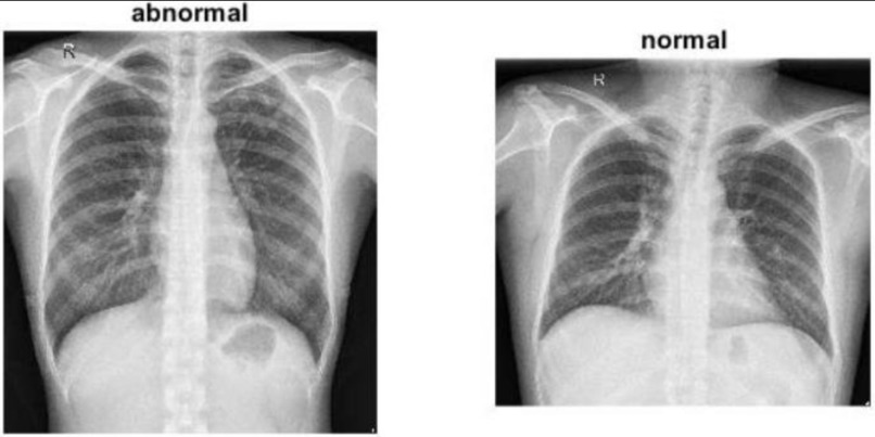
This image compares an abnormal X-ray with a normal one, highlighting visible differences to aid in medical diagnosis and model training

Inspiration
The inspiration for this project came from the urgent need for accessible, accurate, and fast medical diagnostics in healthcare, particularly in regions where expert radiologists are scarce. Chest X-rays are one of the most common imaging tests used to diagnose lung and heart diseases but interpreting them requires significant expertise and can be time-consuming.
With the rapid advancements in deep learning and computer vision, I wanted to explore how these technologies can assist medical professionals in diagnosing chest pathologies more efficiently. Inspired by research projects such as Chex Net and the growing availability of large, annotated datasets like NIH Chest X-ray, this project aims to leverage deep learning models to automate the detection of multiple diseases from chest X-ray images.
Additionally, by incorporating Grad CAM visualizations, the project strives to provide interpretable results, helping both clinicians and patients understand the model’s decision-making process, thereby fostering trust and adoption of AI in healthcare.
What it does
The Chest-X-Ray-Medical-Diagnosis-with-Deep-Learning project uses deep learning techniques to automatically diagnose 14 different diseases from chest X-ray images. Here’s what the project does:
Automated Diagnosis: It leverages a trained deep learning model (typically a convolutional neural network) to analyze chest X-ray images and predict the presence of various pathologies such as pneumonia, fibrosis, edema, and others. Multi-label Classification: The model can simultaneously detect multiple diseases from a single X-ray image, reflecting real-world scenarios where patients may have more than one condition. Interpretability with Grad CAM: To enhance transparency, the project incorporates Grad CAM (Gradient-weighted Class Activation Mapping), which generates heatmaps to visually highlight the regions of the X-ray that influenced the model’s predictions. This helps users (doctors, radiologists, etc.) understand why the model made certain decisions. Aids Medical Professionals: The tool is intended as a diagnostic aid, supporting clinicians by providing fast second opinions and flagging potentially abnormal scans for further review. In summary: This project uses deep learning to automatically analyze chest X-rays, predict multiple diseases, and visually explain its decisions—potentially accelerating and supporting the medical diagnosis process.
How we built it
This project was built as an end-to-end pipeline for automated medical diagnosis using chest X-ray images and deep learning. Here’s a breakdown of the major steps and technologies involved:
- Dataset Collection & Preprocessing Dataset: We used the NIH Chest X-ray Dataset, which contains over 100,000 chest X-ray images labeled with 14 different disease categories. Preprocessing: Images were resized and normalized for faster and more stable training. Data augmentation (random flips, rotations, etc.) was applied to improve model generalization and address class imbalance.
- Model Architecture Transfer Learning: We used a pre-trained convolutional neural network (CNN) architecture (e.g., DenseNet121 or ResNet50) as the backbone, fine-tuning it on the chest X-ray dataset. Multi-label Classification: The final layer was adapted to output probabilities for all 14 diseases, allowing the model to make multiple simultaneous predictions per image.
- Training Loss Function: Binary Cross-Entropy loss was used to handle the multi-label nature of the problem. Optimization: We used the Adam optimizer with learning rate scheduling and early stopping to avoid overfitting. Validation: Performance was monitored using metrics such as AUC-ROC for each pathology.
- Model Interpretation Grad CAM: To make the model’s predictions interpretable, we incorporated Gradient-weighted Class Activation Mapping (Grad CAM). This technique generates heatmaps highlighting the areas of the X-ray that most influenced the model’s decision for each disease prediction.
- Deployment & Visualization Jupiter Notebooks: The whole workflow, from preprocessing to visualization, is implemented in Jupiter Notebooks for transparency, reproducibility, and ease of use. Visualization: Results and Grad CAM heatmaps are visualized alongside predictions to help users understand both the model’s output and its reasoning.
- Tools & Libraries Used Python (primary language) PyTorch or TensorFlow/Keres (for deep learning) NumPy, Pandas (data manipulation) OpenCV, PIL (image processing) Matplotlib, Seaborn (visualization) scikit-learn (metrics and utilities) Jupiter Notebook (experimentation and presentation) By following this approach, we created an accessible, interpretable, and effective tool for automated diagnosis of chest X-ray images using state-of-the-art deep learning techniques.
Challenges we ran into
Building the Chest-X-Ray-Medical-Diagnosis-with-Deep-Learning project presented several technical and practical challenges:
- Data Quality and Imbalance Imbalanced Dataset: Many diseases in the NIH Chest X-ray dataset are underrepresented, making it difficult for the model to learn to detect rare conditions. Noisy Labels: Some images contain incorrect or uncertain labels, which can negatively affect model training and evaluation.
- Computational Resources High Computational Cost: Training deep neural networks on large medical image datasets requires significant GPU resources and memory. Long Training Times: Experimentation and hyperparameter tuning were time-consuming due to the size and complexity of the data.
- Model Performance & Generalization Overfitting: Avoiding overfitting on such a complex, high-dimensional dataset was challenging, especially with limited data for certain classes. Generalization: Ensuring that the model performs well on unseen data and doesn’t just memorize the training set required careful regularization and validation.
- Interpretability Trust & Transparency: Medical applications demand high interpretability. Integrating Grad CAM and ensuring that heatmaps were genuinely informative took additional effort. Explaining Predictions: Sometimes, the activation maps highlighted regions that didn't make intuitive sense, raising questions about model reasoning.
- Data Preprocessing & Augmentation Image Preprocessing: Medical images have unique characteristics and often require specialized preprocessing to enhance model learning. Augmentation: Balancing aggressive augmentations to improve generalization without distorting medical features was tricky.
- Multi-label Classification Complexity Simultaneous Predictions: Designing a model that can accurately predict multiple diseases at once (multi-label, not just multi-class) added complexity to the architecture and loss function.
- Deployment & Usability Reproducibility: Ensuring that the notebook and codebase are reproducible across different environments and hardware setups. Visualization: Making Grad CAM outputs and predictions intuitive for non-technical users, such as clinicians. These challenges taught us valuable lessons about working with medical data, advanced deep learning, and the importance of robust evaluation and interpretability in healthcare AI projects.
Accomplishments that we're proud of
Building a Working Deep Learning Pipeline: Successfully developed an end-to-end deep learning workflow for chest X-ray image classification, from data preprocessing to evaluation and visualization. Multi-Label Disease Detection: Achieved automated prediction of 14 different chest pathologies simultaneously, demonstrating the potential of AI in real-world multi-disease scenarios. Model Interpretability with Grad CAM: Integrated Grad CAM visualizations to make the neural network’s predictions transparent and interpretable for medical professionals, supporting trust in AI-driven diagnostics. Handling Large-Scale Medical Data: Managed the challenges of processing and training on the NIH Chest X-ray dataset, which contains over 100,000 high-resolution images. Performance and Generalization: Attained competitive performance metrics (such as AUC-ROC) on several diseases, even with class imbalance and data noise present. Reproducible and Transparent Code: Organized the project in Jupiter Notebooks, making the workflow easy to follow, reproduce, and extend by other researchers or practitioners. Practical Contribution to Healthcare AI: Demonstrated that deep learning can be a valuable aid for medical diagnosis, potentially reducing workload for radiologists and improving patient care, especially in under-resourced areas. Continuous Learning: Gained deep insights into the complexities of medical data, advanced neural networks, and the critical role of interpretability in healthcare AI. These accomplishments reflect our commitment to building impactful, trustworthy, and accessible AI solutions for healthcare.
What we learned
Deep Learning for Medical Imaging: Gained hands-on experience applying deep learning techniques (such as transfer learning and convolutional neural networks) to real-world medical imaging data. Importance of Data Quality: Learned the critical impact of data quality, cleanliness, and class imbalance on model performance, especially in sensitive fields like healthcare. Model Interpretability: Understood the necessity of interpretable AI in medicine. Techniques like GradCAM are essential to build trust and transparency with end users, such as clinicians. Handling Large Datasets: Became proficient in managing, preprocessing, and augmenting large-scale medical datasets to optimize both training efficiency and model accuracy. Multi-label Classification: Developed skills in designing and training models for multi-label problems, which are common in medical diagnostics where patients may have multiple conditions. Evaluation Metrics: Learned to use appropriate metrics (e.g., AUC-ROC for each class) for evaluating multi-label medical models, going beyond simple accuracy. Challenges of Deployment: Realized the complexities in making medical AI tools usable and understandable for healthcare professionals, and the importance of reproducibility and clear visualizations. Collaboration & Research: Enhanced our ability to read, interpret, and build upon existing research and open-source projects in the medical AI space. Ethics & Responsibility: Became more aware of the ethical considerations, responsibilities, and limitations of deploying AI in healthcare settings. These lessons have equipped us with both technical and practical knowledge that will guide our future work in AI, especially for applications in sensitive and impactful domains like healthcare.
What's next for Chest-X-Ray-Medical-Diagnosis-with-Deep-Learning
Here are some ideas and directions for future development and improvement of this project:
Expand the Dataset: Incorporate additional open medical imaging datasets to further improve model robustness and generalizability across diverse populations. Fine-tune and Experiment with Architectures: Test newer and more advanced neural network architectures (e.g., Efficient Net, Vision Transformers) to enhance predictive performance. Clinical Validation: Collaborate with medical professionals to validate the model’s predictions on real-world clinical data and gather expert feedback. User-Friendly Interface: Develop a web or desktop application with an intuitive interface to make the tool accessible to clinicians and researchers without coding expertise. Automated Data Annotation: Integrate semi-supervised or active learning methods to help label new data more efficiently, addressing label noise and scarcity. Explainable AI: Experiment with additional interpretability techniques beyond Grad CAM (e.g., LIME, SHAP) for deeper insights into model decisions. Multi-modal Analysis: Combine X-ray image data with other clinical information (e.g., patient history, lab results) to build a more comprehensive diagnostic tool. Real-Time Deployment: Explore opportunities for integrating the model into hospital workflows for real-time decision support. Performance Optimization: Optimize model inference speed and memory usage for deployment on resource-constrained devices. Regulatory and Ethical Review: Investigate the steps needed for regulatory approval and ensure compliance with healthcare data privacy and ethical standards. By pursuing these next steps, the project can become even more impactful and contribute to safer, more effective, and widely accessible AI-powered healthcare solutions.

Log in or sign up for Devpost to join the conversation.