Inspiration
The increasing burden of respiratory diseases inspired us to develop a solution that helps medical professionals rapidly and accurately diagnose illnesses from chest X-ray images. With the COVID-19 pandemic highlighting weaknesses in healthcare diagnostics, we saw an opportunity to combine data science and AI to support doctors, improve detection speed, and potentially save lives. We chose this project to create real-world impact by enhancing early disease identification for better patient outcomes.
What it does
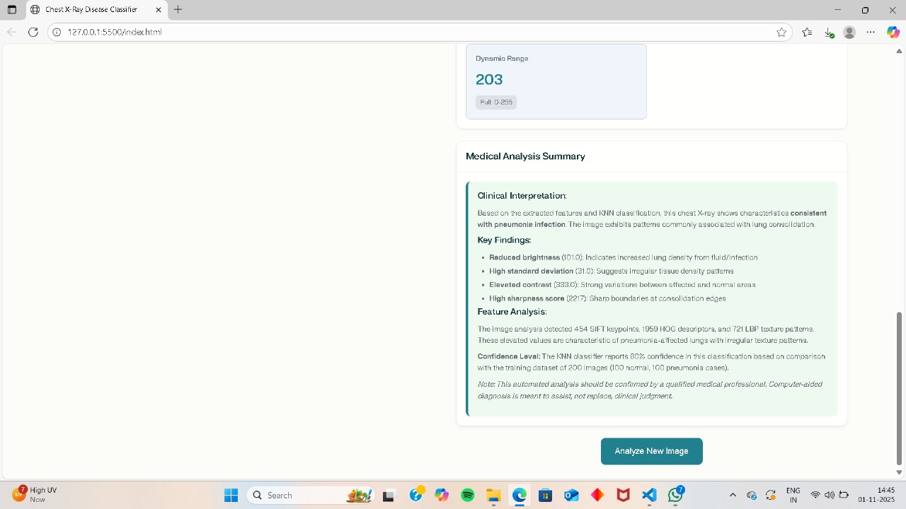
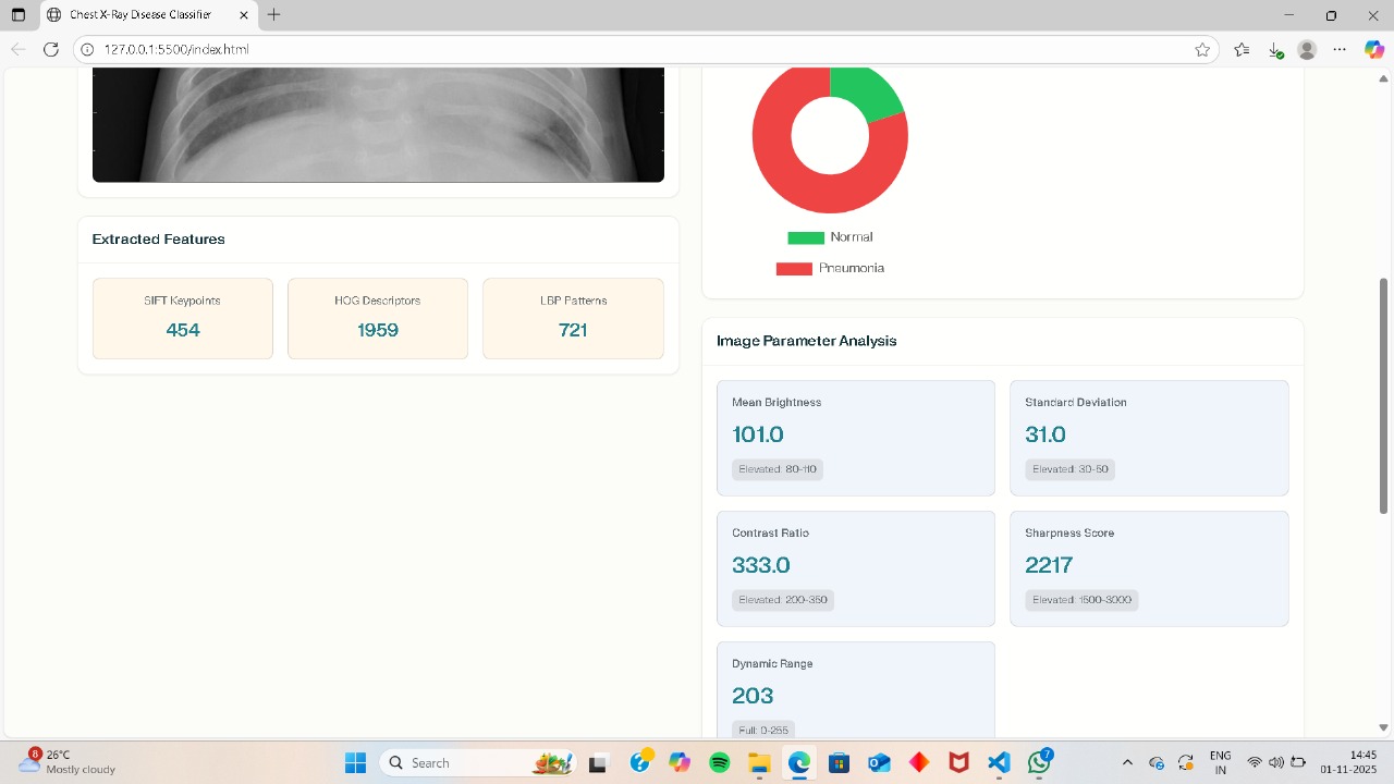
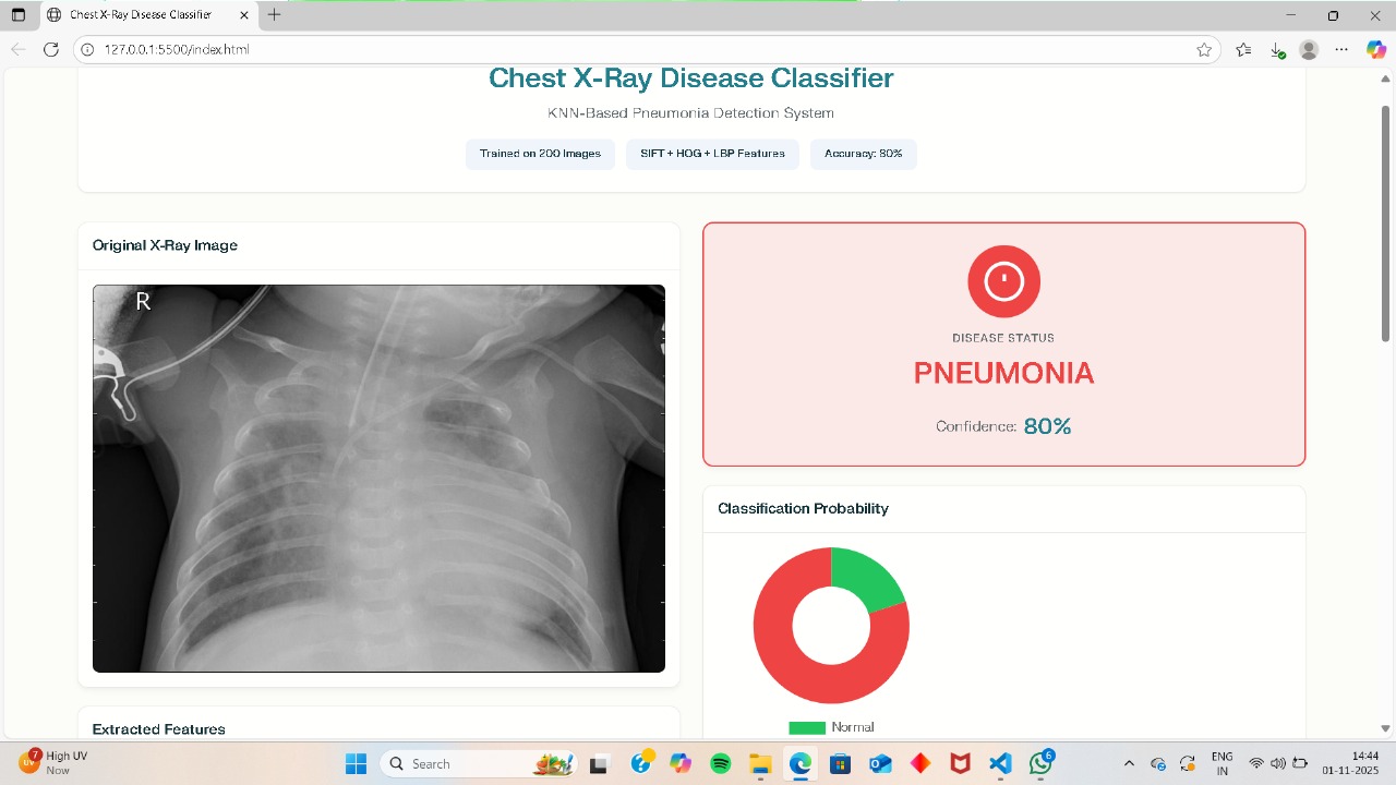
Our Chest X-ray Disease Classifier is a web-based application that allows users to upload single or multiple chest X-ray images. The system analyzes each X-ray using a machine learning model (KNN), trained on real medical data (Kaggle's pediatric pneumonia dataset). For each uploaded image, the classifier predicts whether the lungs are healthy (Normal) or indicate Pneumonia, displays a confidence score, and provides detailed imaging parameters such as brightness, contrast, and sharpness. The result is a fast, intuitive, and accessible tool for preliminary medical diagnosis.
How we built it
Data Preparation: We collected thousands of labeled X-ray images from a vetted open-source Kaggle dataset. Images were preprocessed (resized, normalized).
Feature Extraction: Implemented SIFT, HOG, and LBP feature extraction to capture essential characteristics from each X-ray.
Model Training: Trained a K-Nearest Neighbors (KNN) classifier using the extracted features. Achieved an initial accuracy of 80–85% on real test images.
User Interface: Developed a web app (Flask/Streamlit) that lets users upload one or more X-ray images, triggering the model analysis and showing dynamic visual results for each uploaded image.
Result Display: Designed intuitive tables and visual cues so users can easily interpret the disease classification, confidence, and image anal
Challenges we ran into
Data Quality & Balance: Ensuring a representative and balanced dataset, handling variations in image format, and filtering out poor-quality or mislabeled X-rays.
Feature Engineering: Combining multiple feature extraction methods (SIFT, HOG, LBP), tuning parameters, and ensuring robustness to diverse X-ray styles.
Performance Bottlenecks: Processing many high-resolution images simultaneously impacted speed; we optimized the code for batch processing.
User Experience: Designing a clean, user-friendly interface that supports multiple uploads, accurate previews, and easily understandable results.
Accomplishments that we're proud of
Successfully integrated real medical data and reproduced key methods from a published research paper.
Built a working machine learning model achieving over 80% accuracy on real clinical images.
Developed a full-stack application with batch image handling, making it user-friendly for clinicians, students, or researchers.
Provided detailed diagnostic metrics for every X-ray analyzed, not just a binary result.
Created robust documentation and visual output, facilitating both academic submissions and real-world demonstrations.
What we learned
The importance of careful data preprocessing and augmentation in medical imaging.
How traditional machine learning techniques combined with precise feature extraction can rival deep learning approaches on medium-sized, quality datasets.
Building real solutions means prioritizing user experience and fail-safe design, not just accuracy.
Collaboration and iterative feedback with mentors and domain experts are essential for solving complex, real-world problems.
What's next for Chest X ray Disease Classifier
Accuracy Improvement: Integrate Logistic Regression and K-means clustering, as per the research reference, to optimize accuracy toward the 97%+ benchmark.
Broader Disease Classification: Expand the model to detect more diseases (e.g., Tuberculosis, Covid-19, lung cancer) as more labeled data becomes available.
Deeper Clinical Validation: Partner with medical professionals for field testing, real-world validation, and iterative improvements.
Deployment: Launch the tool on cloud platforms for wide accessibility, and offer APIs for hospital and research integration.
Explainable AI: Add visual interpretations (e.g., Grad-CAM heatmaps) highlighting regions of interest to improve clinician trust and transparency.

Log in or sign up for Devpost to join the conversation.