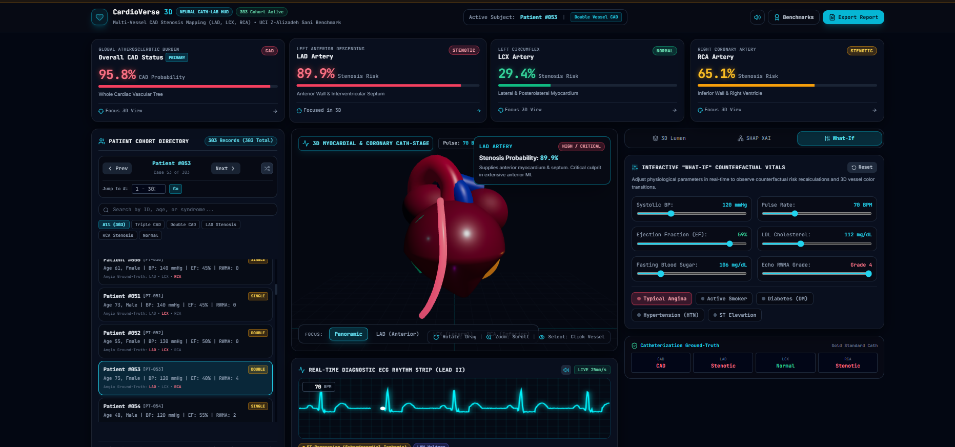
Inspiration Raw risk percentages don't show where heart disease is developing. We wanted to transform abstract clinical numbers into an intuitive, interactive 3D digital twin of coronary anatomy.
What it does Predicts overall CAD and vessel-specific stenosis across the LAD, LCX, and RCA arteries from clinical vitals, dynamically mapping risk onto an interactive, beating 3D human heart with live blood flow particles, an intravascular lumen cross-section simulator, a real-time Lead II ECG oscilloscope, and SHAP explainability.
How we built it ML & XAI: Trained calibrated Extra Trees/Random Forest models on the 303-patient UCI dataset using 54 non-invasive features, with strict target leakage safeguards and TreeSHAP feature attribution. Backend: FastAPI serving sub-15ms predictions, patient profiles, and counterfactuals. Frontend & 3D: React 19, Tailwind CSS, Three.js / WebGL, and HTML5 Canvas in a cyber-clinical Cath-Lab HUD. Challenges we ran into Preventing target leakage by strictly decoupling all post-catheterization imaging features. Mathematically syncing 3D coronary artery splines and blood flow particles with dynamic, beating myocardial contractions at 60 FPS. Accomplishments that we're proud of Achieving 0.932 ROC-AUC for CAD and 0.847 ROC-AUC for LAD stenosis without target leakage. Delivering a tactile, non-generic HUD with a complete 303-patient explorer and live "What-If" risk recalculation. What we learned How echocardiographic wall motion abnormalities (RWMA) translate into territory-specific vascular risk. The vital importance of probability calibration for trustworthy medical AI.
