-
-
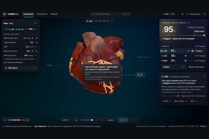
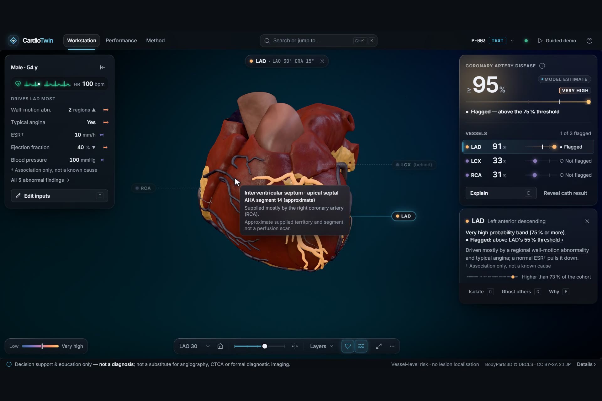
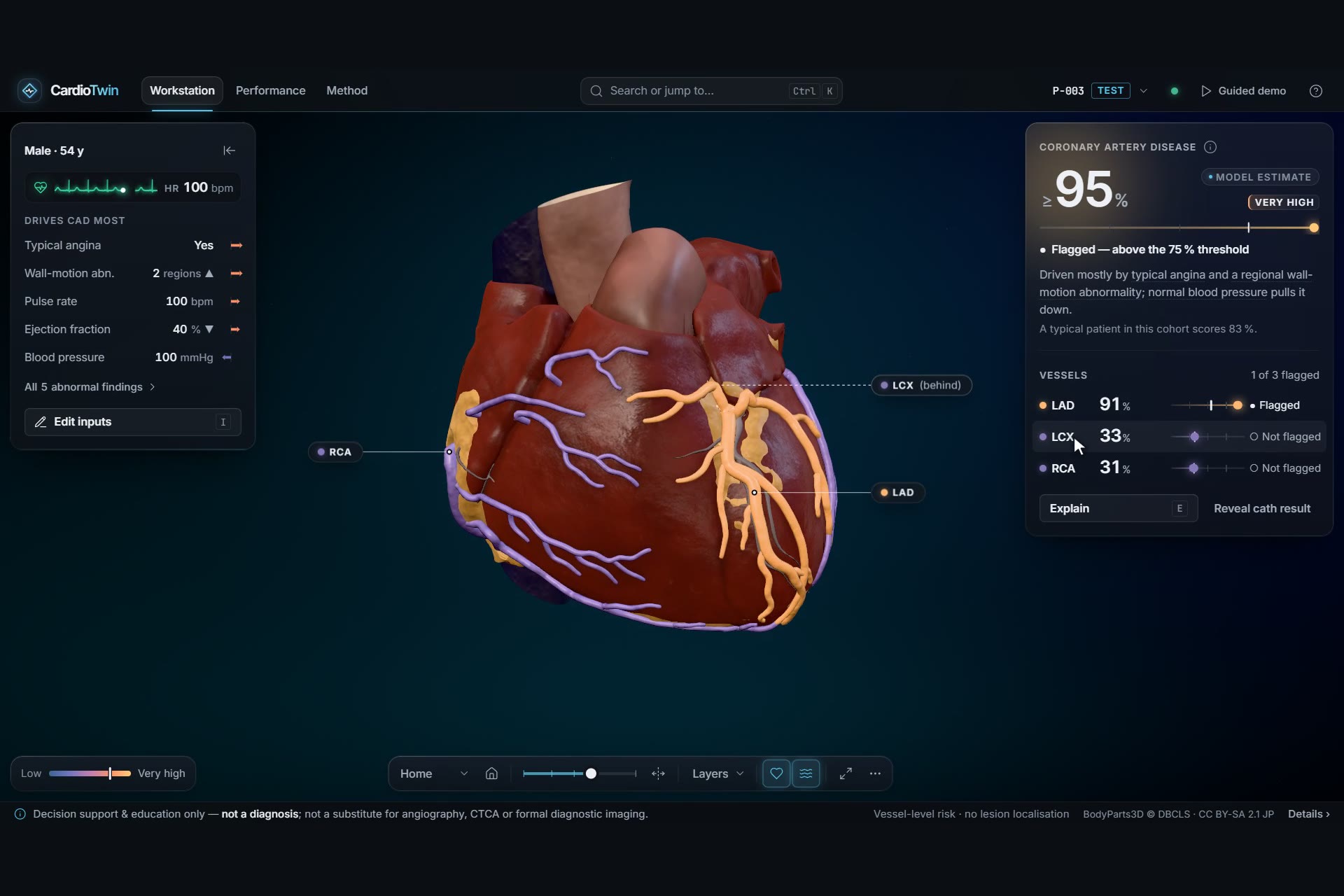
Each coronary artery coloured by its own predicted probability on a BodyParts3D heart, with a vessel inspector for the LAD
-
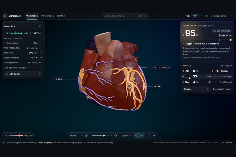
Four calibrated probabilities at a glance: overall CAD plus LAD, LCX and RCA stenosis, each against its decision threshold
-
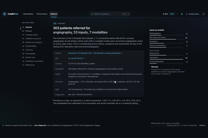
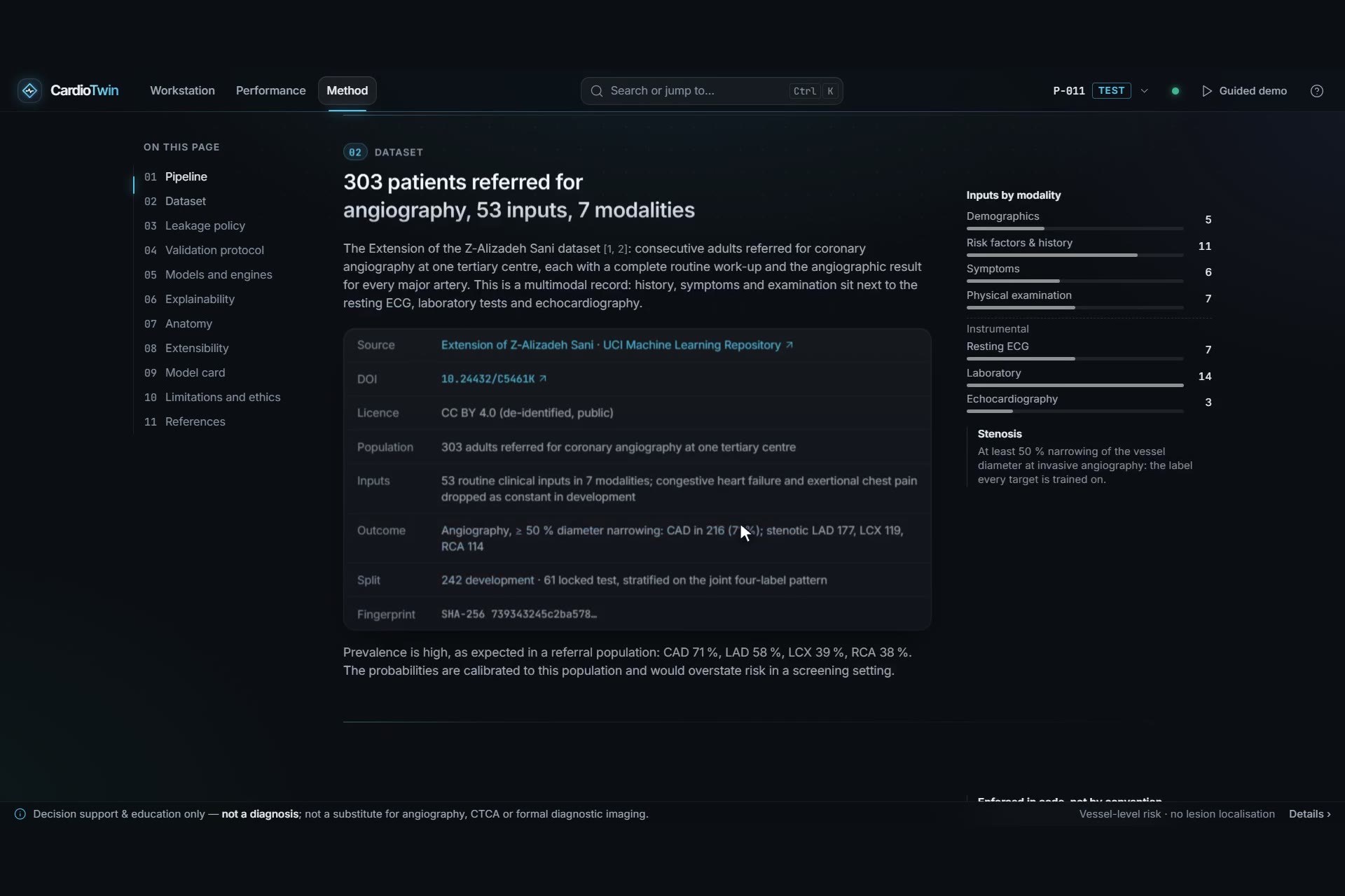
Method page: 303 patients, 53 routine inputs across 7 modalities, with the dataset licence and leakage policy
-
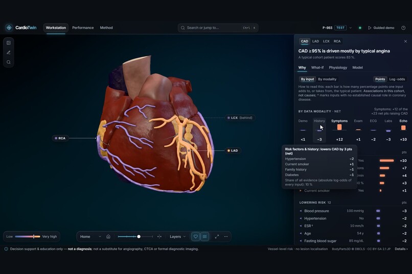
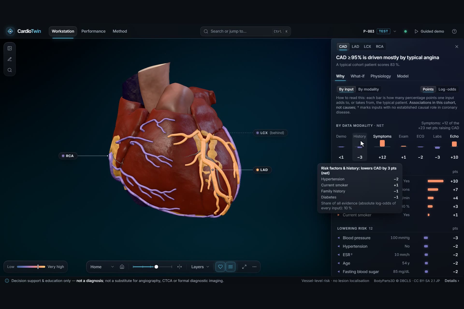
Exact SHAP explanations in percentage points, grouped by clinical modality; contributions add up to the model output
-
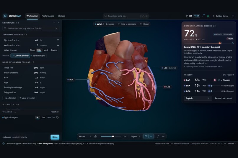
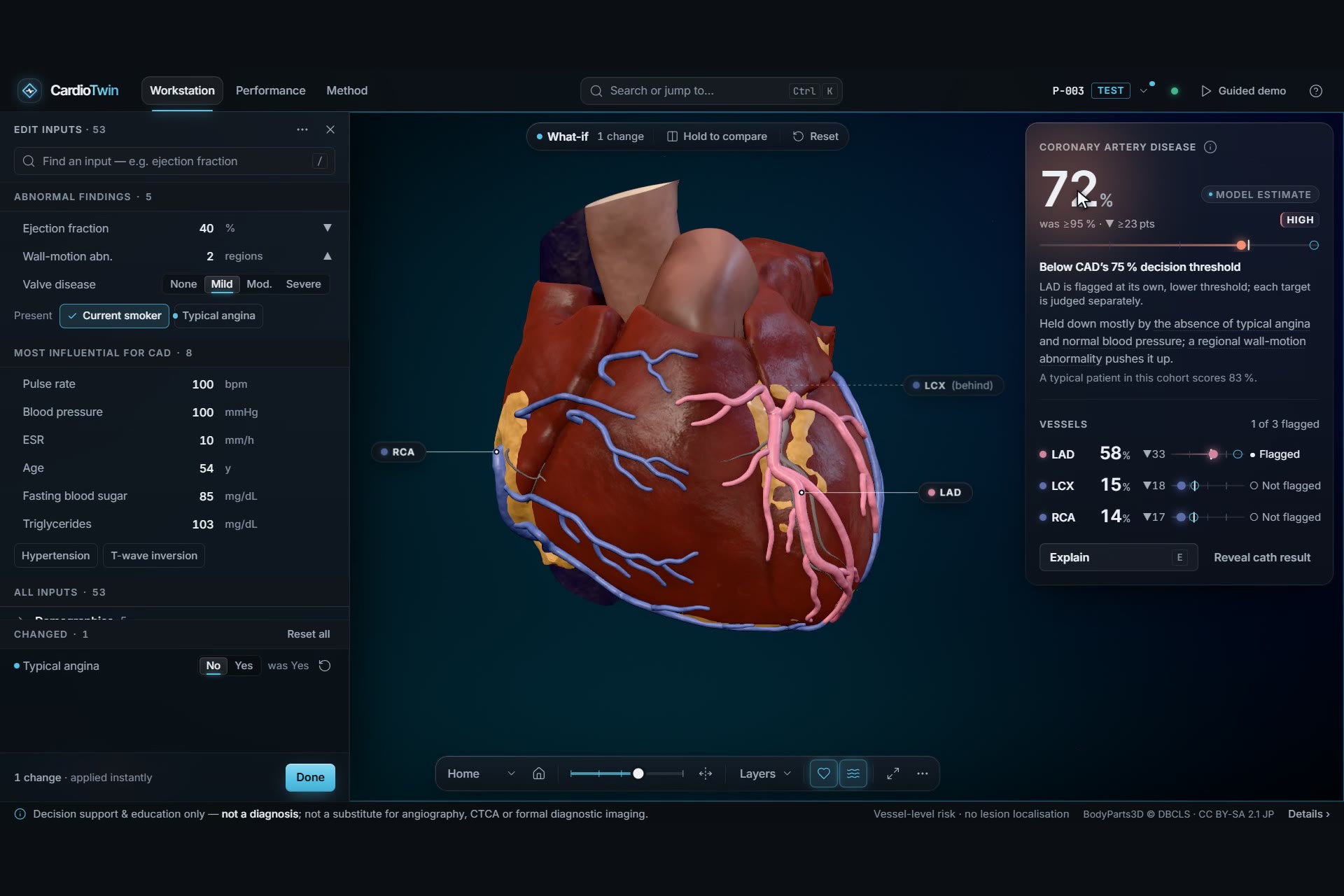
What-if, live: edit one input and every probability, explanation and 3D colour updates together (CAD ≥95 % → 72 %)
-
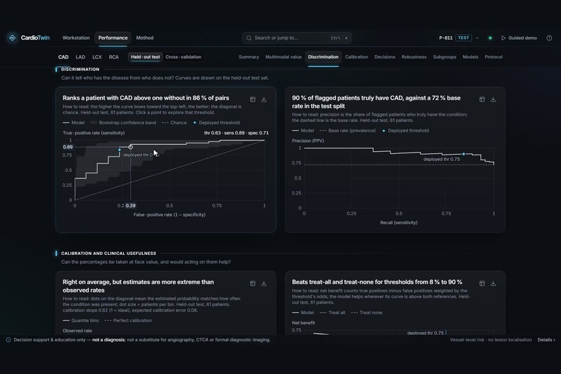
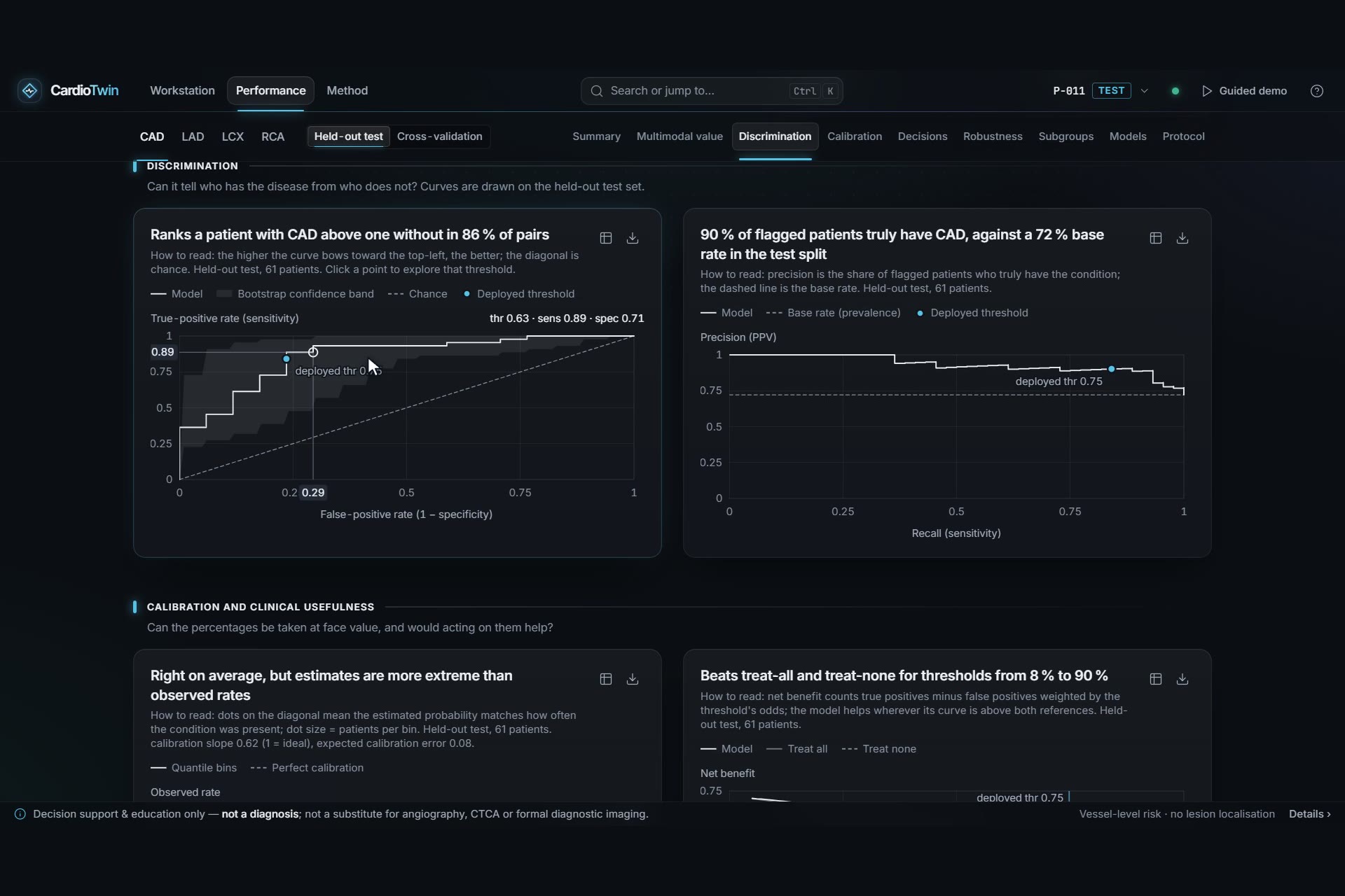
Validation on a locked test set: ROC, precision-recall, calibration and decision curves, each with 95 % confidence intervals
Live demo: https://cardio-twin-tau.vercel.app (runs fully in the browser) · Demo video: https://youtu.be/eplDOnx9qDc · Code: https://github.com/Mohammad-Umar7/CardioTwin
Team: Mohammad Umar, Bilal Feroz, Fuad Ahamed, Awaiz Ahmed
The five numbers
- 0.858 (95 % CI 0.743–0.955): CAD ROC-AUC on 61 patients the model never saw, with F1 0.871 and precision 0.902
- 0.934: Median CAD ROC-AUC over 200 random re-splits with the whole recipe re-run (LAD 0.844). The locked test split was among the hardest 3 %
- +0.069 (95 % CI +0.024 to +0.115): LAD ROC-AUC gained by adding ECG, labs and echo to bedside data
- 2.2e-16: Largest probability difference between the Python server and the in-browser engine, with identical SHAP values. The browser answers in 1.2 ms (p50)
- 60 / 78: Cited anatomical reference checks the 3D heart passes (up from 35 / 70 at baseline; 8 minor, 10 still fail), across 41 structures and 18 SCCT coronary segments
Inspiration
Patients sent for coronary angiography already come with a history, an ECG, blood tests and an echo. Yet the numbers clinicians get from them are whole-heart scores. The scores say nothing about which artery is likely narrowed, and nothing about why. In the public cohort we worked with, every one of the 303 patients was referred for an invasive angiogram, and 29 % of them turned out not to have coronary artery disease.
We wanted a tool that answers three questions at a glance: how likely is disease, in which vessel, and what in this patient's record drives it? It had to be honest about uncertainty, and it had to run on an ordinary laptop.
What it does
- Predicts four targets from 53 routine inputs (demographics, history, symptoms, examination, ECG, labs, echo): overall CAD and ≥ 50 % stenosis of the LAD, LCX and RCA. The angiography columns are never inputs.
- Explains every estimate exactly. Each input's signed contribution is shown in percentage points, grouped by modality, with its reference range in a physiology view.
- Maps risk onto real anatomy. A 3D thorax and heart built from BodyParts3D colours each artery by its own probability. You can rotate, zoom and use C-arm projections. Selecting the LAD flies the camera to its angiographic best view and opens a vessel inspector. The heart can be peeled open, beats at the patient's pulse rate, shows blood flow along real centrelines, and tints each artery's perfusion territory.
- What-if in real time. Edit any input and all four probabilities, the explanations and the 3D colours update together. ICE strips show how the risk moves across an input's range.
- Checks itself. For the 61 unseen test patients you can reveal the actual angiogram result. A performance page shows every metric with its confidence interval, plus calibration, decision curves, robustness and subgroups.
- Stays safe. A clinical-safety disclaimer is always on screen and printed on the two-page report. Out-of-range inputs are refused rather than extrapolated.
How we built it
- ML (Python, scikit-learn, XGBoost).
- A 20 % test split was locked before any modelling. Eleven candidate models were compared with repeated, nested cross-validation tuned on log-loss.
- Each target uses a margin ensemble of logistic regression and XGBoost with Platt calibration and a Youden threshold, all fitted on out-of-fold predictions.
- The ensemble's CV estimate is cross-fitted, so no reported number helped choose the model it scores.
- A separate analysis re-runs the whole recipe on 200 random splits and ablates each clinical modality with corrected-t intervals.
- Exact explainability.
- Linear SHAP for the logistic part and our own float64 TreeSHAP for XGBoost, whose float32 output was not additive enough. The two combine linearly, and base + Σ SHAP equals the model output to 1.8e-15.
- A calibrated-space copy of the SHAP values lets the UI speak in percentage points.
- Portable model.
- The trained model is exported as one JSON file.
- A line-by-line TypeScript port evaluates it in a Web Worker, mirroring XGBoost's float32 split comparisons.
- The FastAPI server and the browser therefore agree to the last bit, and the app still works with no backend (the live Vercel deployment).
- 3D anatomy (Blender 5.1, headless, fully scripted).
- BodyParts3D meshes are cleaned, smoothed, decimated, and bisected so the heart can be opened.
- Missing structures (aortic root and valve, epicardial fat, in-vivo vein calibres) are synthesised.
- Perfusion territories are baked into vertex colours, blended with the AHA 17-segment map.
- Coronary centrelines are extracted and labelled with SCCT 2014 segments, and the result is exported as a meshopt-compressed glTF.
- Web app (React, TypeScript, React Three Fiber).
- One persistent WebGL canvas and a single risk colour ramp shared by shaders, charts and legend.
- Adaptive quality tiers keep the app responsive on integrated GPUs, with a 2D fallback when WebGL is unavailable.
- Engineering.
- One command starts everything (
dev.ps1/make), and one process can serve app and API on one URL. - Docker with a hardened compose file.
- CI covers all four subsystems, with byte-identical retraining, an end-to-end check against the running server and container, and a clean-checkout run on Windows and Linux.
- One command starts everything (
Challenges we ran into
- The test set disagreed with cross-validation. CAD and LAD scored 0.08–0.13 ROC-AUC lower on the locked test set than in CV. Rather than tune until it looked better, we re-ran the full recipe on 200 random splits. The locked split turned out to sit at the 3rd percentile for CAD and the 2nd for LAD, and the clinical baseline dropped on it too. The gap came from a hard split, not from overfitting, and we report both numbers.
- A calibration bug found in review. An independent review of v1.0 found that Platt scaling had been fitted on margins from differently tuned fold models. The fix was specified on development data only, the test set was scored exactly once more, and the change log is public in the model card.
- Bit-exact parity across two languages. XGBoost compares features in float32. One wrong rounding misroutes a patient at a split threshold. We built a bit-level oracle and probed all 444 thresholds (4,130 probes) until every one routed the same way in TypeScript and Python.
- Anatomy that is actually right. BodyParts3D comes from one cadaver: the veins are collapsed, and there is no aortic valve or root. We compiled a cited anatomical reference, turned it into 78 automated checks, and closed gaps one by one. Some checks still fail, and we list them.
Accomplishments that we're proud of
- Every number in the app, the README and the report is traceable to a generated artifact or a reproducible check, and every test metric has a 95 % CI.
- The same model gives the same answer to 2.2e-16 on a server and in a browser, with explanations that add up exactly.
- A 3D heart where the colour of the LAD can only ever show P(LAD). One registry drives the model, the API and the 3D mapping, and the end-to-end check fails if they diverge.
- Honest reporting: the model does not significantly beat a 5-feature clinical baseline on the small test set, and the project says so.
What we learned
- A single 61-patient test split is a noisy judge. Repeated re-splits are cheap insurance against both over- and under-claiming.
- Proper scoring rules matter for tuning. Tuning on ROC-AUC drove regularisation to the edge of its range, and log-loss did not.
- "Explainable" is only useful when the explanation is exact and uses the clinician's units (percentage points, with reference ranges), not raw log-odds.
- For medical 3D, anatomical correctness needs a written, cited reference and automated checks, just like model metrics.
What's next
- External, multi-centre validation with recalibration to local prevalence, and decision thresholds set by the clinical costs of each setting.
- Imaging inputs (CT coronary angiography, perfusion) to move from vessel-level to lesion-level targets.
- Closing the remaining anatomical checks: the coronary-sinus course, vessel seating in the grooves, and the BodyParts3D-sourced pulmonary/tricuspid annuli and SVC length.
- A FHIR import, so a real (de-identified) record can be opened in the workstation.
Data and anatomy. UCI Extension of Z-Alizadeh Sani dataset (CC BY 4.0, doi:10.24432/C5461K) · BodyParts3D, © The Database Center for Life Science (CC BY-SA 2.1 JP). The code is MIT-licensed.
Clinical safety. Research and educational decision support only. Not a diagnosis, and not a substitute for angiography, CT coronary angiography or clinical judgement.
Built With
- blender
- docker
- fastapi
- github-actions
- python
- react
- scikit-learn
- tailwindcss
- three.js
- typescript
- vercel
- vite
- xgboost


Log in or sign up for Devpost to join the conversation.