-
-
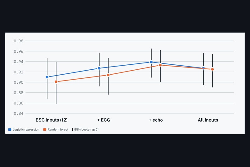
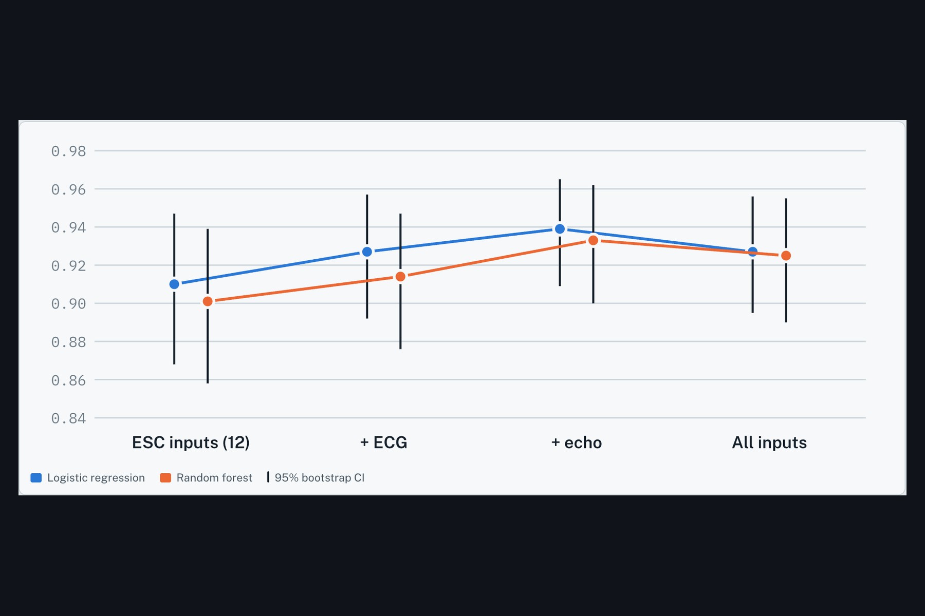
what each modality adds for CAD
-
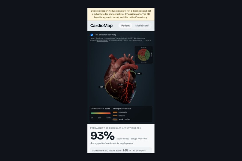
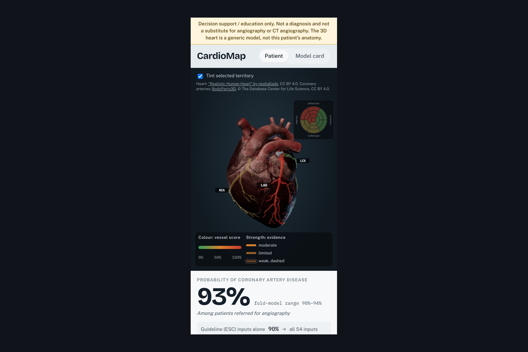
mobile layout
-
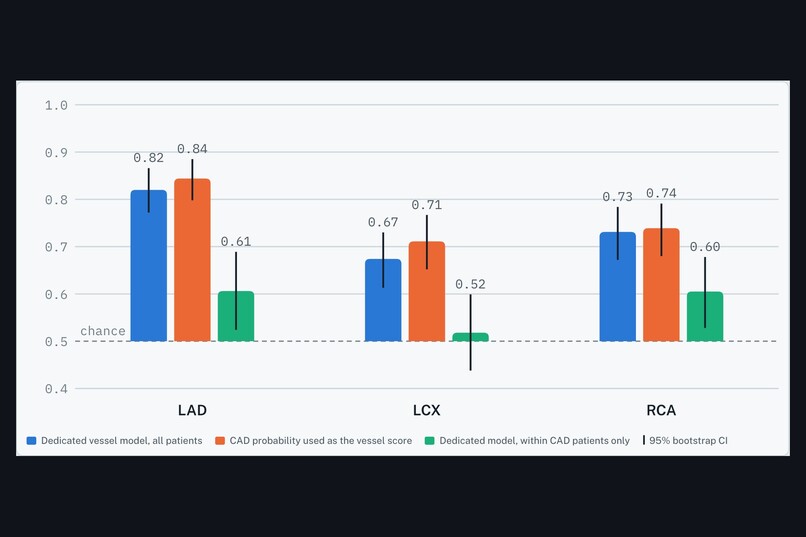
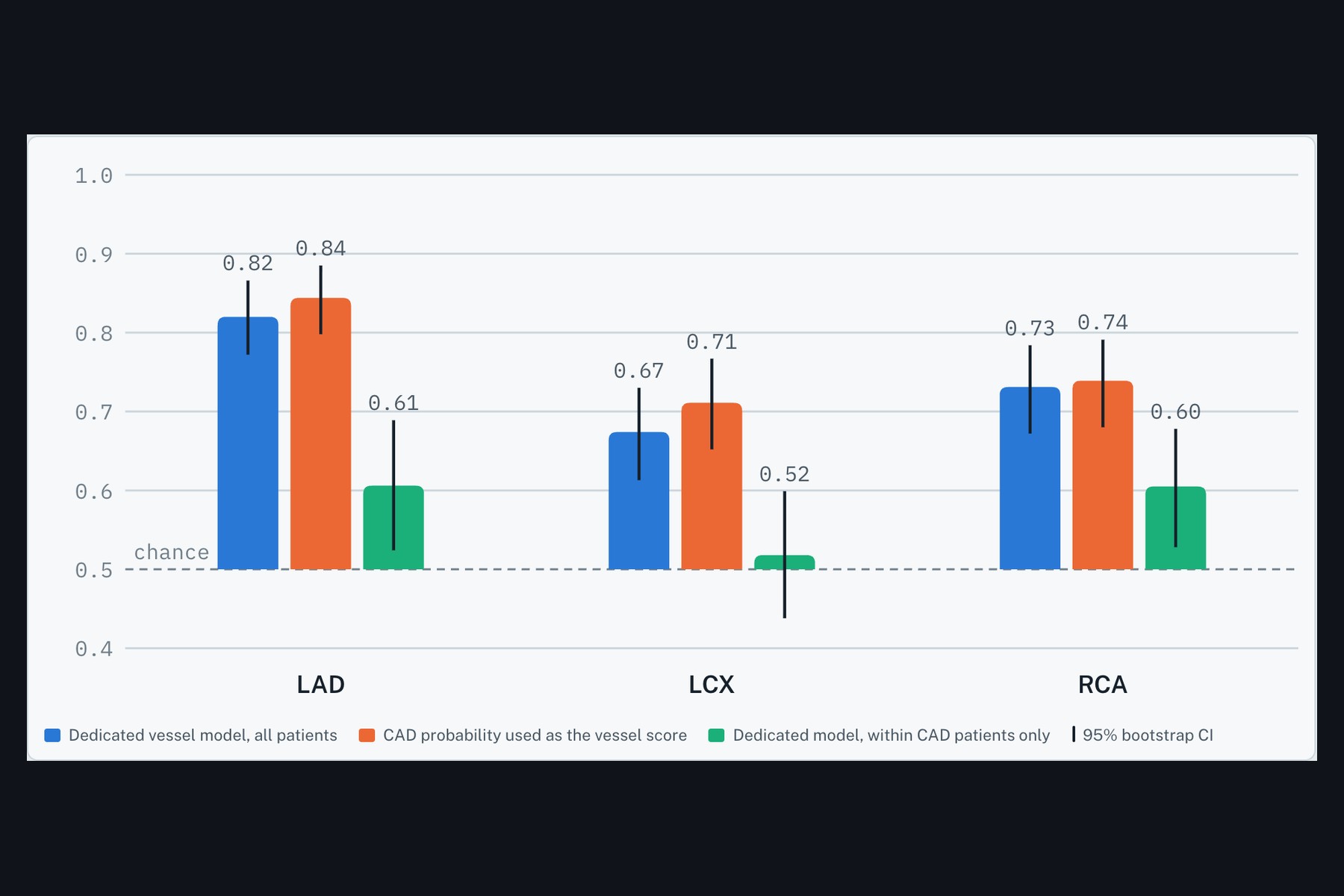
vessel AUC: dedicated vs CAD probability vs within CAD
-
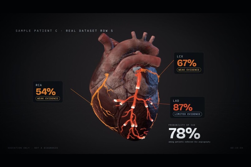
CardioMap, vessel scores beside P(CAD)
-
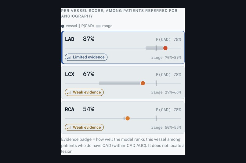
vessel scores with evidence badges
-
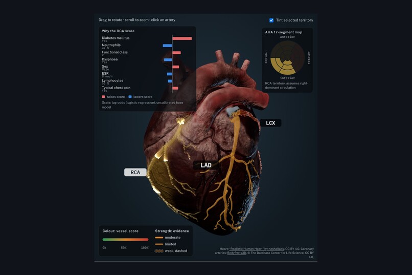
RCA territory tint and AHA bullseye
-
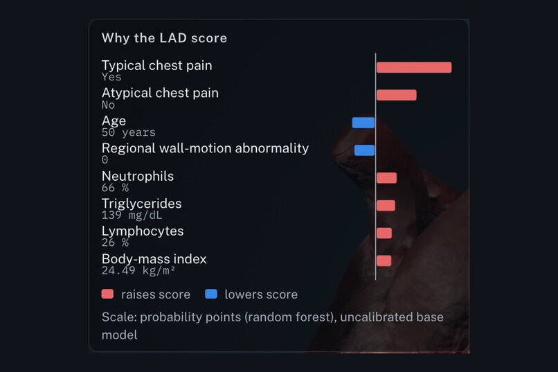
SHAP explanation in clinical units
-
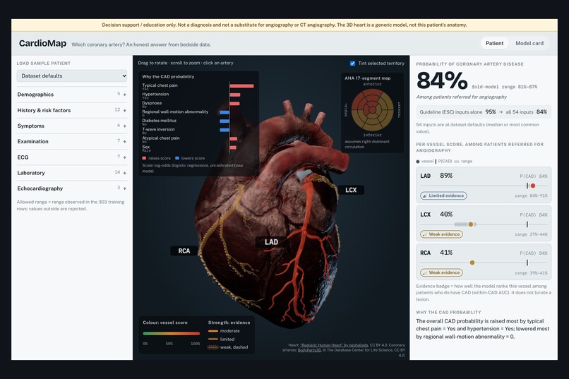
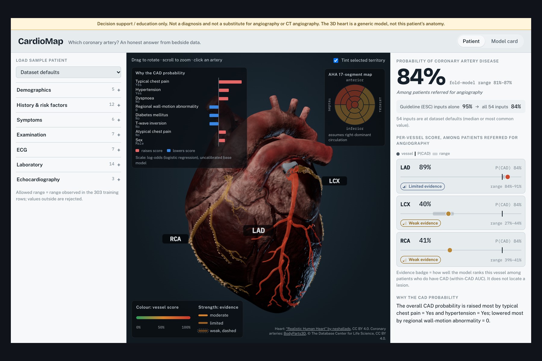
dataset-default patient
-
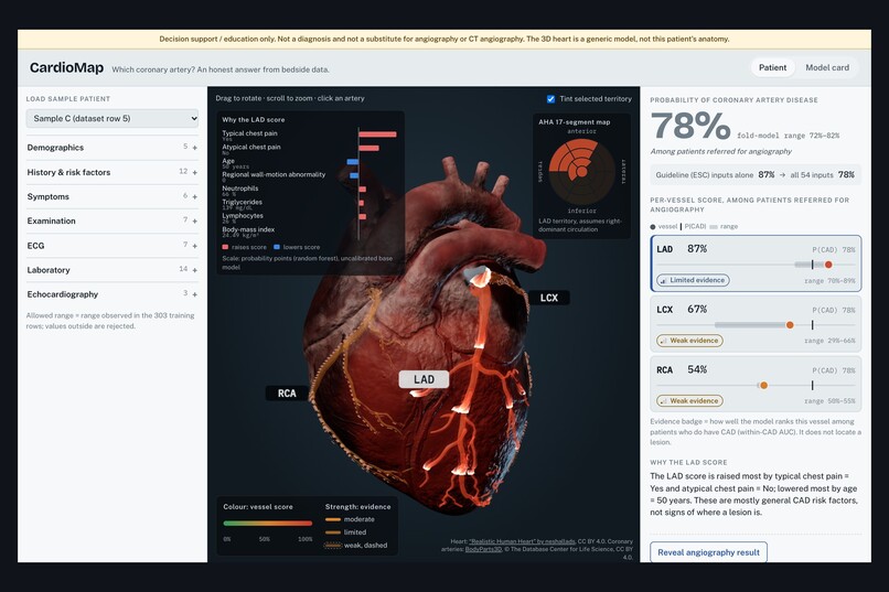
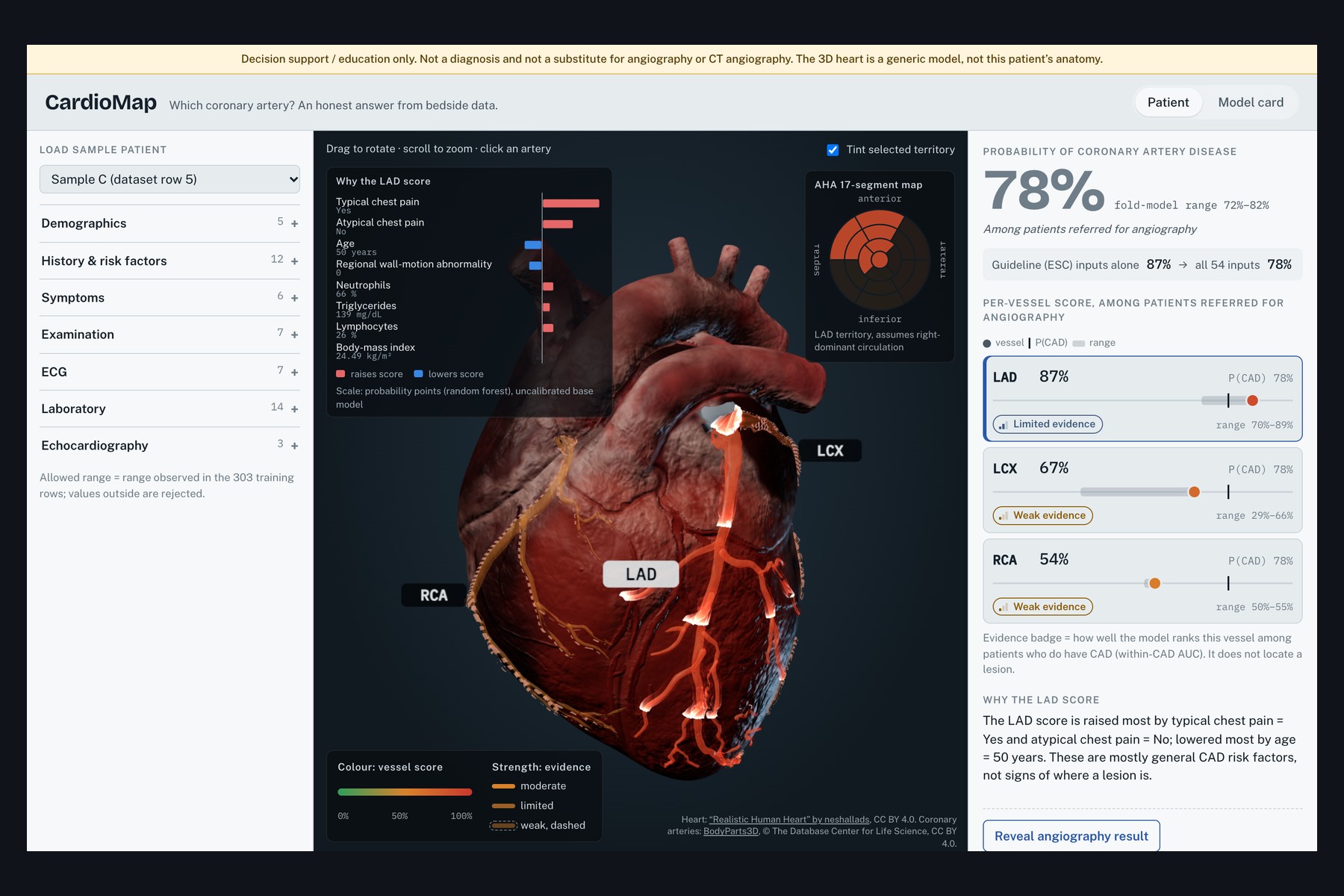
full patient view, LAD selected
Devpost submission fields
About the project (paste into "Project Story")
Inspiration
Ask a model trained on bedside data "which coronary artery is blocked?" and it will happily paint the LAD red. The question behind CardioMap: is that red LAD real information about where the disease is, or just overall CAD risk wearing a different label?
The Z-Alizadeh Sani extension dataset (303 patients referred for angiography, 55 bedside variables, per-vessel stenosis labels for LAD, LCX and RCA) makes the question testable. The answer shaped the whole product.
What it does
CardioMap takes history, symptoms, exam, ECG, labs and echo, then shows:
- Probability of CAD with a fold-model range, next to a guideline baseline built from the 12 ESC inputs alone ("Guideline inputs alone: 87% → all 54 inputs: 78%").
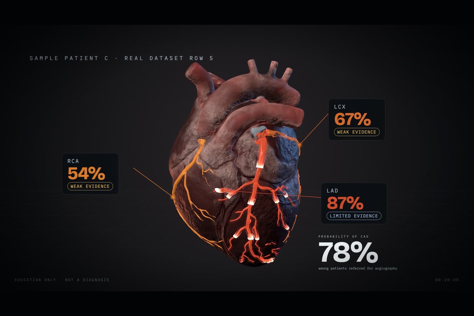
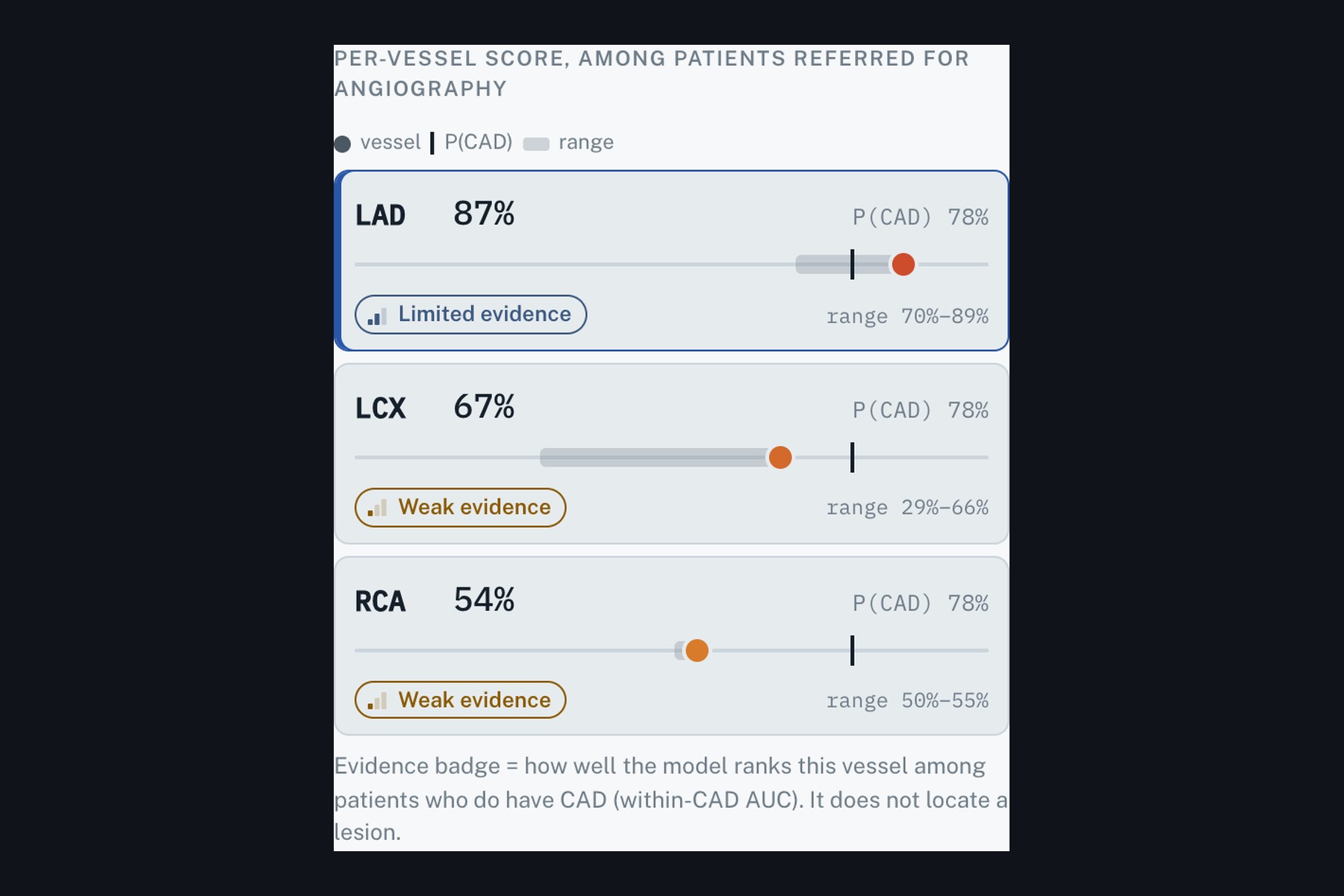
- A score per vessel (LAD, LCX, RCA), always printed beside $P(\text{CAD})$, with an evidence badge (weak / limited / moderate) earned from how well that vessel model ranks patients who already have CAD.
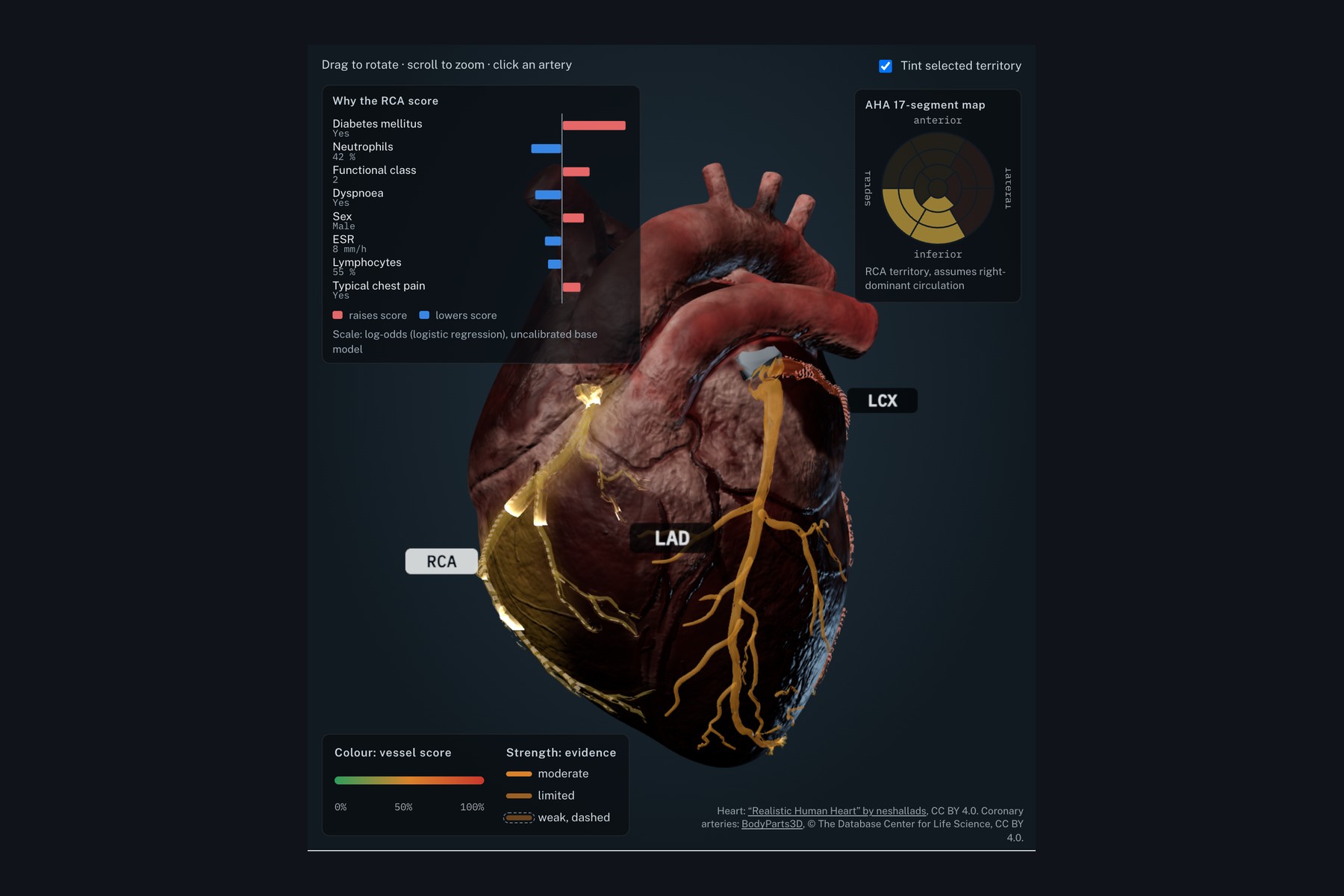
- An interactive 3D heart. Each artery is coloured by its score; opacity and a dashed outline encode the evidence badge, so a weak signal looks weak. Click an artery to tint its myocardial territory, light up an AHA 17-segment bullseye and send a glowing stream along the vessel.
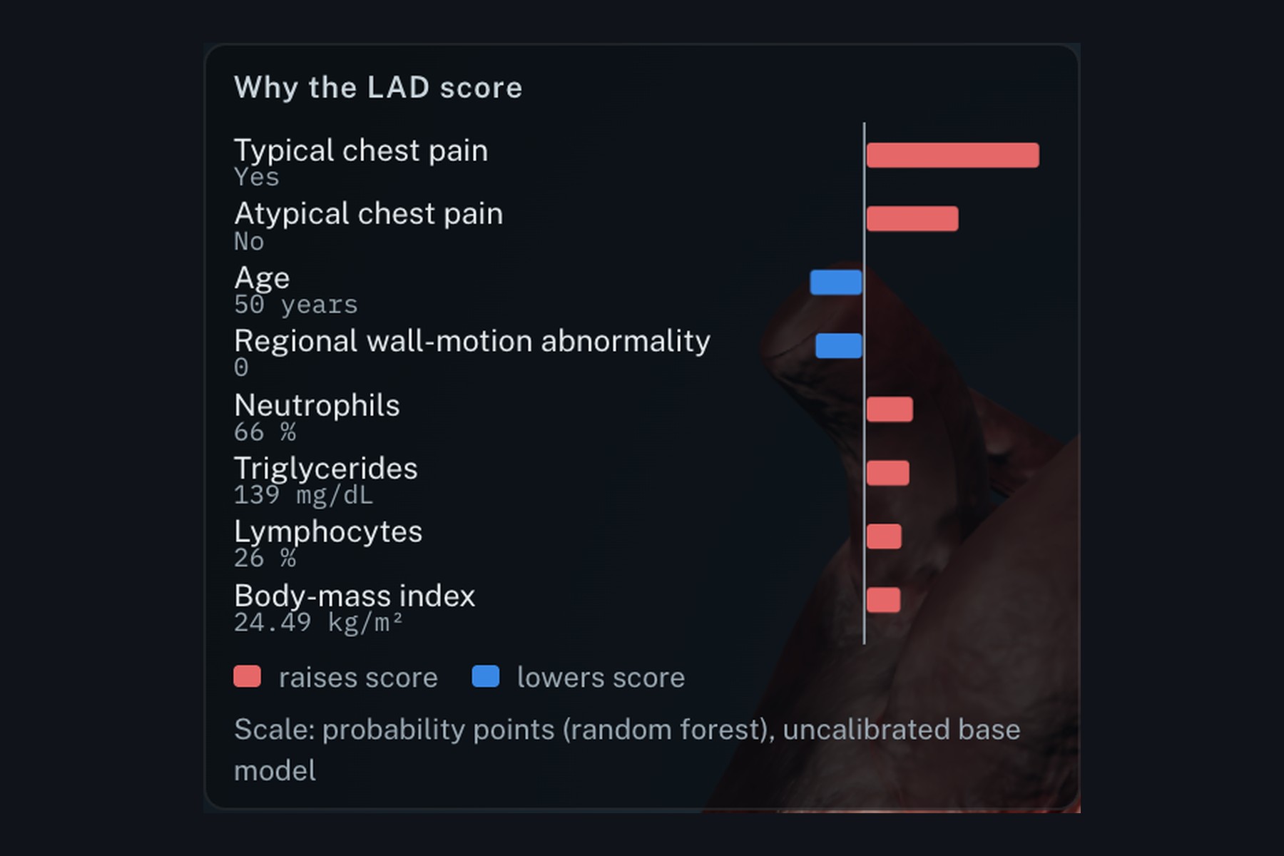
- A SHAP chart in clinical units ("Typical chest pain = Yes", "Age = 50 years") plus one plain-language sentence.
- A model card with every metric and its 95% CI, the vessel AUC comparison, the modality ablation and the limitations.
Every percentage is labelled among patients referred for angiography, and every screen carries the disclaimer: decision support / education only, not a diagnosis.
The finding
Paired cross-validation (5×10 repeated stratified CV, 2,000 patient-level bootstrap resamples):
| Vessel (served model) | Dedicated model AUC | CAD probability used as the vessel score | Within CAD patients |
|---|---|---|---|
| LAD (RF) | 0.85 | 0.85 | 0.67 |
| LCX (RF) | 0.75 | 0.72 | 0.62 |
| RCA (LR) | 0.73 | 0.74 | 0.61 |
- No dedicated vessel model beats simply reusing $P(\text{CAD})$ as the vessel score. Every difference CI includes zero (one LR case only just excludes it, in the direction of the dedicated model being worse).
- Among patients who do have CAD, vessel AUCs fall to 0.52 to 0.67: barely above chance for telling which artery.
- A hierarchical model $P(\text{CAD}) \times P(\text{vessel} \mid \text{CAD})$ gave no gain.
- What bedside data does do well: CAD itself. ESC inputs alone reach AUC 0.910; adding ECG and echo reaches 0.939, a gain of $+0.029$ (95% CI 0.013 to 0.048).
So the UI never lets a red artery stand alone. Evidence badge rule, from the mean within-CAD AUC of the LR and RF dedicated models:
$$ \text{badge} = \begin{cases} \text{weak} & \overline{\text{AUC}}{\text{within CAD}} < 0.60 \ \text{limited} & 0.60 \le \overline{\text{AUC}}{\text{within CAD}} \le 0.70 \ \text{moderate} & \overline{\text{AUC}}_{\text{within CAD}} > 0.70 \end{cases} $$
Result: LAD limited, LCX weak, RCA weak.
Classification metrics at a threshold of 0.5 fixed in advance (out-of-fold, random forest): CAD accuracy 0.87, precision 0.87, recall 0.95, F1 0.91. Vessel recall is low (LCX 0.34, RCA 0.32) because vessel scores are rarely confident, which is exactly the point.
How we built it
- ML (Python, scikit-learn): for each target, logistic regression vs random forest chosen by an inner 5-fold CV on training data only, never by reported results. Final models wrapped in
CalibratedClassifierCV(method="sigmoid"); five fold models give the variability band. Label columns (LAD,LCX,RCA,Cath) are asserted absent from every feature set and rejected by the API with a 400. - Explanations: exact SHAP on the uncalibrated base model. For logistic regression, $\phi_j = \beta_j \, (x_j - \bar{x}_j)$ in standardised space; for random forests, path-dependent TreeSHAP. Both are implemented in numpy and tested equal to the
shaplibrary (max error about $10^{-16}$), which kept the serverless function small. - Backend: FastAPI. One function,
prob_to_color(p), turns probabilities into colours; the frontend never recomputes them, and a Playwright test checks that the colour rendered on each artery mesh equals the API's colour. - Frontend: React, TypeScript, three.js via react-three-fiber. Custom shaders for the territory tint (smooth per-vertex weights), the dashed weak-evidence outline and the additive glowing stream.
frameloop="demand"keeps it light on laptops without a dedicated GPU. - 3D heart: the textured "Realistic Human Heart" model (neshallads, CC BY 4.0) with real coronary arteries from BodyParts3D (CC BY 4.0) fitted onto it: rigid ICP, then non-rigid NRICP registration, then a seating pass so no artery segment floats above or sinks below the surface. Territory weights are balanced to the AHA 17-segment shares (LAD 7 : LCX 5 : RCA 5).
- Reproducibility: dataset SHA-256 checked at load, seeds fixed, 98 tests, a Dockerfile that retrains and tests at build time, and a 6-page write-up.
Challenges we ran into
- Honesty vs. a flashy demo. A glowing red artery is persuasive. Designing the evidence encoding (badge, opacity, dashed outline, $P(\text{CAD})$ always beside the vessel score) so the visual could not overclaim took more iterations than any model.
- Fitting two anatomies together. The arteries from BodyParts3D and the textured surface came from different hearts. Early fits left vessels floating in mid-air or buried in muscle; non-rigid registration plus a seating pass brought both to 0%.
- A glow that vanished on Apple GPUs. The artery meshes had no normals, so the shader normalised a zero vector and produced NaN on Metal. Fixed by exporting normals, recomputing them when missing and making the shader NaN-safe.
- Serverless size limits. pandas + shap + scipy + scikit-learn exceeded the function size limit. Rewriting SHAP in numpy (verified exact) and making the inference path pandas-free cut the dependencies to what serving actually needs.
Accomplishments we're proud of
- A negative result turned into a product feature instead of being hidden.
- Every number in the UI is read from artifacts produced by the evaluation script, never hard-coded.
- Tests that guard the claims: no label leakage, rendered colour equals API colour, arteries seated on the surface, SHAP exact.
What we learned
- Per-vessel prediction from bedside data is mostly overall risk in disguise. Measuring within-CAD discrimination is the test that exposes it.
- Calibration and uncertainty display matter as much as AUC when the output is a colour on an organ.
- Guideline inputs are a strong baseline; ECG and echo add a small but real gain.
What's next for CardioMap
- External validation on a second centre and a population not pre-selected for angiography.
- Inputs that carry real location signal (regional wall-motion by segment, ECG lead territories, CT calcium by vessel), with the evidence badges re-earned on that data.
- Patient-specific anatomy from CT angiography instead of a generic heart.
Team: Samarth Verma, Yashi Rajoriya, Anshuman Dubey. AI assistance (Claude Code, Claude) is disclosed in the README.
"Try it out" links
- Live app:
- Demo video: https://www.youtube.com/watch?v=l9Fn8tcCOsU
- Source code: https://github.com/samarthverma4/CardioMap
- Write-up (PDF): https://github.com/samarthverma4/CardioMap/blob/main/docs/writeup.pdf
Image gallery (docs/devpost/, 1800×1200, 3:2)
00_poster.jpg: CardioMap, vessel scores beside P(CAD)01_patient_view.jpg: full patient view, LAD selected02_defaults.jpg: dataset-default patient03_rca_territory.jpg: RCA territory tint and AHA bullseye04_shap_lad.jpg: SHAP explanation in clinical units05_vessel_rows.jpg: vessel scores with evidence badges06_vessel_auc.jpg: vessel AUC: dedicated vs CAD probability vs within CAD07_ablation.jpg: what each modality adds for CAD08_mobile.jpg: mobile layout
Video demo link
Built With
- bodyparts3d
- docker
- drei
- explainable-ai
- fastapi
- glsl
- machine-learning
- numpy
- pandas
- playwright
- pytest
- python
- react
- react-three-fiber
- scikit-learn
- shap
- sketchfab
- three.js
- trimesh
- typescript
- uvicorn
- vercel
- vite
- webgl

Log in or sign up for Devpost to join the conversation.