-
-
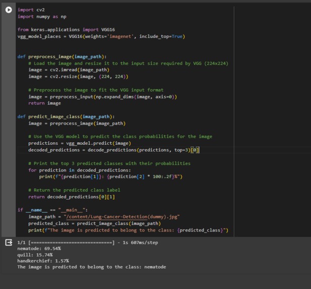
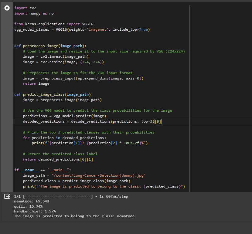
project code
Inspiration
The cancer patients I've observed in my day-to-day life encountered significant issues during result retrieval, facing delays in receiving their reports
What it does
It helps me empathize with the patients and understand the challenges they face. It teaches me about their struggles and failures, highlighting that success often emerges from setbacks. It also demonstrates that while success might seem simple in words, it often requires overcoming various obstacles and failures along the way.
How we built it
I developed the project using specific datasets and tested it within the Python environment. The project was tailored to address the needs of the users, focusing on time management and optimizing processes to minimize the time required.
Challenges we ran into
One of the primary challenges we encountered revolved around handling machine learning models and image datasets. Specifically, the time-consuming aspect was in the process of constructing the model and achieving the desired accuracy rates. This was notably prominent when dealing with image datasets, necessitating more time and effort to attain the desired level of accuracy.
Accomplishments that we're proud of
Model Development: Successfully constructing a robust machine learning model tailored for time management, especially when dealing with image datasets, demonstrates significant technical expertise.
Optimization: Streamlining the process to minimize the time required for both model building and achieving high accuracy rates indicates efficiency and innovation.
Addressing User Needs: Developing a solution that directly addresses the time management needs of users showcases a deep understanding of their requirements and challenges.
Overcoming Challenges: Conquering hurdles, particularly those related to handling machine learning models and large image datasets, signifies resilience, problem-solving skills, and dedication.
Impact: If applicable, any positive impact observed or feedback received from users or stakeholders regarding the effectiveness of the solution is a notable accomplishment.
What we learned
Dataset Challenges: Dealing with image datasets and machine learning models provided insights into the complexities and time-intensive nature of handling such data, offering lessons on efficient data preprocessing and model optimization techniques.
Technical Proficiency: Building and optimizing machine learning models in Python honed your programming skills and understanding of various ML algorithms and libraries, contributing to your technical expertise.
User-Centric Approach: Understanding the importance of addressing user needs, especially in the context of time management, emphasized the significance of aligning technological solutions with real-world problems.
Problem-Solving: Overcoming challenges, whether in data handling or model accuracy, enhanced your problem-solving abilities, fostering resilience and adaptability in the face of complexities.
Project Management: Managing a project involving data, model development, and optimization provided insights into effective project planning, time management, and the iterative nature of development.
Continuous Improvement: Recognizing that success often comes from iterative improvements, acknowledging failures as learning opportunities, and continuously refining the model for better performance.
What's next for Cancer Cell Detection and Segmentation
Enhanced Accuracy: Continual refinement and advancement of machine learning models can lead to even higher accuracy rates in detecting and segmenting cancer cells. This could involve exploring more sophisticated algorithms, leveraging larger and diverse datasets, and optimizing model architectures.
Real-time Applications: Progressing towards real-time analysis and diagnosis using these models can significantly impact patient care by enabling quicker assessments and treatment decisions.
Automation and Integration: Integrating these models into existing healthcare systems and workflows can streamline processes, aiding pathologists and oncologists in their analysis and decision-making processes.
Multi-modal Fusion: Integrating information from various imaging modalities (e.g., MRI, CT, histopathology slides) can provide a more comprehensive view, potentially improving accuracy and diagnostic capabilities.
Personalized Medicine: Advancements may lead to tailoring treatments based on precise cell-level information, enabling more personalized and effective therapies for cancer patients.
Ethical Considerations: Further exploration and adherence to ethical guidelines regarding patient data privacy, interpretability of models, and responsible deployment in healthcare settings will continue to be critical.
Collaboration and Research: Continued collaboration between medical professionals, data scientists, and researchers is crucial for advancing this field, encouraging the exchange of knowledge, datasets, and methodologies.


Log in or sign up for Devpost to join the conversation.