-
-
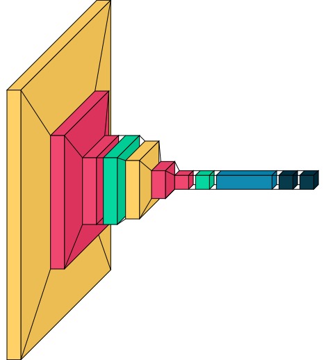
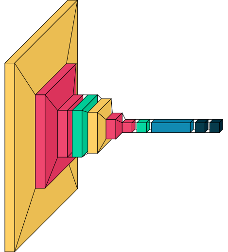
Neural Network Architecture used
-

A sample
-

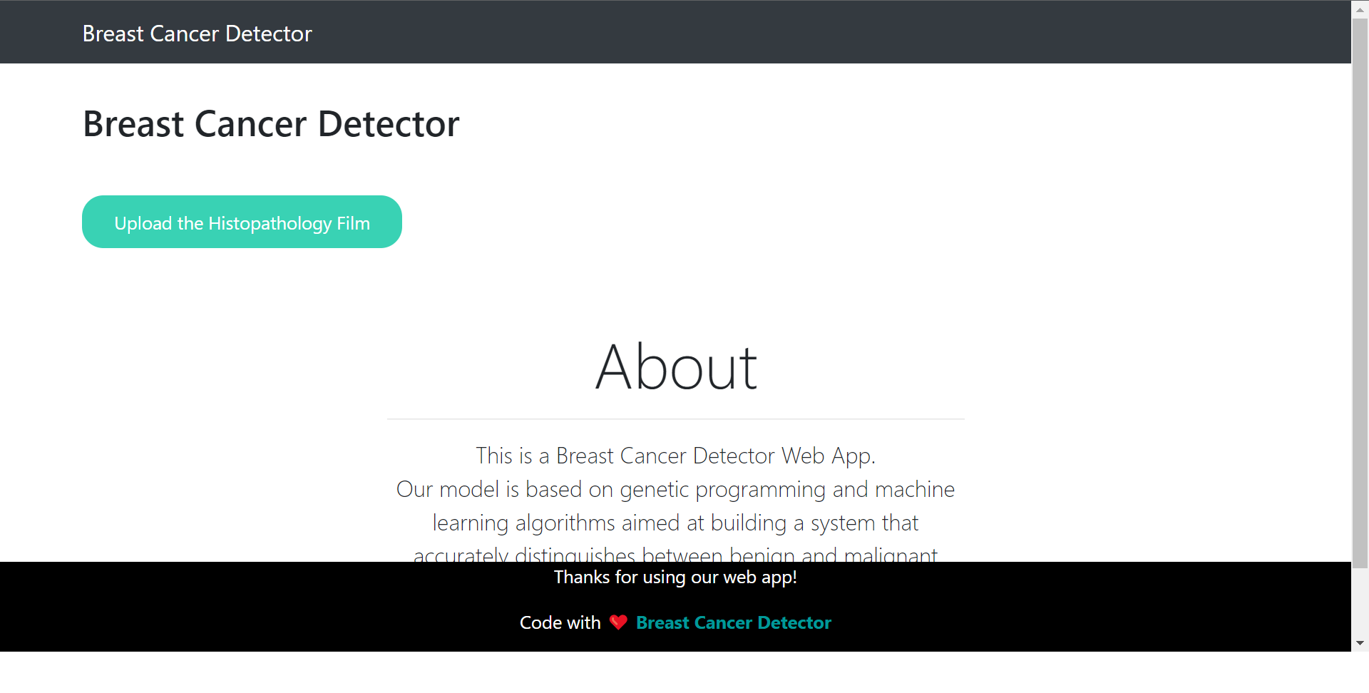
home page

Inspiration
Breast cancer is a common cause of death and is the only cancer that is widespread in women around the world . Many diagnostic imaging methods have been developed for early detection and treatment of breast cancer and reduced mortality , and many methods are used to assist in the diagnosis of breast cancer to improve diagnostic accuracy. There are several empirical studies on breast cancer using machine learning. Here, we applied genetic programming techniques to select the best properties and complete parameter values for the machine learning classifier.
To facilitate interpretation and analysis, histopathology film pretreatment helps improve the visibility and intensity distribution of the surrounding area, and several methods have been reported to assist in this process Feature extraction is an important step in the detection of breast cancer as it helps distinguish between benign and malignant tumors. After extraction, image properties such as smoothness, roughness, depth, and regularity are extracted by segmentation .
What it does
Our model is based on genetic programming and machine learning algorithms aimed at building a system that accurately distinguishes between benign (non cancerous) and malignant (cancerous) breast tumors. Different transformation-based texture analysis techniques are applied to transform an image into a new shape using the spatial frequency property of pixel intensity changes. Different techniques can be used to reduce the dimension of feature representation. Many studies have been attempted to automate the diagnosis of breast cancer based on machine learning algorithms.
How we built it
We used techniques of Computer Vision to achieve our goal. We used a neural network with CNNs and some Dense Layers. We collected the data from Kaggle. First we convert the image into a 3D array of numbers. The array of images and labels are passed into the neural network to find accuracy. We achieved accuracy of 92.2% on our test dataset.
Our model involves data mining (from kaggle) and machine learning techniques to detect and classify breast cancer . It can be divided into three main phases:
- Pre processing
- Feature extraction
- Classification
Challenges we ran into
The dataset was very huge to deal with so we had subdivided into multiple datasets and then we performed with all the subdivided datasets individually and found out the most accurate dataset among them.
Accomplishments that we're proud of
We are glad to tell that our model can not only be deployed on local system but also on global internet. Which makes it more easy to use and understand.
What we learned
We learnt how we can deal with huge datasets without altering the results and give optimum result in minimum time. We also learnt how machine learning models can be made user friendly by deploying them globally or locally.
What's next for Breast Cancer Detector App
Currently we are predicting over a single patch of the complete image but later we shall be improving it to make the prediction over the complete histopathology film at a time.
We are planning to make it more interactive by integrating with various medical APIs to make the website more useful and to make it a complete website and not only a detector one. We also plan to create a mobile application too.
Built With
- css
- flask
- h5
- heroku
- html
- javascript
- keras
- machine-learning
- matplotlib
- python
- sklearn
- tensorflow
- tf


Log in or sign up for Devpost to join the conversation.