-
-
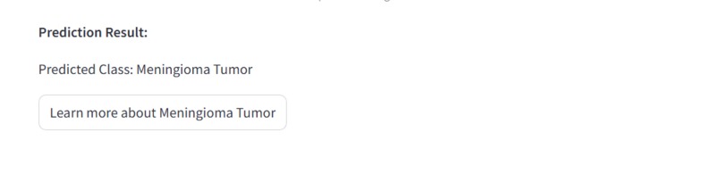
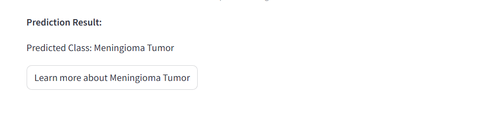
Prediction results screen
-
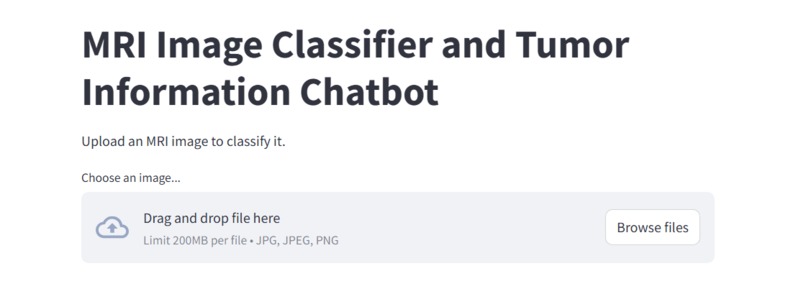
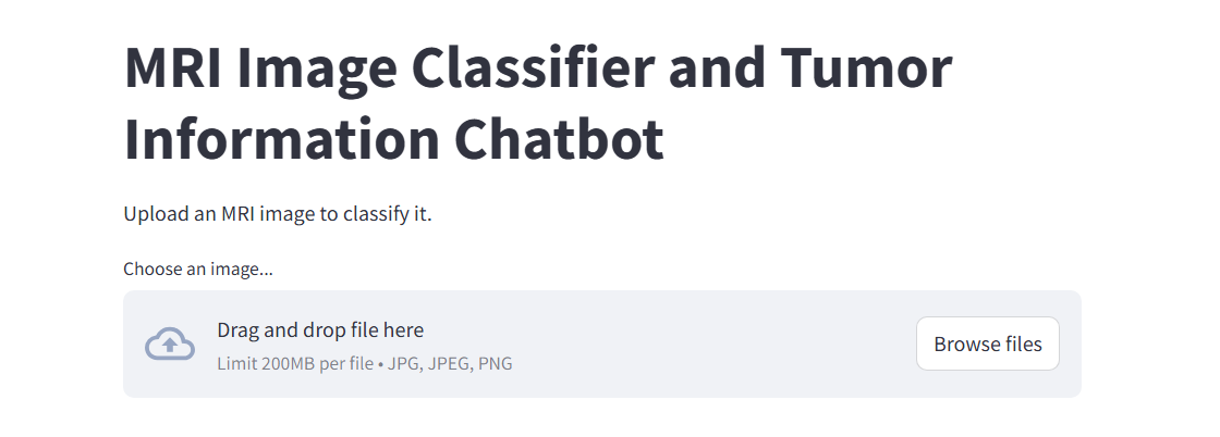
Initial pop-up screen
-
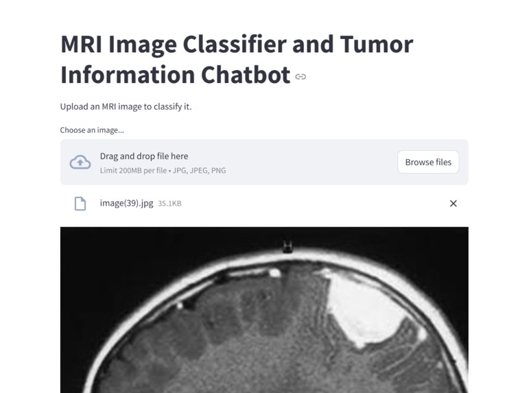
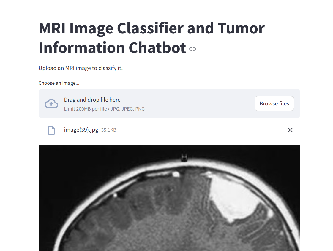
Loading an MRI picture screen
-
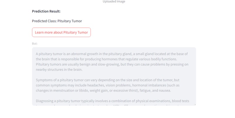
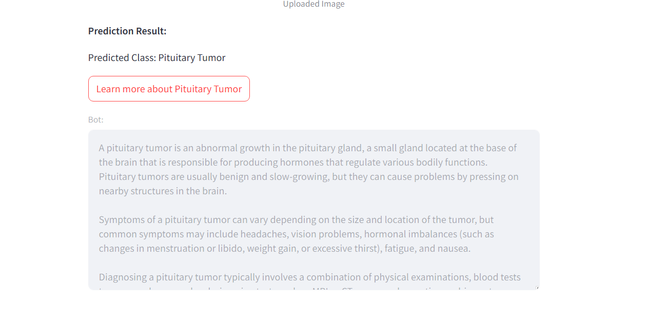
Chatbot Screen After Prediction
Inspiration
Our team was deeply inspired to create an innovative solution to a modern medical problem, particularly by the personal experience of one of our members whose grandfather tragically passed away due to rapidly spreading tumors. The lack of early detection and accessible information made us realize the urgent need for tools that could make a difference. This project is not just about technology, but about giving people a fighting chance—had something like this existed, it might have helped extend his life and provided crucial insight into his condition sooner
What it does
This web app serves as an MRI image classifier and chatbot, designed to assist with the early detection of brain tumors. By uploading an MRI scan, the app uses a trained machine learning model to analyze the image and classify the type of tumor, if present. Additionally, it includes a chatbot feature that provides users with detailed information about the detected tumor type, helping them better understand their condition. This combination of technology and AI-driven insights offers a user-friendly tool aimed at improving early diagnosis and awareness of brain tumors.
How we built it
We began our project by sourcing a high-quality brain MRI dataset from Kaggle, which included images categorized into four classes: glioma, meningioma, pituitary tumors, and no tumor. Using this dataset, we used a pre-trained convolutional neural network (CNN) model (VGG 16) to classify the MRI images. Training the model was an intensive process, taking approximately two hours to complete and fine-tune the network for accurate predictions. Once the model was trained, we integrated it into a user-friendly web interface using Streamlit, allowing users to upload MRI images for real-time classification. We also added a chatbot feature to provide detailed information about each tumor type, creating a comprehensive tool aimed at improving understanding and early detection of brain tumors.
Challenges we ran into
Throughout the development of our web app, we faced several challenges that required creative problem-solving. One of the major hurdles was organizing the MRI images into different categories or "buckets." MRI scans are often taken from different angles, and failing to account for this variation could negatively impact our model's performance. By categorizing the images based on angles, we were able to increase the accuracy of our predictions, ensuring more reliable results. Another challenge we encountered was related to the chatbot feature, which provides users with additional information about tumor types. Unfortunately, the chatbot service is not free, and integrating it posed some financial constraints for our team. Balancing the need for advanced functionality while staying within budget was a key consideration, and we had to make difficult decisions on how to allocate resources effectively.
Accomplishments that we're proud of
We are very proud that we sticked through! This was definitely one of the most difficult projects we have embarked on and for the most part unexplored territory. It is all coming together as we are so very proud of our work.
What we learned
hrough this project, we learned valuable lessons that will shape our future work. We gained a deep understanding of connecting a front-end user interface with a backend, creating a seamless experience between the two. One key takeaway was learning how to store a trained model efficiently in an .h5 file, allowing us to access the model for future use. We also discovered just how awesome Streamlit is for building and deploying web apps quickly and effectively. Another lesson was how computationally intensive comparing MRI images can be—it took much longer than expected to process the data. Finally, we learned critical techniques for improving accuracy, such as organizing images into more granular categories and addressing variations in the dataset. These insights will guide us in refining our model and overall approach to medical imaging.
What's next for Braizen
Next for us is to continue improving the app, refining our model to achieve even higher accuracy rates. We'll explore feeding more variable data into the system—images with different colors, sizes, angles, and exposure levels—to better handle real-world variability. We’re also eager to integrate more advanced AI techniques into our processes, exploring deeper models and smarter algorithms. By doing so, we aim to create an even more robust and reliable tool for early tumor detection, ultimately pushing the boundaries of what’s possible in medical imaging.


Log in or sign up for Devpost to join the conversation.