-
-
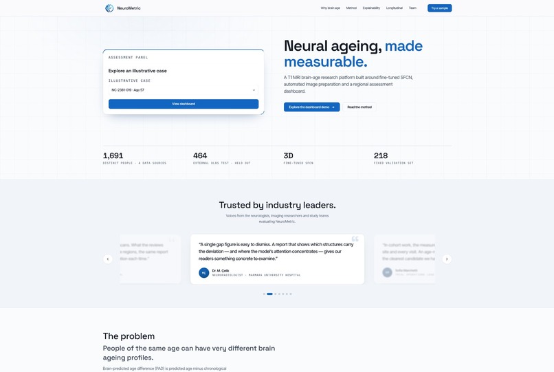
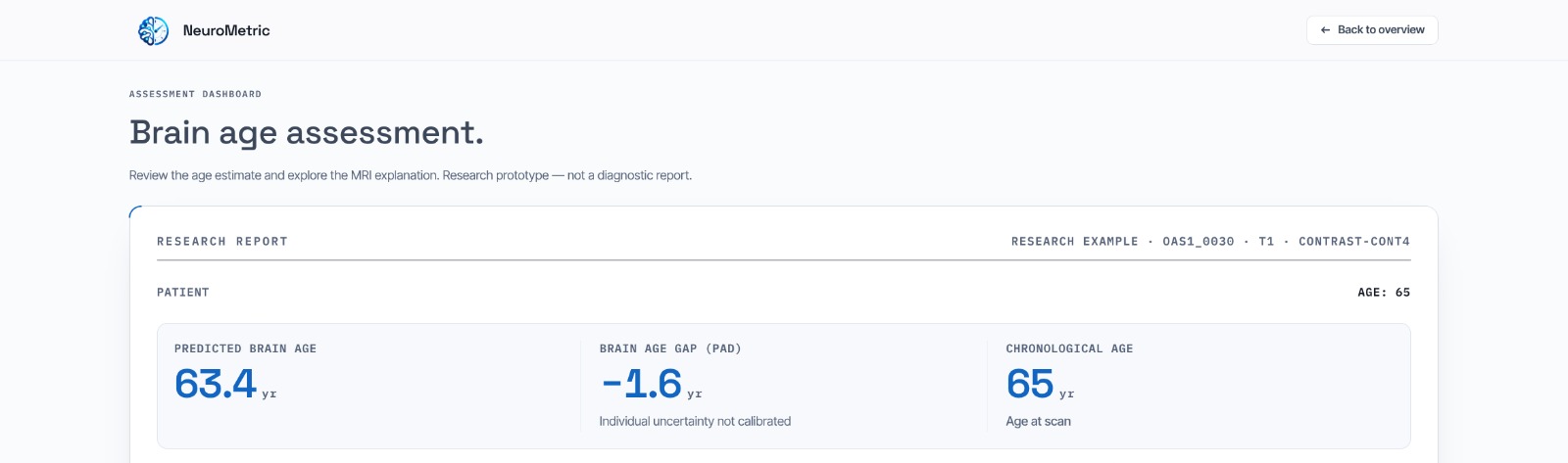
Landing Page
-
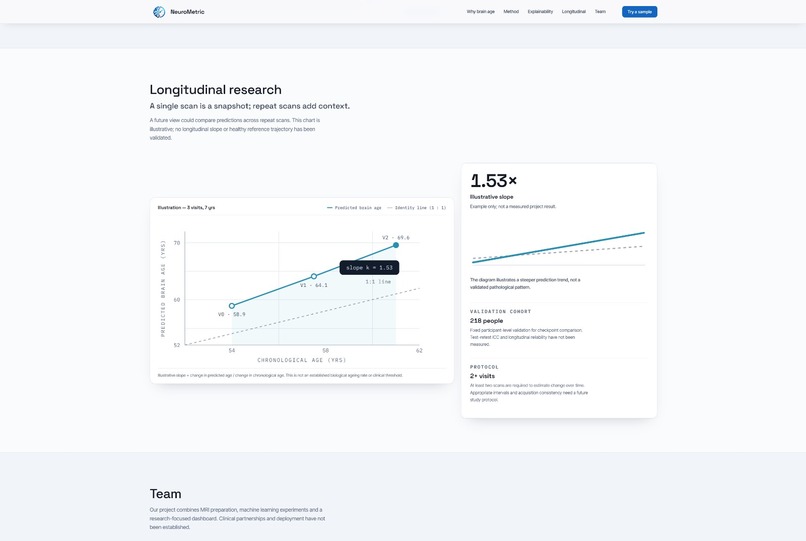
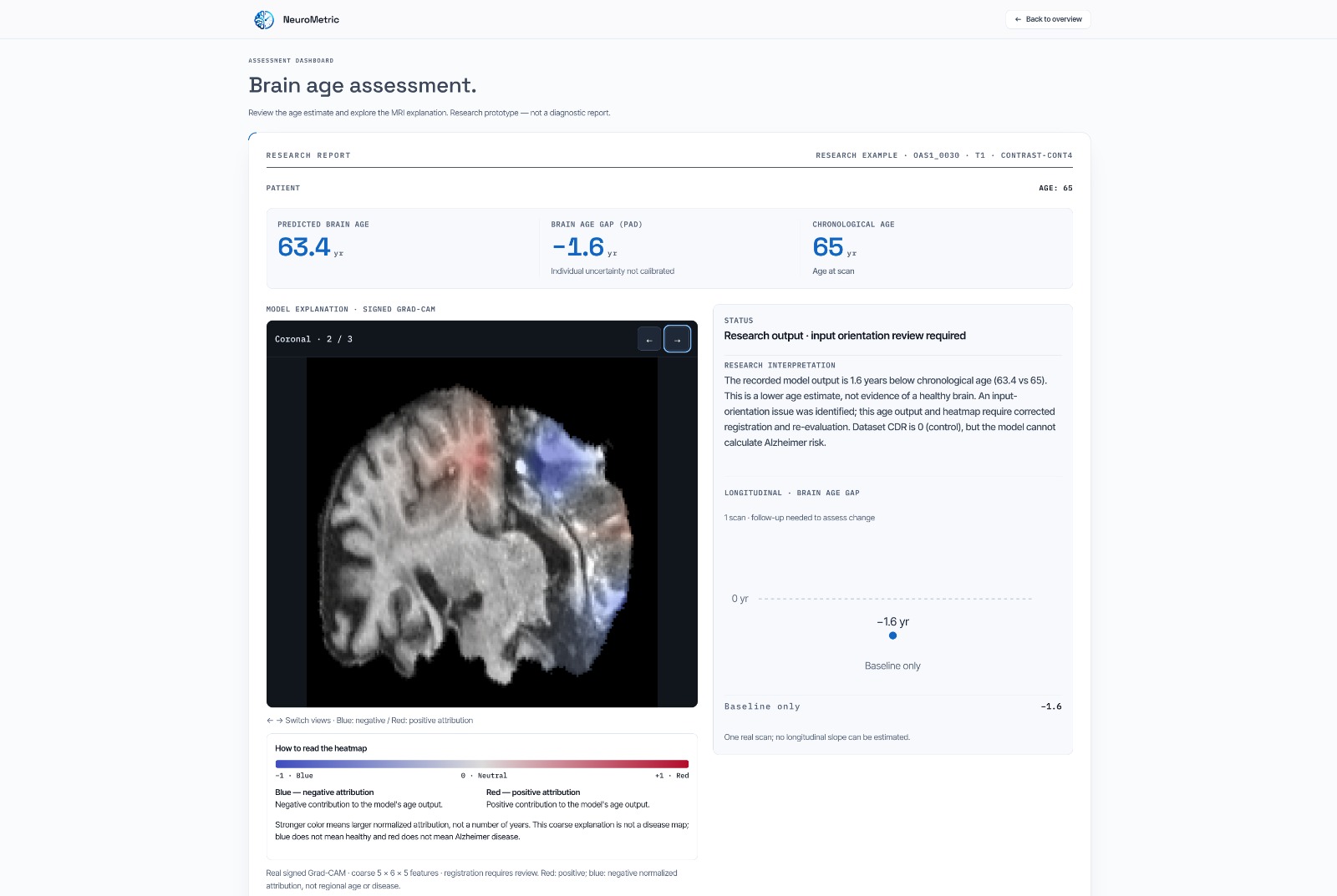
Longitudinal Graph
-
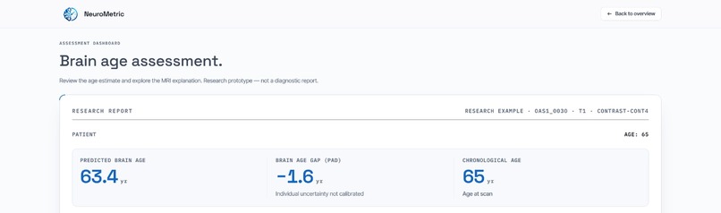
Brain Age Prediction
-
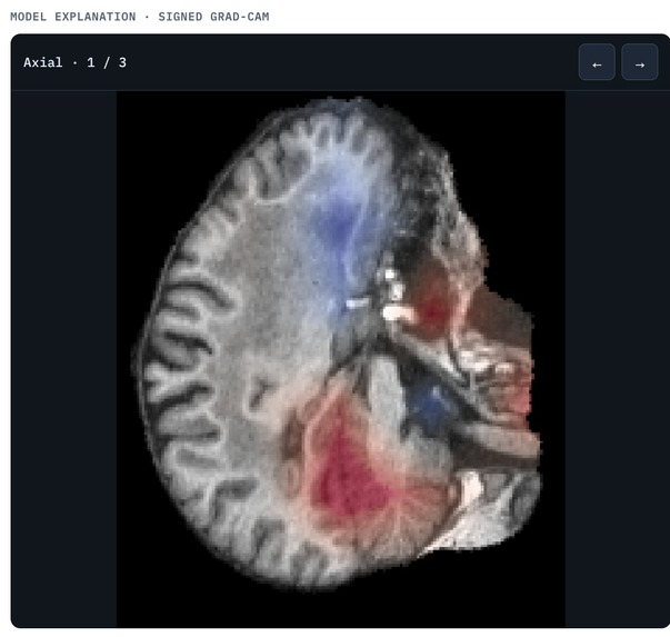
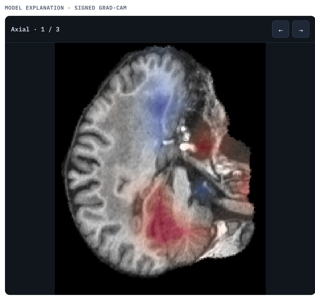
Model Attention Heat Map
-
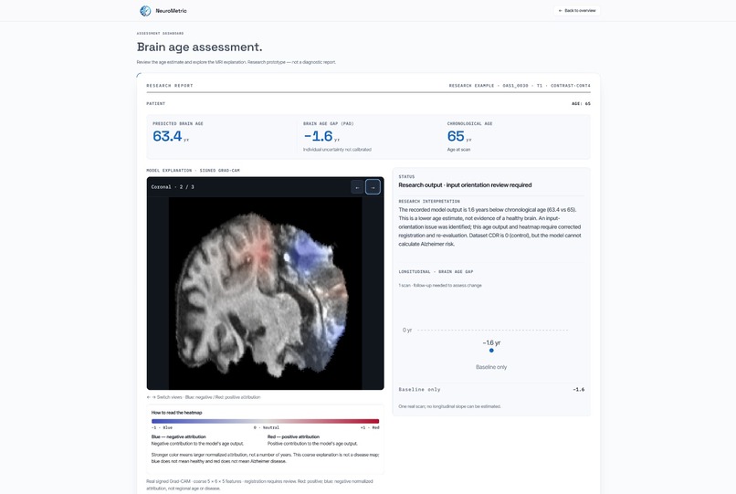
Clinical Dashboard
-
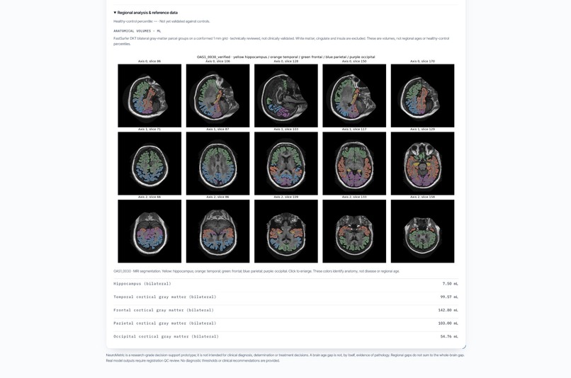
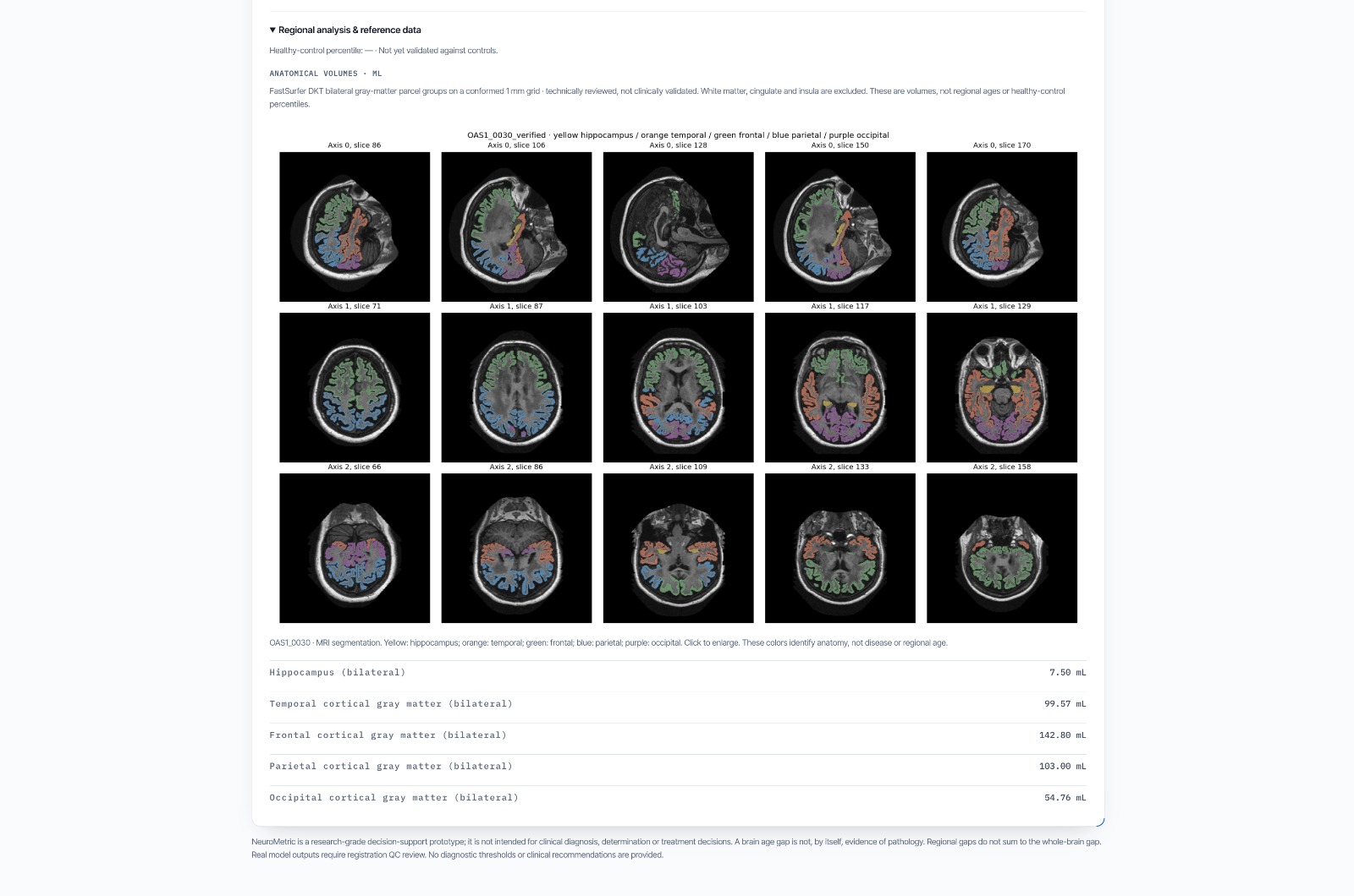
Regional Volume Analysis
-

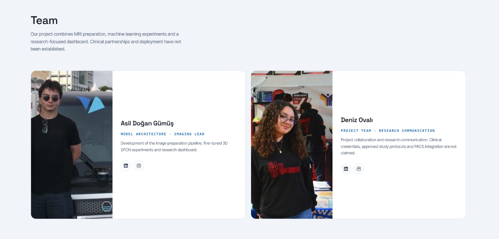
Team
Inspiration
A person can be 50 years old, yet their brain structure may appear younger or older than that of an average 50-year-old. An atypical pattern of brain aging may be associated with neurodegenerative or psychiatric conditions. We became interested in brain aging after learning that the interpretation of structural changes in the brain can vary between clinicians. Previous research has shown that two radiologists assessing the same MRI scan can reach significantly different conclusions. This made us question how much the assessment of brain aging depends on individual clinical judgment.
We wanted to explore whether there could be a more objective way to assess brain aging—one that could identify patterns beyond what can be consistently captured through visual inspection alone. This question became the starting point for NeuroMetric.
What it does
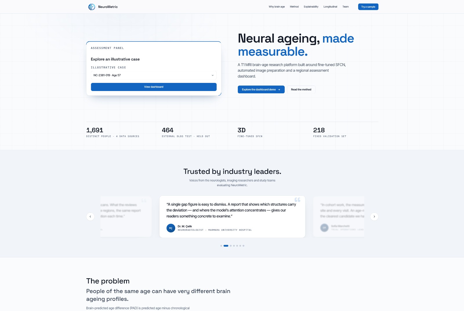
NeuroMetric estimates biological brain age from MRI scans and compares it with the patient’s chronological age, providing an interpretable measure of brain aging.
Beyond the prediction itself, our clinical dashboard transforms these results into an intuitive interface designed for clinicians. It allows doctors to view brain-age estimates, compare biological and chronological age, examine the age gap, measure regional brain volumes, visualize brain regions through heat maps, and access patient-level results in a structured clinical environment.
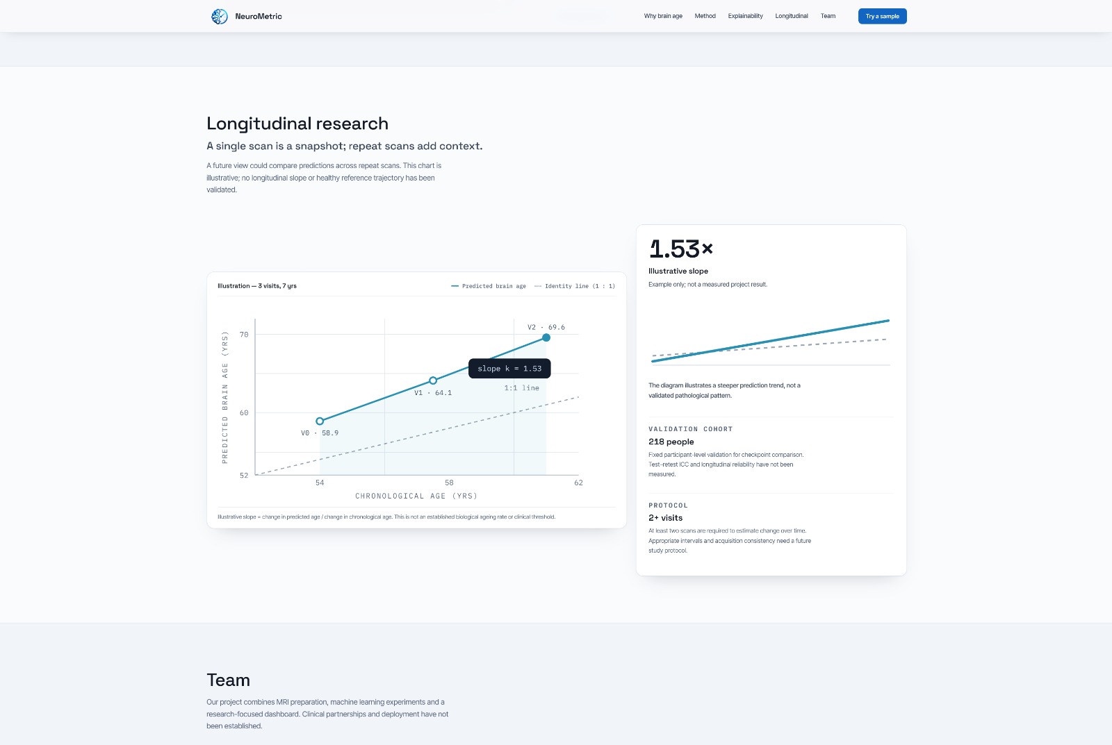
The dashboard also supports longitudinal monitoring by tracking brain-age estimates and regional brain volumes over time. By visualizing these changes as trends, NeuroMetric can identify an increasing rate of brain-age progression and flag these changes for clinical attention.
By bringing AI-based analysis into a clinician-oriented interface, NeuroMetric aims to make brain-age assessment more accessible, consistent, and easier to interpret, while supporting physicians in evaluating brain aging alongside other clinical information.
How we built it
We first defined NeuroMetric as an end-to-end platform for estimating brain age from MRI scans and comparing it with chronological age. We evaluated pretrained 3D ResNet and SFCN architectures and selected a fine-tuning approach to adapt them to our data.
We prepared subject-level training, validation, and independent test sets using MRI datasets from IXI, SALD, NIMH, and DLBS. We developed an MRI preprocessing pipeline including non-brain tissue removal, spatial alignment to a standard space, intensity normalization, and model input preparation. We also performed automated quality checks and visual inspections to identify and remove unsuitable scans.
We trained and evaluated both ResNet and SFCN models. After comparing their performance, SFCN showed more promising results, so we continued optimizing and training this model.
We then integrated the model into the NeuroMetric Clinical Dashboard, creating a clinician-oriented interface for viewing brain-age predictions and comparing biological brain age with chronological age. This allowed us to connect the machine learning model with a practical interface rather than keeping it as an isolated prediction system.
Challenges we ran into
One of our biggest challenges was preparing MRI scans from multiple public datasets. Differences in image orientation, resolution, and acquisition protocols required a consistent preprocessing pipeline and careful quality control.
We also found that pretrained models did not perform well out of the box on our data, making fine-tuning necessary. Processing large 3D images added computational demands, while cloud execution issues required debugging and automated monitoring.
Another challenge was measuring success appropriately: brain-age prediction is a regression task, so we tracked mean absolute error and predictions within specific year ranges rather than classification accuracy. Finally, we had to distinguish promising validation results from independently tested performance and avoid presenting experimental outputs as established clinical findings.
Accomplishments that we're proud of
We are proud of turning a brain-age prediction model into a complete platform rather than stopping at the prediction itself. We built an MRI preprocessing pipeline, evaluated and fine-tuned multiple 3D models, selected SFCN based on its performance, and integrated it into a clinician-oriented dashboard with regional volume analysis, heat maps, and longitudinal tracking.
What we learned
We learned that building medical AI requires more than achieving good model performance. Working with MRI data from different datasets taught us the importance of preprocessing, data quality, and independent testing. We also learned how to evaluate regression models appropriately and how important it is to make AI outputs interpretable and useful in a clinical context.
What's next for NeuroMetric
Our plan is to move beyond whole-brain age estimation and develop a region-specific approach for identifying localized patterns of brain aging. NeuroMetric currently provides regional brain volume analysis and heat maps, but it does not yet estimate the biological age of individual brain regions. Our next step is to develop this capability and quantify regional aging relative to age-matched populations.
This could provide more clinically meaningful insights by helping researchers investigate localized patterns associated with neurodegenerative and neurological conditions. Regional brain-age estimates could also support longitudinal monitoring and help identify changes that may not be captured by a single whole-brain estimate.
We also aim to expand the model using larger and more diverse neuroimaging datasets, improve its generalizability across populations, and incorporate explainable AI methods to show which brain regions and imaging features contribute most to each prediction. Ultimately, we envision NeuroMetric evolving from a brain-age prediction tool into an interpretable clinical decision-support platform for personalized brain health assessment.
Built With
- 3dresnet
- css3
- cuda
- fastsurfer
- fsl
- html5
- ixi
- javascript
- kaggle-datasets
- kaggle-notebooks
- matplotlib
- nibabel
- numpy
- nvidia
- pandas
- python
- pytorch
- scikit-learn
- sfcn
- synthseg
- synthstrip

Log in or sign up for Devpost to join the conversation.