-
-

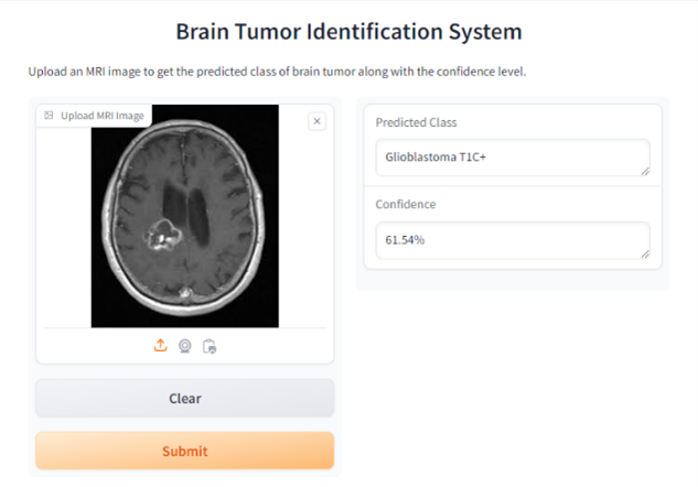
Classification Output Example
Inspiration
According to a survey by the National Higher Education, Science, Research and Innovation Policy Council (NXPO), cancer patients in Thailand die at an average rate of 342 people per day, or 124,866 people per year (data from 2020). Although brain cancer patients make up only a small fraction of all cancer patients, it is a disease with a very high mortality rate, is difficult to cure, and has a survival rate of less than 10%.
Given this data, the project team recognized the danger of this disease and saw the opportunity to involve artificial intelligence in addressing this problem by developing a machine learning model that can be used to diagnose alongside medical personnel. This model has the capability to increase accuracy and reduce the time required for diagnosis, helping patients receive appropriate and timely treatment.
What it does
This model can detect 13 types of brain tumors and one type of small lump caused by chronic inflammation (Granuloma) with an accuracy of over 96% in detecting and classifying tumor types. The 96% accuracy was calculated using K-Fold Cross Validation, a method of validating the model's accuracy by dividing the data into k equally sized groups, then training and testing the model k times. In each iteration, one group of data that the model has never seen is used as test data, while the remaining data is used for training, rotating until all k iterations are complete, making the model's accuracy assessment more reliable. The data used for testing totaled 4,478 samples, and after validation, the program achieved an accuracy of 96.09%.
How we built it
The Brain Tumor Identification System was developed using machine learning method for the detection and identification of brain tumor types. We used EfficientNetV2S pretrained model as the base model. All the data that have been used to train our model is opensource and collected from kaggle.com
Challenges we ran into
Previously, there were some issues with the prediction code. After successfully training the AI, during the prediction phase, when providing images for prediction that were not from the random set in the folder, the model often produced a single answer for all images. However, this problem has now been resolved. The issue was the mismatch between the preprocess function of randomizing images from a folder and the preprocess function from a specific image
Accomplishments that we're proud of
-Successfully developed a machine learning model with an accuracy of 96.09% for detecting and classifying brain tumors, including 13 different types of tumors and granuloma. -Overcame initial problems where the model provided uniform predictions for all input images, resulting in a reliable and diverse output. -Utilized K-Fold Cross Validation to validate the model, ensuring robustness and reliability in the results. This method increased the credibility of the model’s performance metrics. -Integrating use of AI in medical diagnostics, specifically in the challenging area of brain tumor detection, potentially speeding up diagnosis and improving patient outcomes. -Deepened our understanding of machine learning techniques and their practical applications in solving real-world medical problems.
What we learned
We have gained hands-on experience in training and fine-tuning machine learning models, particularly for medical imaging and classification tasks, and an understanding of how AI can be used to solve real-world medical problems. Gained experience in managing a project from inception through to completion, including the stages of development, testing, and deployment.
What's next for Brain Tumor Identification System
Possibility of expanding the model to recognize more types of brain tumors, further increasing its utility and accuracy.


Log in or sign up for Devpost to join the conversation.