-
-
Brain Tumor Detector
Inspiration
The inspiration behind this brain tumor detection project stems from the critical need for early and accurate diagnosis in medical imaging. Brain tumors can be life-threatening, and their early detection significantly improves treatment outcomes. However, traditional diagnostic methods are often time-consuming, prone to human error, and can miss subtle indicators in imaging data.
Witnessing the impact that delayed diagnosis can have on patients and their families motivated the creation of a tool that leverages the power of AI and deep learning. By automating the detection process and ensuring high precision, this project aims to assist healthcare professionals in identifying tumors at their earliest stages, ultimately saving lives and enhancing patient care.
The goal is not just to build a tool, but to contribute to a future where advanced technology aids in more effective, timely medical interventions, making a tangible difference in the fight against cancer.
What it does
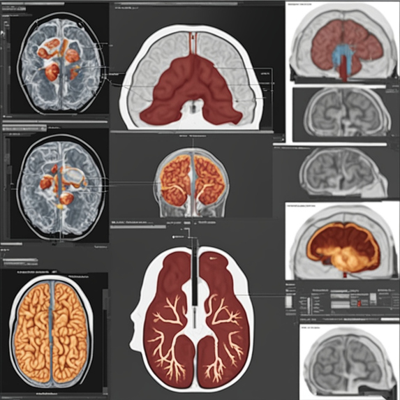
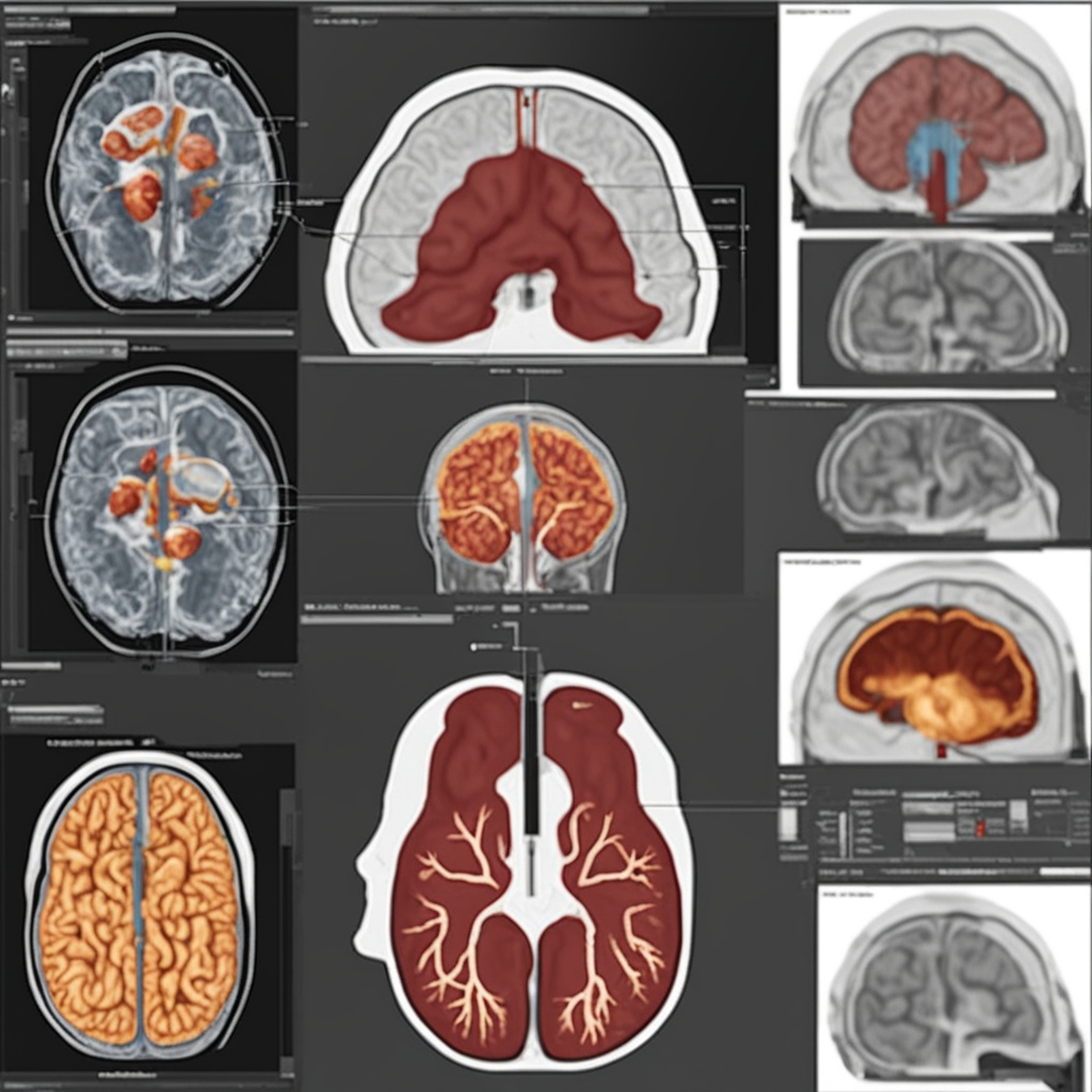
The brain tumor detection tool utilizes advanced AI and deep learning algorithms to analyze MRI images and accurately identify the presence of brain tumors. By processing and interpreting complex medical imaging data, the tool can distinguish between healthy and abnormal tissues, pinpointing tumors with high precision.
Designed to assist healthcare professionals, this tool enhances the diagnostic process by providing rapid, reliable analysis, which can be especially crucial in early-stage detection. It reduces the time and effort required for manual examination, helps minimize human error, and supports doctors in making informed decisions about patient care.
With its ability to deliver consistent and accurate results, this tool plays a vital role in improving treatment outcomes and ultimately saving lives.
How we built it
The brain tumor detection tool was built using a combination of advanced deep learning techniques and robust image processing methods. Here's an overview of the development process:
Data Collection and Preprocessing: We started by gathering a comprehensive dataset of MRI images, which were meticulously labeled to indicate the presence or absence of brain tumors. The dataset was then preprocessed to enhance image quality, normalize pixel values, and ensure consistency in image dimensions.
Model Selection and Training: We chose a convolutional neural network (CNN) architecture due to its proven effectiveness in image classification tasks. The model was designed to automatically extract features from the MRI images, learning to differentiate between healthy and tumor-affected brain tissues. We experimented with various architectures and hyperparameters to optimize performance.
Training Process: The model was trained using the labeled dataset, with a significant portion reserved for validation to monitor its accuracy and prevent overfitting. We employed techniques such as data augmentation, dropout, and batch normalization to improve the model's robustness and generalization capabilities.
Evaluation and Fine-Tuning: After training, the model was rigorously evaluated on a separate test set to assess its accuracy, sensitivity, and specificity. Based on the results, we fine-tuned the model to enhance its precision in detecting tumors, particularly in challenging cases with subtle indicators.
Deployment: Once the model achieved the desired performance metrics, we integrated it into a user-friendly interface that allows healthcare professionals to upload MRI images and receive diagnostic results quickly. The tool was optimized for speed and reliability, ensuring it could be effectively used in clinical settings.
Ongoing Improvement: The tool is designed to continuously learn from new data, enabling ongoing improvements in accuracy and expanding its capabilities to detect various types of brain tumors.
This collaborative effort between medical professionals and AI experts ensures that the tool not only meets the highest standards of accuracy but also aligns with the practical needs of those on the front lines of patient care.
Challenges we ran into
During the development of the brain tumor detection tool, we encountered several challenges that required innovative solutions:
Data Availability and Quality: Obtaining a large, high-quality dataset of labeled MRI images was a significant challenge. Medical imaging data is often scarce, and the labeling process is time-consuming and requires expert knowledge. We addressed this by collaborating with medical institutions and using data augmentation techniques to expand our dataset.
Class Imbalance: The dataset exhibited an imbalance, with fewer images of certain types of tumors. This imbalance could lead to a model biased towards the more prevalent classes, potentially missing rarer but critical cases. To mitigate this, we employed techniques like oversampling, undersampling, and implementing specialized loss functions to ensure the model learned effectively across all classes.
Complexity of MRI Images: Brain MRI images are complex and can vary significantly between patients, making it challenging for the model to learn consistent patterns. We tackled this by using advanced preprocessing techniques to enhance image clarity and applying CNNs capable of capturing intricate features from the images.
Overfitting: During the training process, the model occasionally overfitted to the training data, reducing its ability to generalize to new, unseen data. We countered this with regularization techniques such as dropout, early stopping, and cross-validation, ensuring the model remained robust and could perform well on new data.
Interpretability: While deep learning models are powerful, they often operate as "black boxes," making it difficult to interpret their decisions. This is a critical issue in the medical field, where understanding the reasoning behind a diagnosis is crucial. We addressed this by incorporating techniques such as Grad-CAM, which provides visual explanations of the model’s predictions, helping to build trust with healthcare professionals.
Computational Resources: Training deep learning models on large medical datasets requires significant computational power. We optimized the training process by using cloud-based resources and fine-tuning the model’s architecture to balance performance and efficiency.
Deployment and Integration: Integrating the model into a user-friendly tool that could be easily adopted in clinical settings was another hurdle. We focused on developing a streamlined interface and ensuring the model could deliver rapid, accurate results, even in resource-constrained environments.
Overcoming these challenges required close collaboration between data scientists, engineers, and medical professionals, ensuring that the final product was both technically sound and practically valuable in real-world medical scenarios.
Accomplishments that we're proud of
We’re proud of several key accomplishments in the development of our brain tumor detection tool:
High Accuracy and Precision: Achieving a high level of accuracy and precision in detecting brain tumors, especially in challenging cases with subtle indicators, is one of our proudest accomplishments. The tool consistently delivers reliable results, providing valuable support to healthcare professionals in diagnosing brain tumors early.
Scalable and Efficient Model: We successfully developed a deep learning model that is both scalable and efficient, capable of processing large volumes of MRI images quickly. This ensures the tool can be effectively used in clinical settings without compromising on speed or accuracy.
User-Friendly Interface: We’re proud of the intuitive and user-friendly interface we built for the tool. It allows healthcare professionals to easily upload MRI images and receive diagnostic results without needing extensive technical knowledge, making advanced AI accessible to a broader audience.
Collaboration with Medical Experts: Our close collaboration with medical professionals throughout the project was crucial to its success. By integrating their expertise into the development process, we ensured that the tool meets the practical needs of those on the front lines of patient care.
Overcoming Challenges: We’re particularly proud of how we overcame the various challenges we encountered, from handling data scarcity and class imbalance to ensuring model interpretability. Each obstacle pushed us to innovate and improve the tool, ultimately leading to a more robust and reliable product.
Impact on Patient Care: The potential impact of our tool on patient care is perhaps our greatest accomplishment. By aiding in the early detection of brain tumors, the tool has the potential to improve treatment outcomes, save lives, and contribute to the fight against cancer.
These accomplishments reflect the dedication and collaborative effort of our team, driving us to continue improving the tool and expanding its capabilities.
What we learned
Throughout the development of the brain tumor detection tool, we gained valuable insights and lessons:
Importance of Data Quality: High-quality, well-labeled data is crucial for training effective AI models. We learned that investing time in data collection and preprocessing pays off significantly in terms of model performance.
Handling Class Imbalance: Dealing with class imbalance taught us the importance of implementing techniques like oversampling, undersampling, and adjusting loss functions to ensure that the model learns to recognize less frequent but critical cases effectively.
Complexity of Medical Imaging: The complexity and variability in medical images highlighted the need for sophisticated preprocessing techniques and robust models capable of handling diverse and intricate patterns.
Model Interpretability: We learned that interpretability is essential in the medical field, where understanding the rationale behind AI decisions can impact trust and adoption. Techniques like Grad-CAM helped us provide visual explanations of model predictions, enhancing transparency.
Balancing Performance and Resources: Training deep learning models requires balancing computational resources with performance. Optimizing the model’s architecture and leveraging cloud-based resources were crucial for efficient training and deployment.
User-Centered Design: Developing a user-friendly interface underscored the importance of designing tools that cater to the end-users’ needs. Simplifying the interaction for healthcare professionals ensured that the tool could be seamlessly integrated into clinical workflows.
Collaboration with Experts: Working closely with medical professionals throughout the project reinforced the value of interdisciplinary collaboration. Their insights were essential in aligning the tool with real-world clinical needs and challenges.
Iterative Improvement: The process of refining and iterating on the model and tool taught us that continuous improvement is key. Regular evaluation and feedback help in identifying areas for enhancement and ensuring the tool remains effective and relevant.
These lessons have not only enriched our understanding of AI in medical applications but also laid the foundation for future projects, guiding us in building more effective and impactful tools.
What's next for Brain Tumor Detector
For the brain tumor detection tool, several exciting directions lie ahead:
Integration with Clinical Systems: Expanding the tool’s integration with existing hospital and clinic management systems to streamline workflows and facilitate seamless use in medical environments.
Expanding Capabilities: Incorporating additional features, such as the ability to detect and classify different types of brain tumors or other brain abnormalities, to enhance the tool’s diagnostic utility.
Improving Model Accuracy: Continuously improving the model’s accuracy and precision by incorporating more diverse and comprehensive datasets, including different imaging modalities and patient demographics.
Real-Time Analysis: Developing capabilities for real-time analysis of MRI scans, providing immediate feedback to clinicians during patient evaluations and potentially integrating with MRI machines.
Enhancing Interpretability: Further enhancing model interpretability and visualization tools to offer more detailed insights into how diagnoses are made, helping clinicians understand and trust the AI’s decisions.
User Feedback and Iteration: Collecting feedback from healthcare professionals who use the tool to identify areas for improvement and ensure that the tool remains aligned with clinical needs and preferences.
Regulatory Approvals: Working towards obtaining necessary regulatory approvals and certifications to ensure the tool meets medical standards and can be widely adopted in clinical practice.
Integration with Other AI Tools: Exploring the possibility of integrating the brain tumor detection tool with other AI-based diagnostic systems or platforms to provide a more comprehensive diagnostic solution.
Educational Resources: Developing training and educational resources for medical professionals to effectively use the tool and understand its capabilities and limitations.
Global Expansion: Exploring opportunities to deploy the tool in different regions and healthcare settings worldwide, addressing diverse healthcare needs and contributing to global efforts in cancer diagnosis and treatment.
These next steps aim to enhance the tool’s functionality, usability, and impact, ensuring it continues to provide valuable support in the early and accurate detection of brain tumors.


Log in or sign up for Devpost to join the conversation.