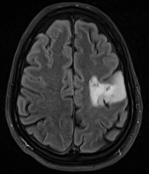
Inspiration

Brain Tumor is hard to detect and can go unnoticed sometimes during initial stage. That is why we decided to build a CNN (Convoluted Neural Network) and train it with images to enable detection of the tumor.

What it does

The CNN is trained with many tumor positive and tumor negtive results. By leveraging tensorflow, the deep learning library in python, we were able to successfully train the model to predict if a MRI scan, a test_dataset , has tumor or not with an accuracy range of 85-95%.

How we built it

The model uses a Convoluted Neural Network of many nodes each having weights affecting the outputs of the node in the next layers and so on to the next input thus making the network predict a scenario better over time with more epochs.

Challenges we ran into

We ran into some errors with the community cloud but the SSF foundation was very helpful and guided us in building this amazing model.

Accomplishments that we're proud of

The model prediction at one point had a max accuracy of 97%

What we learned

Learnt Tensorflow and other deep learning libraries in python.

What's next for Brain Tumor

More optimization needed. There is a need to embed it in a functioning UI style webapp or desktop app.

Built With

Share this project:

Updates

posted an update

In this tough conditions day by day the analysing of tumours in brain is a heavy task.We have built a amazing Machine learning tool to detect the tumours easily which is very helpful for the doctors

Log in or sign up for Devpost to join the conversation.

posted an update

Scientists have found in a recent study that only three different genetic alterations drive the early development of malignant glioblastomas. At least one of these three cancer drivers was present in all tumors investigated. The tumors develop for up to seven years before they become noticeable as symptoms and are diagnosed. However, in contrast to their early development, glioblastomas, which return after therapy, share no concurrent genetic alterations. Abstract This review recounts the history of brain tumor diagnosis from antiquity to the present and, indirectly, the history of neuroradiology. Imaging of the brain has from the beginning held an enormous interest because of the inherent difficulty of this endeavor due to the presence of the skull. Because of this, most techniques when newly developed have always been used in neuroradiology and, although some have proved to be inappropriate for this purpose, many were easily incorporated into the specialty. The first major advance in modern neuroimaging was contrast agent–enhanced computed tomography, which permitted accurate anatomic localization of brain tumors and, by virtue of contrast enhancement, malignant ones. The most important advances in neuroimaging occurred with the development of magnetic resonance imaging and diffusion-weighted sequences that allowed an indirect estimation of tumor cellularity; this was further refined by the development of perfusion and permeability mapping. From its beginnings with indirect and purely anatomic imaging techniques, neuroradiology now uses a combination of anatomic and physiologic techniques that will play a critical role in biologic tumor imaging and radiologic genomics.

Log in or sign up for Devpost to join the conversation.