-
-
-
-
-
-

-
-
-
-
-
-
-
-
-

-
-

-
-

3D Visualization of Glioma cells
-


Inspiration
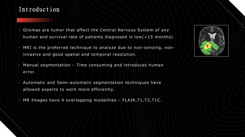
In MRI scans, there are four different imaging modalities, each capturing the brain in 155 slices of 2D images, resulting in a total of 620 images for a radiologist to analyze. Glioma cells are a type of tumor that affects the human nervous system. The task of reviewing 620 images is not only tedious but also susceptible to human error, demanding a high level of expertise that may not always be available.
This challenge inspired me to develop an automated segmentation system for glioma cells. By leveraging deep learning neural networks, this solution aims to assist doctors and radiologists, making the process more efficient and accurate.
What it does
This project is mostly a type of research work and couldn't be developed fully into a portable software but what it does is takes MRI machine output file, sends it thru a pretrained model and gives out the predicted area and tumor shape along with different tumor parts labelled
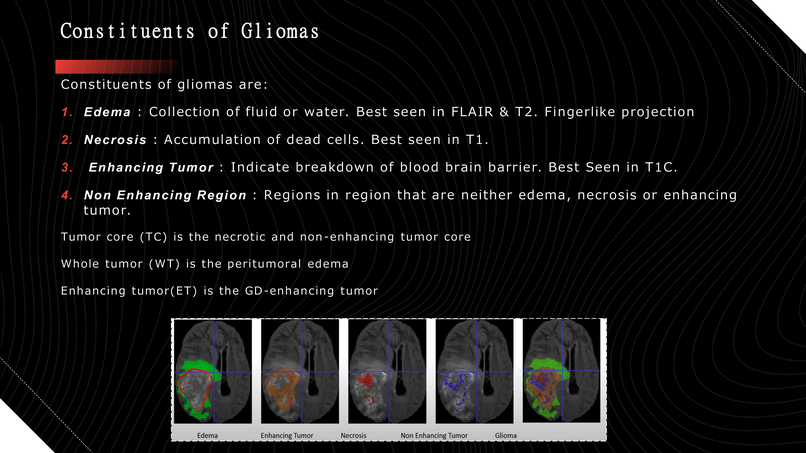
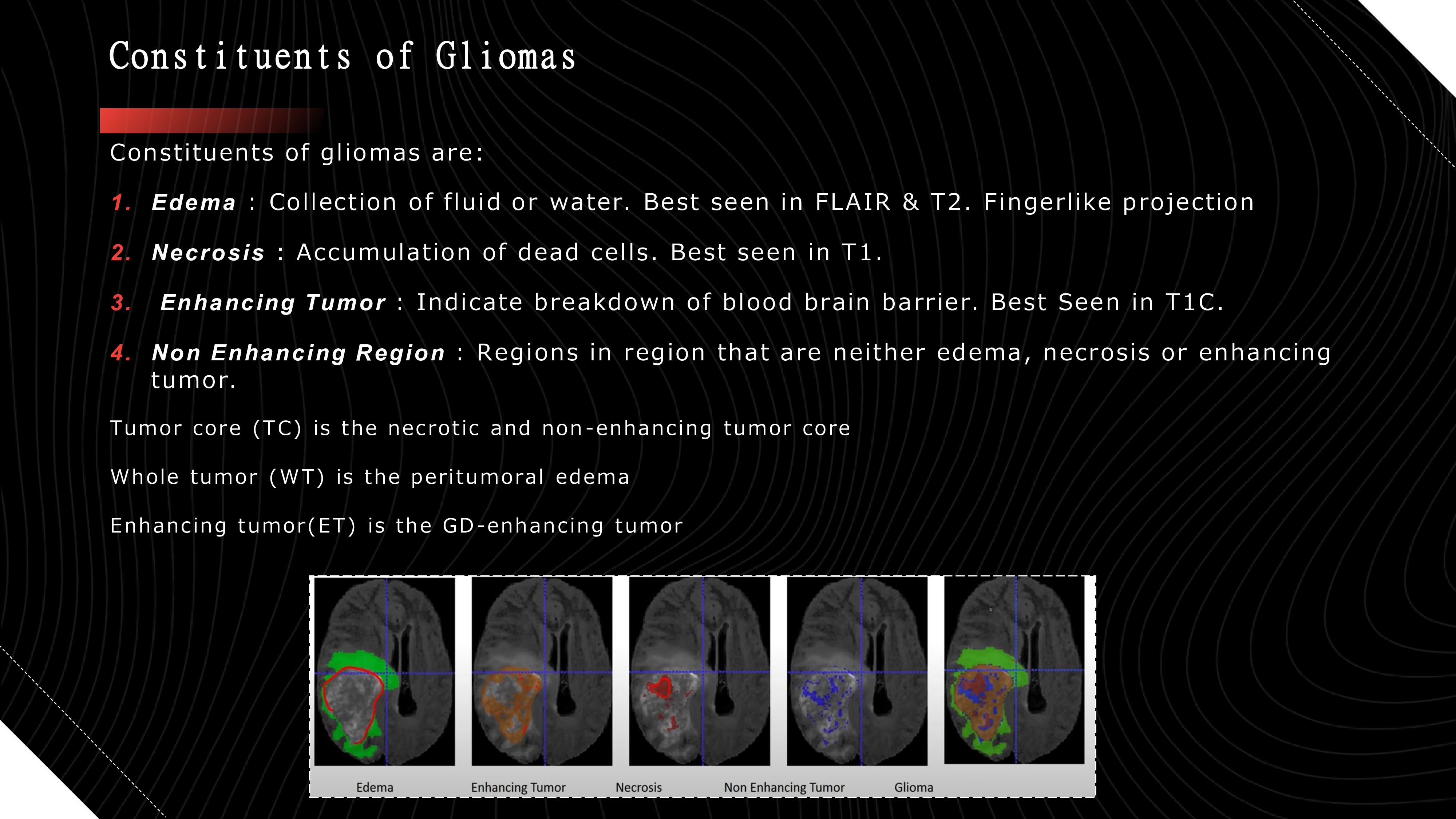
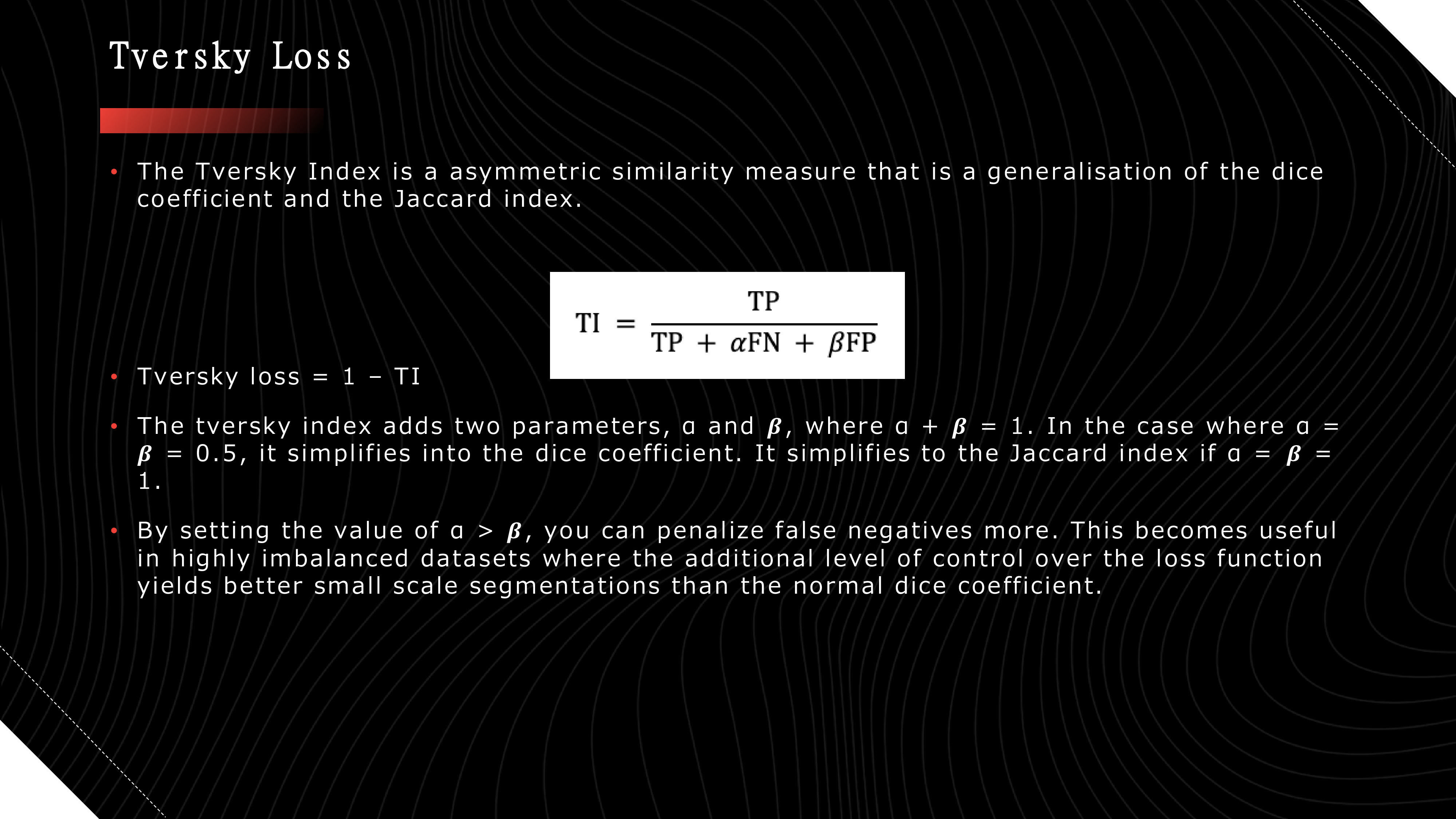
->Tumor core (TC) is the necrotic and non-enhancing tumor core
->Whole tumor (WT) is the peritumoral edema
->Enhancing tumor(ET) is the GD-enhancing tumor
This automated segmentation aids in identifying and delineating tumor regions, providing crucial information for diagnosis and treatment planning.
How we built it
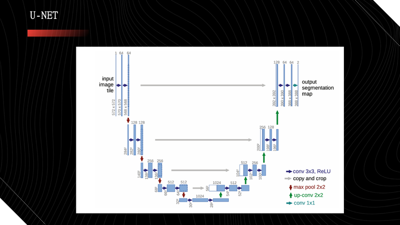
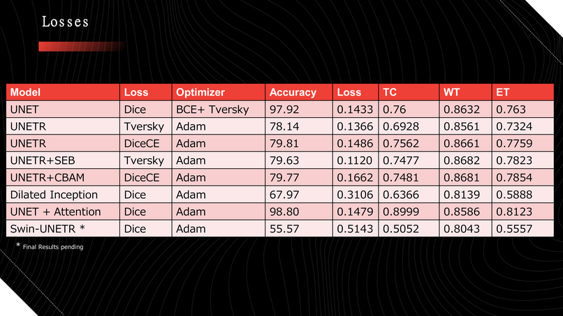
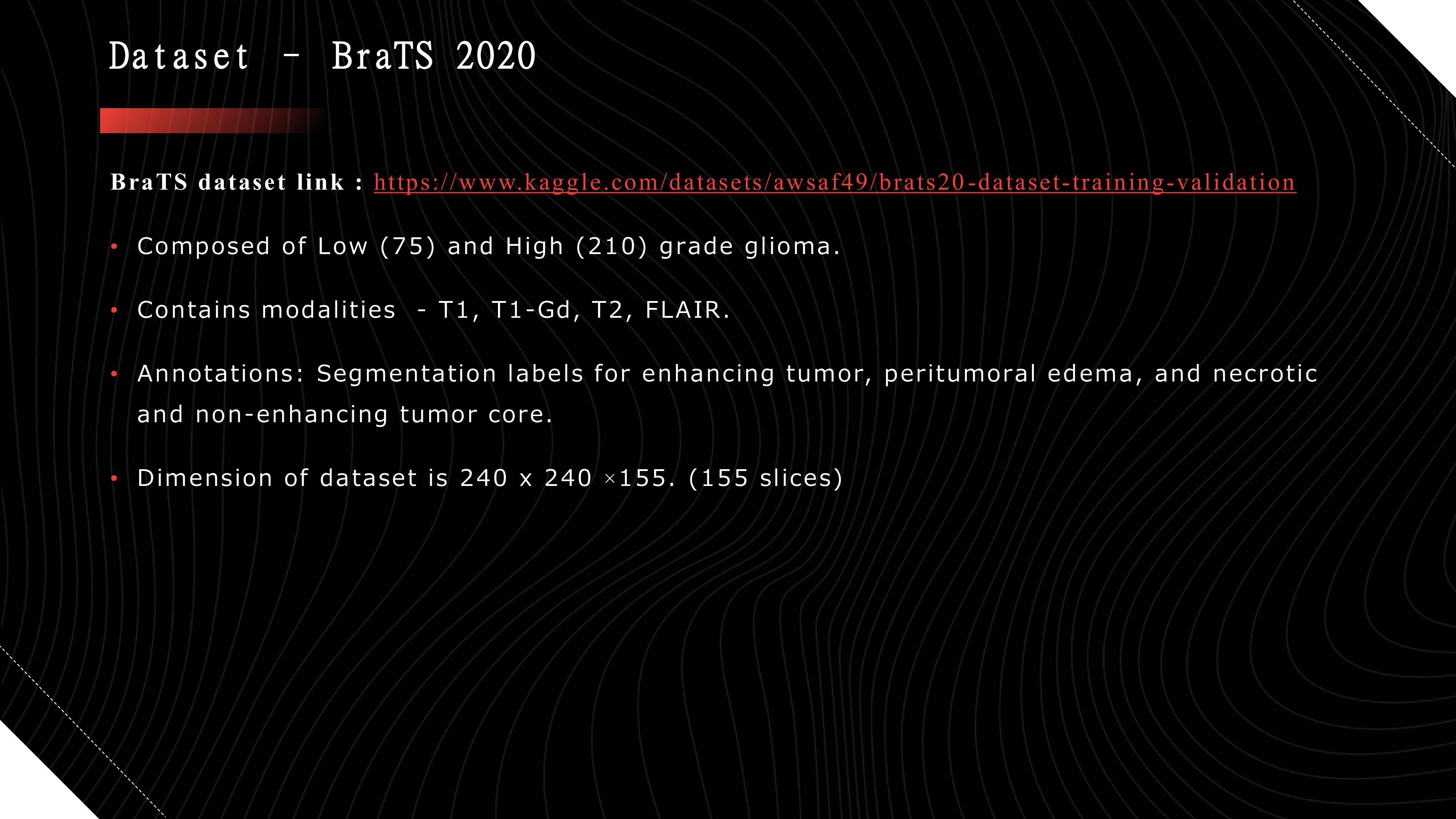
We started with basic fundamental of any computer vision project , finding right dataset it took almost a day to find a right dataset with enough diversity of low and high grade glioma cells Then we converted our data to numbers since on this we can perform any sort of deep learning. We started with a very basic architecture used in segmentation task knows as UNET with results from here we then researched into more potential architecture to increase our results.
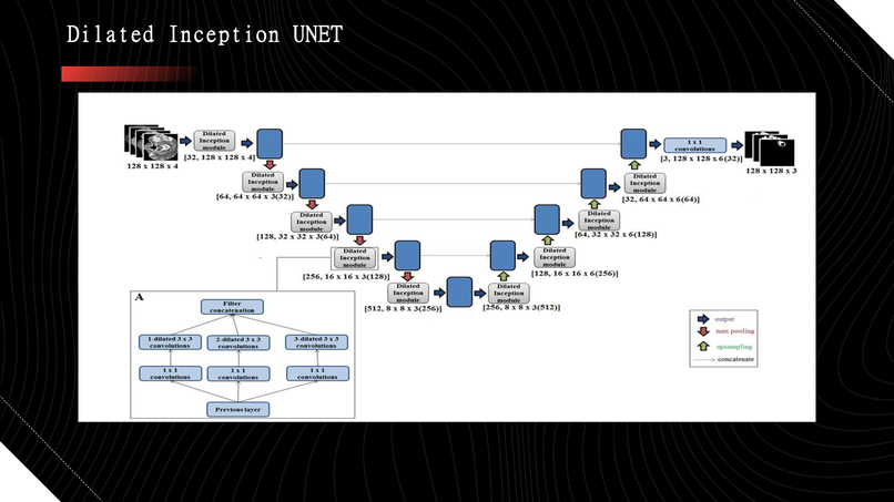
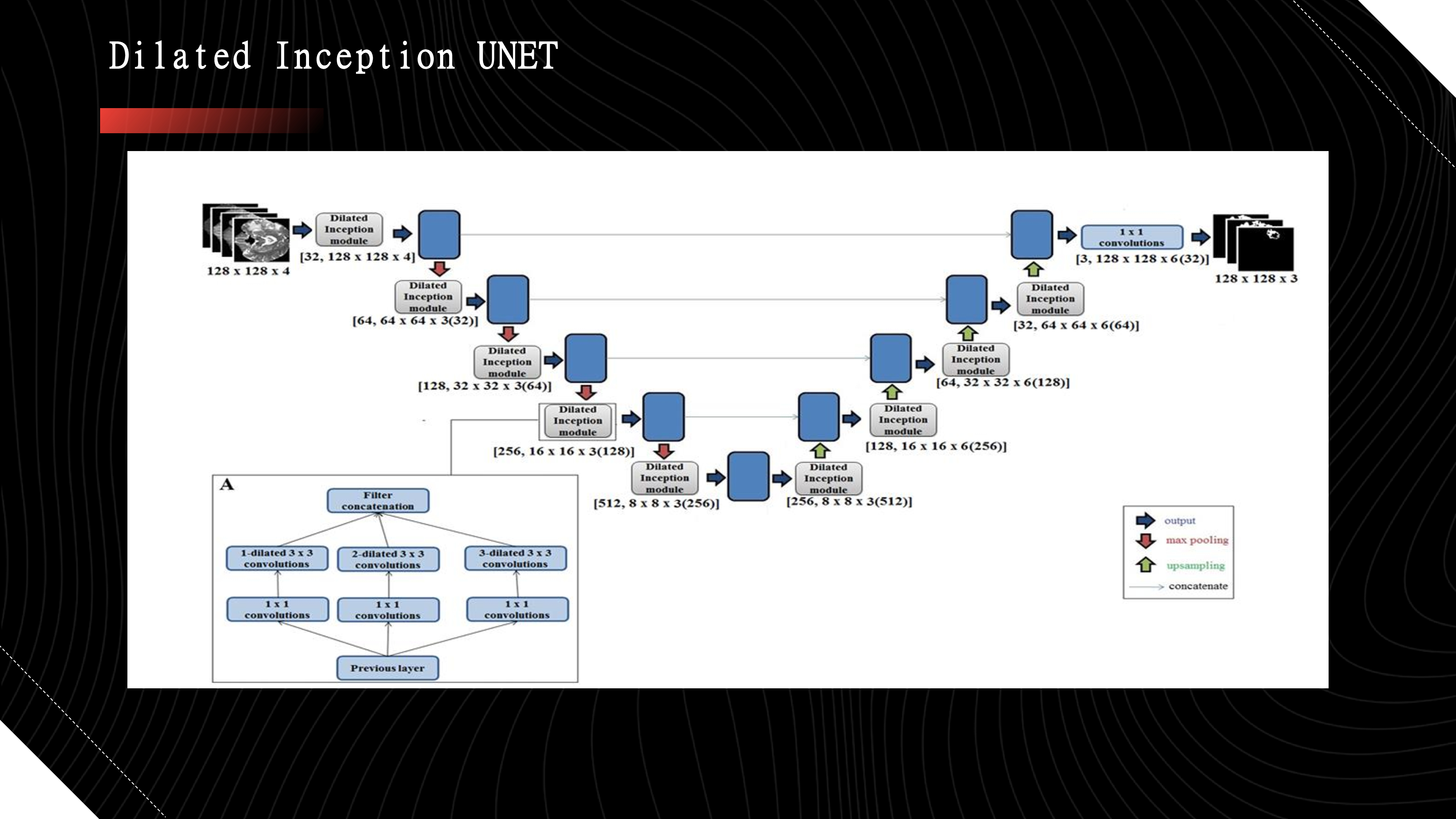
-> Later on we tried incorporating Dilated convolution in inception v2 blocks and arrange them as UNET
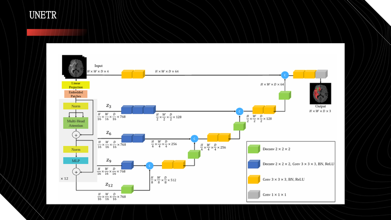
-> UNETR (UNET w Transformer)
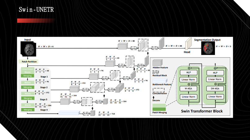
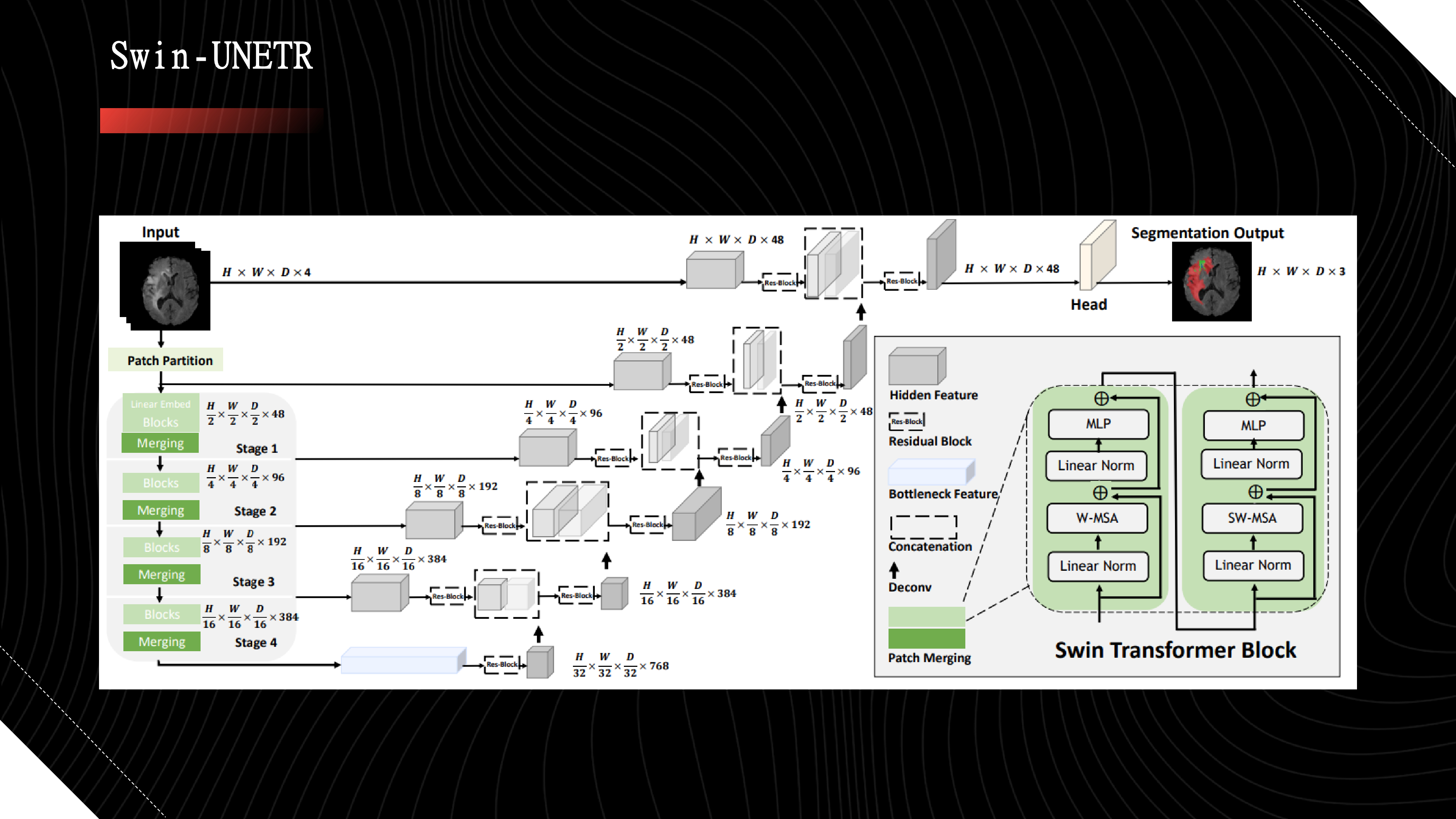
-> Swin-UNET(UNET w Swin Transformer) [this is better for high res images but final results are still pending as we needed to train on a huge amount of data for this model (over 2k)
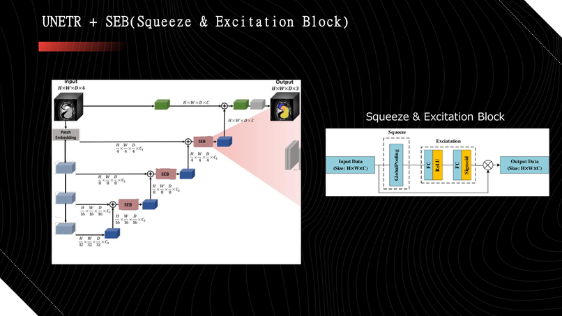
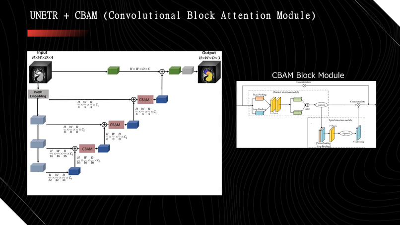
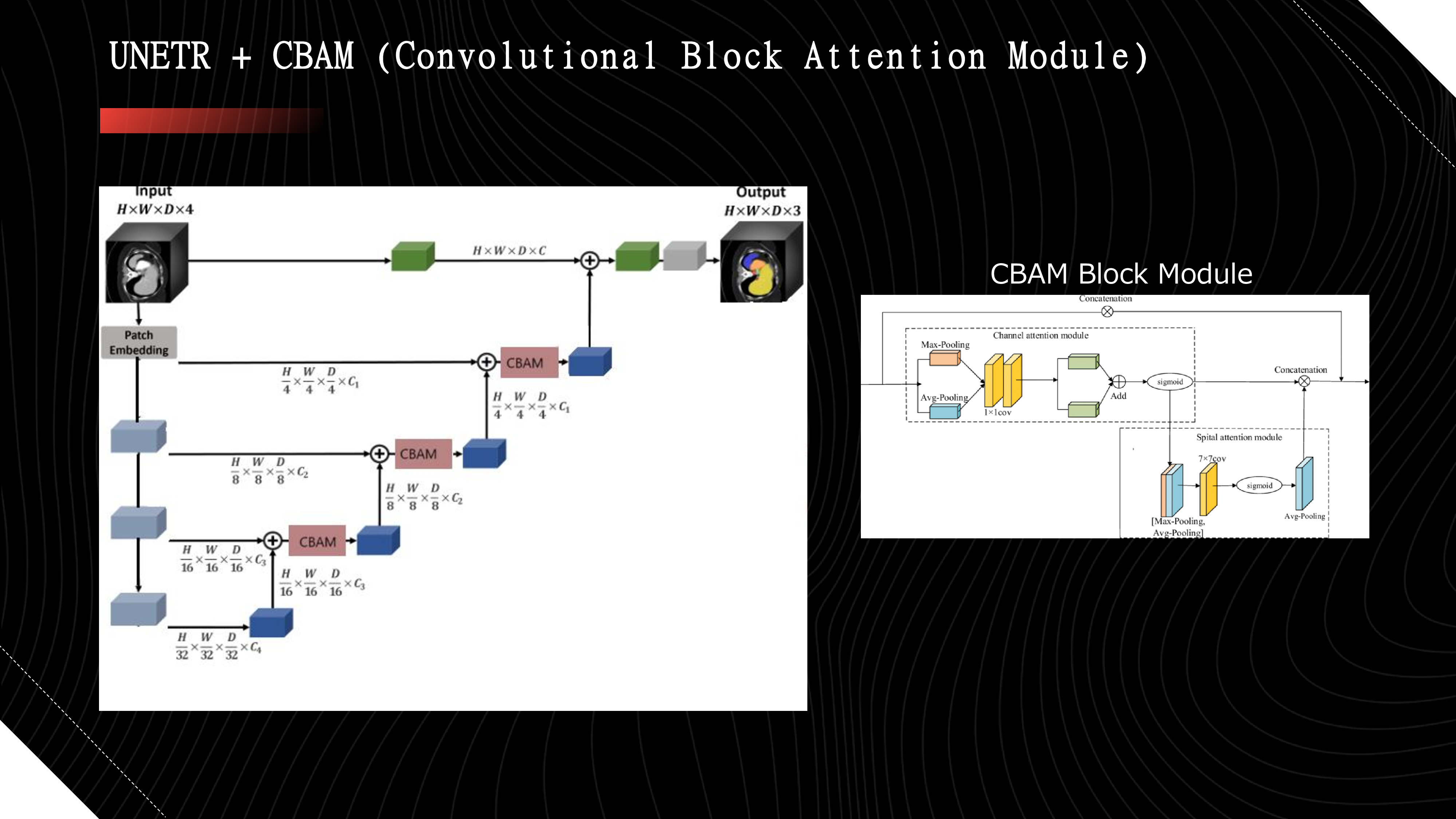
->We innovatively integrated Squeeze & Excitation and CBAM blocks into UNETR, creating new architectures that have not been documented in any research paper. Although the initial results were not optimal,
I believe that with hyperparameter tuning, increased data, and sufficient computing resources, these models can be significantly improved.
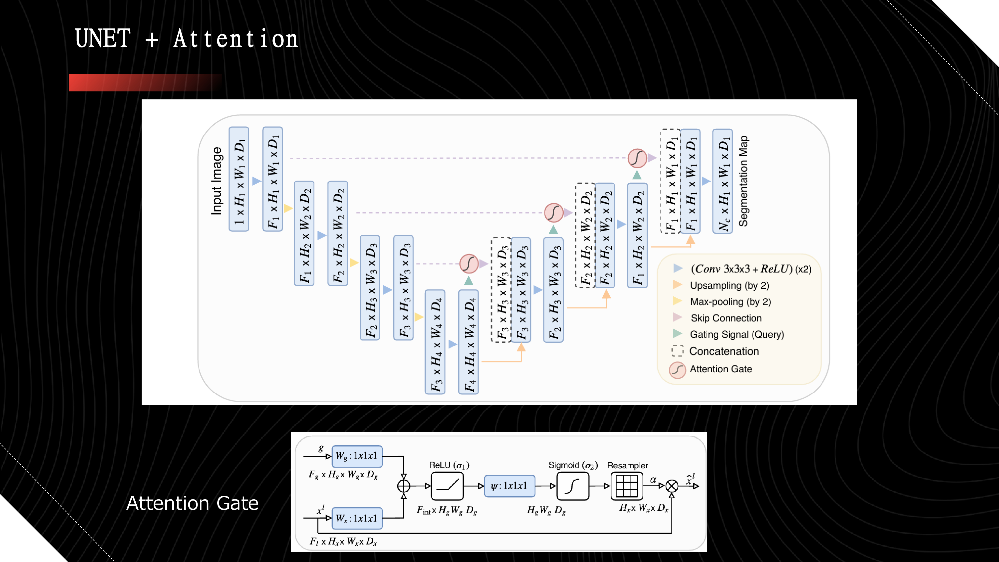
-> Finally, I utilized UNET with Attention, incorporating attention gates into the UNET architecture. This approach yielded the best results among all my models and currently stands as the most effective solution
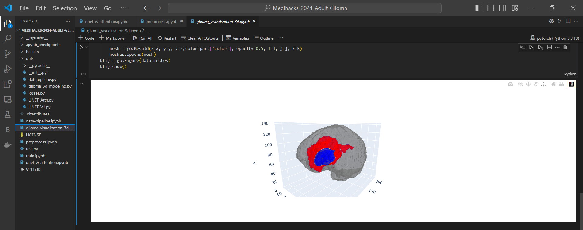
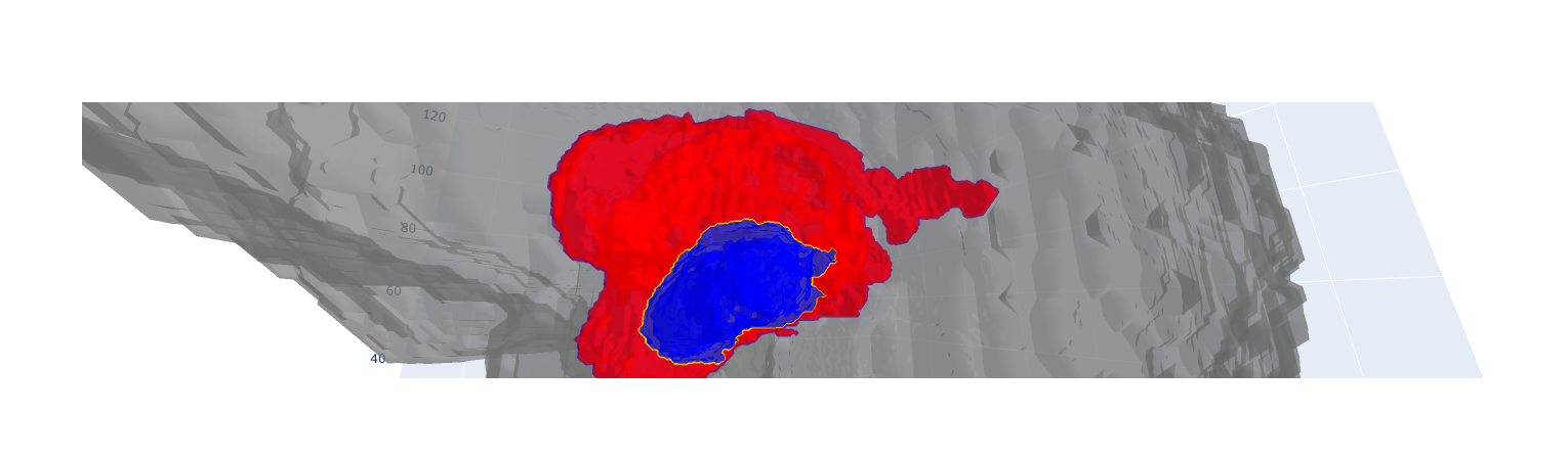
We also built a way to 3d visualize the tumor shape instead of traditional 2D slice image. This will help us examine the shape and location much better as the 3D model can be moved around, zoomed rotated and saved in any order.
Challenges we ran into
The biggest challenge I faced was insufficient computing power . This limitation resulted in a lot of idle time and restricted me from increasing the complexity of my architectures (such as increasing the number of kernels), which could have yielded better results.
To partially overcome this, I utilized multiple Kaggle accounts to speed up the training process, but this solution was also limited in scope.
Accomplishments that we're proud of
The completion of this project in such a short span is a significant accomplishment, but there's still more to be done.
Developing seven different architectures, including two entirely original ones, within this timeframe is something I'm particularly proud of.
This project has opened up a new research area for me, which I am excited to explore further and contribute to in the long term.
What we learned
I learned alot of different segmentation architectures because of the research papers i read during my time of building this project. I also learnt alot of random errors we could run into and how to solve them especially the library incompatibility error whenever designing new models. I also got to know more about uses of AI in medical field such as identifying schizophrenia using structural mri scans by using neural networks and many other interesting things.
What's next for Automatic Efficient Segmentation of Adult Glioma Tumor
Moving forward, my plan is to continue refining and experimenting with various architectures and creating some newer ones of my own until achieving higher accuracy scores.
Additionally, I aim to transition from focusing solely on the deep learning aspect to developing a complete software solution. While the library files and Jupyter notebooks are currently available on my GitHub, I intend to create a full-fledged desktop application. This will enable users, regardless of their AI/ML/CS expertise, to easily utilize the segmentation capabilities with pre-trained models and efficient workflows. This step will make the technology more accessible and impactful in clinical settings.
We also plan on adding the 3D visualizer of segmented results into an interactive frontend screen instead of it being displayed on browser or within the notebook The implementation of 3D visualizer would help significantly in the identifying the detailed shape and location of each 3 individual part.


Log in or sign up for Devpost to join the conversation.