Inspiration
Brain tumors are one of the most life-threatening diseases, and early diagnosis plays a crucial role in saving lives. Manual detection from MRI scans is time-consuming and prone to human error. This inspired us to develop an automated, AI-driven system that can assist radiologists in identifying tumors more efficiently and accurately.
What It Does
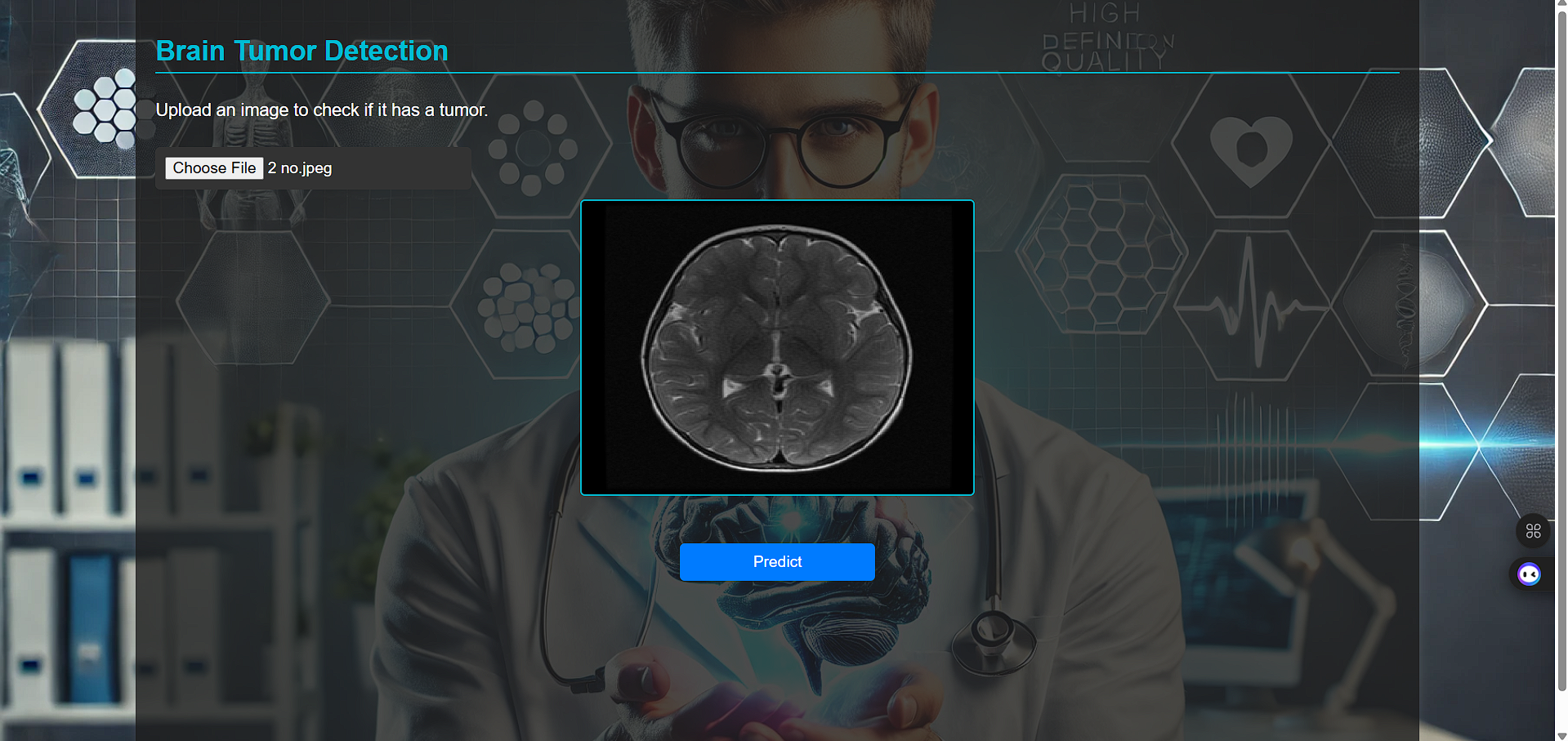
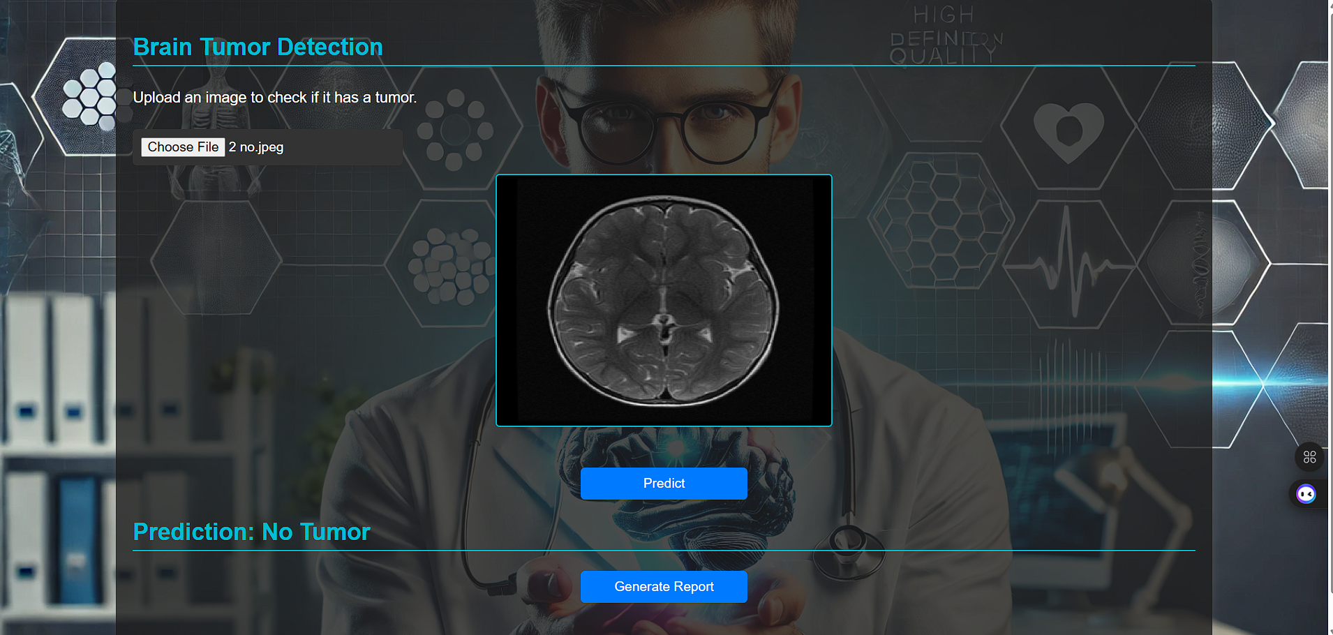
Our project uses deep learning techniques to analyze brain MRI images and detect the presence of tumors. The system classifies MRI scans as “tumor” or “no tumor” and highlights affected regions for better interpretability.
How We Built It
We used Python along with TensorFlow and Keras to build a convolutional neural network (CNN) model. The dataset was preprocessed using OpenCV and NumPy for resizing, normalization, and augmentation. Model training and evaluation were done in Google Colab, and visualizations were created using Matplotlib and Seaborn.
Challenges We Ran Into
- Limited, unbalanced datasets required careful augmentation and normalization.
- Training deep models on high-resolution MRI data was computationally intensive.
- Achieving high accuracy without overfitting needed extensive tuning of hyperparameters.
Accomplishments That We're Proud Of
- Achieved over 95% accuracy on the test dataset.
- Successfully integrated preprocessing, model training, and prediction pipelines.
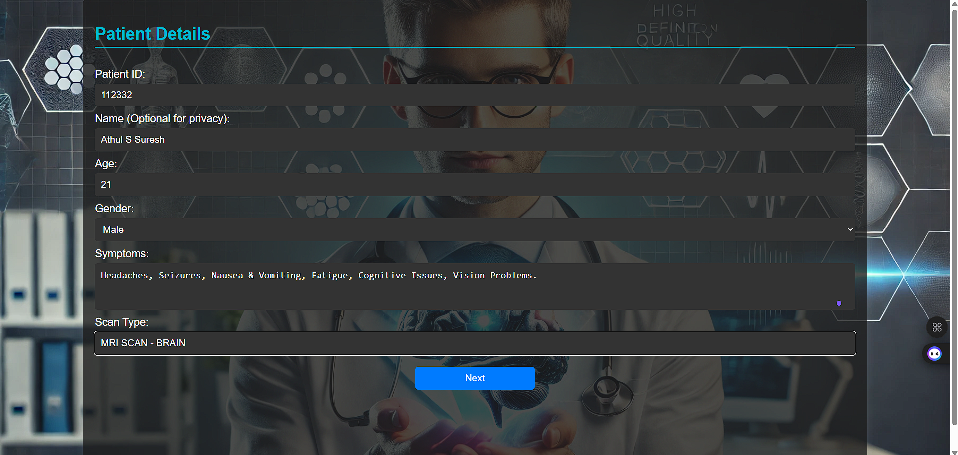
- Built a user-friendly interface for uploading and classifying MRI images.
What We Learned
We learned how to apply CNN architectures for medical imaging, optimize model performance, and handle real-world data imperfections. We also gained experience in teamwork, debugging, and working under time constraints.
What's Next
We plan to extend the model for multi-class tumor classification (meningioma, glioma, pituitary) and integrate it with a web-based medical dashboard for clinical use.
Built With
- keras
- matplotlib
- numpy
- opencv
- pandas
- python
- scikit-learn
- tensorflow

Log in or sign up for Devpost to join the conversation.